A Unique Branching Pattern of the Axillary Artery: A Case Report
Ishwar B. Bagoji1, Gavishiddappa A. Hadimani2, Balappa M. Bannur3, B.G. Patil4, Ambadasu Bharatha5
1 Lecturer, Department of Anatomy, Shri BM Patil Medical College, BLDE UniversityBijapur, Karnataka, India.
2 Lecturer, Department of Anatomy, Shri BM Patil Medical College, BLDE UniversityBijapur, Karnataka, India.
3 Professor, Department of Anatomy, Shri BM Patil Medical College, BLDE UniversityBijapur, Karnataka, India.
4 Professor, Department of Anatomy, Shri BM Patil Medical College, BLDE UniversityBijapur, Karnataka, India.
5 Lecturer, Department of Pharmacology, Shri BM Patil Medical College, BLDE UniversityBijapur, Karnataka, India.
NAME, ADDRESS, E-MAIL ID OF THE CORRESPONDING AUTHOR: Dr. Ambadasu Bharatha, Lecturer, Department of Pharmacology, Shri BM Patil Medical College, BLDE University Bijapur, Karnataka, India.
Phone: 9243342599,
E-mail: ambu2mail@gmail.com
During routine dissection classes for under graduate students, we found a unique and unusual case regarding the anomalous branching in the third part of the axillary artery was terminated into subscapular arterial trunk, superficial brachial artery and deep brachial artery. The subscapular arterial trunk was origin of several important arteries as the circumflex scapular, thoracodorsal, posterior circumflex humeral, thoraco-acromial and lateral thoracic arteries. The deep brachial artery in the arm gave anterior circumflex humeral artery at the surgical neck of humerus, which terminated in the cubital fossa by dividing into radial and ulnar arteries. The superficial brachial artery gave two profunda brachii arteries, both of which passed through spiral groove, along with radial nerve and three muscular branches, to brachialis muscle. This variation is very rare. As per our knowledge, we did not find any literature which explained variations which were similar to this. The normal and abnormal anatomy of the axillary region has practical importance among vascular radiologists and surgeons and it should be known for making an accurate diagnostic interpretation.
Axillary artery, Brachial artery, Subscapular arterial trunk, Variation
Introduction
Axillary artery is the continuation of sub-clavian artery, from the outer border of the first rib and it continues as the brachial artery at the inferior border of teres major. Pectoralis minor muscle divides the artery into three parts, as the first part (proximal), second part (posterior) and third part (distal) to the muscle. As has been classically described in anatomical texts, axillary artery gives six branches, first part giving rise to superior thoracic artery, second part giving rise to thoracoacromial and lateral thoracic arteries and the third part giving rise to sub-scapular, anterior circumflex humeral and posterior circumflex humeral arteries. The brachial artery is the continuation of the axillary artery at the distal (inferior) border of the tendon of teres major and it ends about a centimetre distal to the elbow joint, at the level of the neck of the radius, by dividing into radial and ulnar arteries [1]. Various authors have described the variations in the branching patterns of the axillary and brachial arteries, but this finding was different. The variant observed was not only rare but it seemed to be relevant and significant for understanding the formation of the arteries of the arm.
Case Report
During routine dissection classes conducted for 1st MBBS students in the Department of Anatomy of Shri B M Patil Medical College, Hospital and Research Centre, BLDE University Bijapur, India. We encountered a rare variation in right upper limb of a male cadaver.
The third part of the axillary artery terminated by trifurcating into three large branches (a) subscapular arterial trunk (b) superficial brachial and (c) deep brachial artery.
Common Subscapular arterial trunk: A large collateral branch arose from the axillary artery, at the lateral border of the pectoralis minor muscle. This collateral branch is called as common subscapular trunk. This arterial trunk gave many branches, which otherwise arise from the second and third parts of the axillary artery. They are thoracodorsal, lateral thoracic, circumflex scapular, posterior circumflex and additional thoracic arteries. The common subscapular arterial trunk was crossed superficially by thoracodorsal nerve. The posterior circumflex humeral artery descended superficial to the axillary nerve and passed through the quadrangular space. The circumflex scapular artery passed dorsally through upper triangular space. The thoracodorsal artery accompanied the thoracodorsal nerve, along the distal border of the sub-scapularis muscle. Thoracodorsal artery provided two additional branches to the thoracic wall [Table/Fig-1].
Showing branching pattern of the axillary artery, 1 Axillary artery, 2. Acromio-thoraciac artery, 3. Lateral thoracic artery, 4.Subscapular common trunk, 5.Superficial brachial artery, 6.Deep brachial artery, 7.Additional subscapular branch, 8 Thoraco dorsal artery, 9.Circumplex scapular artery, 10. Posterior circumflex humeral artery, 11a. & b. additional thoracic artery, 12. Anterior circumflex humeral artery 13. Muscular & profunda brachial branches, 14. Anterior ulnar recurrent artery, 15. Ulnar artery, 16.Radial artery
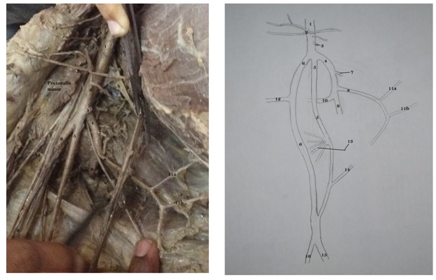
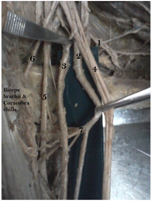
Superficial Brachial artery: Superficial brachial artery extended vertically downwards on the floor of sub-scapularis muscle in the axilla. In the brachium, artery ran between the biceps brachii and brachialis muscle. In the middle of the brachium, superficial brachial artery provided five branches; three were muscular branches supplied the muscles of the brachium, and two branches continued along the radial nerve as the profundabrachii artery, in the spiral groove [Table/Fig-2]. Superficial brachial artery coursed up to the cubital fossa and provided the anterior ulnar recurrent branch which pierced the medial intermuscular septum and anastomosis with the arterial anastomosis, around the elbow joint [Table/Fig-3]. Finally, the superficial brachial artery fused with the deep brachial artery in the cubital fossa.
Shows 1. Subscapular arterial trunk, 2. Superficial brachial artery, 3. Deep brachial artery, 4. Median nerve. 5. Radial nerve, 6. Musculocutaneus nerve. 7. Muscular & profunda brachial arteries
Shows 1. Deep brachial artery, 2. Superficial brachial artery, 3. Anterior ulnar collateral artery, 4. Ulnar artery, 5. Radial artery, 6. Bicipital aponeurosis. 7. Median nerve
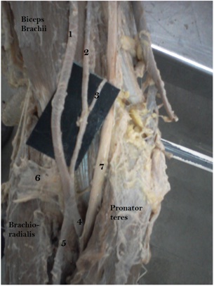
Deep Brachial artery: Deep brachial artery extended lateral to the superficial brachial artery, from the third part of the axillary artery. Immediately after its 1.8 cm course, the artery provided anterior circumflex humeral artery, deep to the median nerve and musculocutaneus nerve. Then, deep brachial artery ran downwards and forwards in the brachium, between the biceps brachii and brachialis muscle. In the cubital fossa, the deep brachial artery terminated into radial and ulnar arteries respectively. Above the cubital fossa, the superficial brachial artery joined the deep brachial artery. So, the arterial pattern in the cubital fossa was in the form of [Table/Fig-3].
Discussion
Variations of the arterial patterns in the human upper limb have been the subject of many anatomical studies. It is not uncommon to find these variations in the branching pattern of axillary artery. Review of literature showed many variations in axillary artery [2–4]. In present case, we encountered a variation in the axillary artery, showing collateral branch and double brachial artery together. During embryogenesis, the lateral branch of the seventh cervical intersegmental artery becomes enlarged to form the axial artery of the upper limb, which on further development, becomes axillary, brachial, radial and ulnar arteries [5]. The arterial anomalies in the upper limb are caused by defects in the embryonic development of the vascular plexus of the upper limb bud. This may be due to arrest at any stage of development which shows regression, retention or reappearance and it may lead to variations in the arterial origins and courses of the major upper limb vessels [6]. Vasudha Saralaya et al., proposed that the large collateral branches from the axillary artery formed common sub scapular trunk [7]. Samuel et al., reported that the third part of the axillary artery gave a common arterial trunk which further branched into anterior and posterior circumflex humeral, sub scapular, radial collateral, middle collateral and superior ulnar collateral arteries, with absence of profunda brachii artery [2]. Our present report differs from this earlier report in branching pattern as well as course of these branches.
Adachi named the superficial branch as a ssuperficial brachial artery; it is so called because it runs superficial to median nerve, whereas usually the brachial artery runs deep into median nerve [8]. Keen subdivided superficial brachial artery (found in 12.3% dissections) into 3 types: (a) those superficial brachial arteries which continued in cubital fossa and bifurcated as usual into radial and ulnar arteries (3.6%). (b) Superficial brachial artery which continues as radial artery and is known as ‘high origin of radial artery’ (5.9%). (c) Superficial brachial artery which continues as ulnar artery and is known as ‘high origin of ulnar artery’ (2.8%) [9]. In the present case, superficial brachial artery again joined the deep brachial artery in cubital fossa, before branching into radial and ulnar arteries.
Conclusion
The knowledge on these variations is necessary for the surgeons, considering the frequency of procedures performed in this region. Variation of large common trunk, as a branch of axillary artery, is worth considering during antegrade cerebral perfusions carried out in aortic surgeries, which creates bypass between axillary artery and sub-clavian artery in cases of sub-clavian occlusions, while treating aneurysms of axillary artery, and during reconstructions of axillary artery after trauma or catheterization or cannulation of axillary artery for various purposes. The existence of such a superficial and deep brachial artery at upper extremity is also clinically important. It may complicate intravenous drug administration and venipuncture in general, also percutaneous brachial catheterization. It may be mistaken for a vein. Its superficial course makes it more prone to injury, which may result in bleeding. Furthermore, the presence of superficial brachial artery can cause misinterpretations of incomplete angiographic images.
[1]. Johnson D, Ellis H, Collins P, Upper Arm. In: Standring S, Ellis H, Healy JC, Johnson D, Williams A, Collins P, edsGray’s Anatomy 2005 39th EdPhiladelphiaElsevier Churchill Livingstone:856 [Google Scholar]
[2]. Samuel VP, Vollala VR, Nayak S, Rao M, Bolla SR, Pammidi N, A rare variation in the branching pattern of the axillary arteryIndian J. Plast. Surg 2006 39:222-3. [Google Scholar]
[3]. Venieratos D, Lolis ED, Abnormal ramification of the axillary artery: sub-scapular common trunkMorphologie 2001 85(270):23-4. [Google Scholar]
[4]. De Garis CF, Swartley WB, The axillary artery in white and negro stocksAm. J. Anat 1928 41:353-97. [Google Scholar]
[5]. Yang HJ, Gil YC, Jung WS, Lee HY, Variations of the superficial brachial artery in Korean cadaversJ Korean Med Sci 2008 23:884-7. [Google Scholar]
[6]. Patnaik VVG, Kalsey G, Singla RK, Branching pattern of axillary artery – a morphological studyJ Anat Soc India 2000 49:127-32. [Google Scholar]
[7]. Saralaya V, Joy T, Madhyastha S, Vadgaonkar R, Saralaya S, Abnormal branching of the axillary artery: sub-scapular common trunk. A case reportInt J Morphol 2008 26:963-6. [Google Scholar]
[8]. Adachi B, Das Arteriensystem des japanerKyoto 1928 1:205-10. [Google Scholar]
[9]. Keen J.A, A study of arterial variations in the limbs with special reference to symmetry of vascular patternAmerican Journal of Anatomy 1961 108:245-61. [Google Scholar]