Head and Neck Squamous Cell Carcinoma (HNSCC) is one of the most common cancers that affect Indians. It accounts for 90% of the cancers arising in the upper aero-digestive tract, making it the most common type of cancer and cause of cancer deaths among patients with head and neck cancer (HNC) [1]. The five-year survival shows wide variations from 20-90%, depending upon the site of origin and extent of disease [2]. The disproportionately higher prevalence of HNC in relation to those of other malignancies in India may be due to the use of tobacco in various forms, consumption of alcohol and low socio-economic conditions, related to poor hygiene, poor diet or infections of viral origin. Only one-third of the patients present with early-stage diseases, which are amenable to cure with surgery or radiotherapy. Two-thirds of these patients have locally advanced diseases at the first presentation itself [1]. Although, histopathology is the gold standard investigation for HNC, an easy to perform, non-invasive and cost-effective technique enabling early detection, is more likely to have significant impact on patient care. Among various cancer markers, glycoconjugates are of great importance, because many of these altered glycoproteins are expressed by cancer cells and they are associated with tumour progression and metastasis [3,4]. Elevation of glycoproteins above the normal levels reflects processes of tissue destruction at the site and release of preformed glycoproteins from the tissue or it may be caused by local synthesis and liberation of glycoproteins by tumour cells. Elevated serum glycoprotein L-fucose has been reported in breast cancer, ovarian cancer, colorectal adenocarcinoma, leukaemia, brain tumours, as well as, in non-neoplastic conditions like cirrhosis of liver and meningitis, rickets, osteomalacia, tuberculosis, cardiovascular disorders [5–10]. However, it has not been evaluated effectively in HNC. Therefore, this study was done to determine the significance of serum L-fucose glycoprotein levels in head and neck malignancies without distant metastases.
Material and Methods
A comparative study was carried out at a tertiary care hospital in South India, in the Department of Otorhinolaryngology, from October 2009 to July 2011. Institutional ethical committee’s clearance was obtained. Informed consents were obtained from all individuals who participated in the study. Fifty adults aged above 18 years, with histopathologically confirmed head and neck malignancies without distant metastases, were compared with 50 age- and sex- matched healthy controls for serum L-fucose glycoprotein levels. It was a single blinded study. The pathologists were unaware of the serum L-fucose levels of HNC patients and laboratory personnel involved were unaware of the identifications of cases and controls. Cases were diagnosed clinically and they were confirmed by doing histopathological investigations. AJCC’s (American Joint Committee on Cancer 2002) staging system was followed for staging of HNC viz. lip and oral cavity cancers, oropharyngeal cancers, hypopharyngeal cancers and laryngeal cancers. The treatment was administered depending on the stage of the tumour. Distant metastases to bone, lung and liver were excluded using plain chest X-rays, abdominal ultrasound. Contrast enhanced CT scan was taken in those patients who showed suspicious lesions on chest X-ray or abdominal ultrasound. As per the exclusion criteria, patients who are known or found to have distant metastases, recurrent head and neck malignancies, associated malignancies other than those which occurred in head and neck region, cardiovascular disease, diabetes mellitus, rheumatoid arthritis, renal diseases, hepatic diseases, cystic fibrosis, other chronic inflammatory conditions and were aged less than 18 years old were excluded from the study group. Similarly, individuals with habits of tobacco chewing, smoking, alcohol consumption, betel nut chewing and were aged less than 18 years were excluded from the control group. Five ml of venous blood was collected, centrifuged and serum was separated and stored at 4OC for analysis. Levels of serum L-fucose were estimated in spectrophotometer (Spectronic 20, Thermo Fisher Scientific, USA) as per Winzler’s method [11].
All clinical and laboratory data were entered in Microsoft Excel 2007 spreadsheet and statistical analysis was done by using SPSS statistical software, version 17.0. Student’s t-test was used to compare L-fucose glycoprotein levels in study and control groups. All p-values which were < 0.05 were considered to be statistically significant.
Results
This study was conducted on 50 patients with clinically and histopathologically confirmed head and neck malignancies and on 50 age- and sex-matched healthy controls. Both, the study group and control group, comprised of 40 males and 10 females. Out of 50 participants in each group, a majority (36%) belonged to 41-50 year age-group, followed by 61-70 year age-group (34%), 51-60 year age-group (24%) and 31-40 year age-group (6%). Mean ages of cases and controls were 55.92 ±10.17 years and 53.32±10.05 years respectively.
Among 50 cases of HNC, oral cavity cancers were most common (34%), whereas malignancies involving larynx, hypopharynx and oropharynx accounted for 28%, 26% and 12% cases, respectively. Stages III and IV were considered as late stages or advanced stages and they represented 44% and 38% cases, respectively [Table/Fig-1].
Tumour Stage among patients with head and neck cancer
| Tumor stage | Number of patients (n=50) | % |
|---|
| Stage I | 5 | 10.0 |
| Stage II | 4 | 8.0 |
| Stage III | 22 | 44.0 |
| Stage IV | 19 | 38.0 |
The mean value of serum glycoprotein L-fucose in cases was 11.33±7.39 mg%, while in healthy controls, it was 4.74±1.55 mg%. The mean values of serum glycoprotein L-fucose in early and late stages of disease and in HNCs involving different sites were higher than those in the control group [Table/Fig-2].
Comparison of serum glycoprotein L-fucose levels in cases and controls according to tumour stage and site
| Cases (mg%) | Controls (mg%) | Two tailed p-value |
|---|
| Early stage | 11.53±10.16 | 4.74±1.55 | 0.0799 |
| Late stage | 11.28±6.79 | 0.00000028 |
| Oral cavity | 11.33±8.28 | 0.0046 |
| Oropharynx | 12.20±4.79 | 0.0124 |
| Hypopharynx | 8.80±4.59 | 0.00778 |
| Larynx | 13.32±9.06 | 0.0036 |
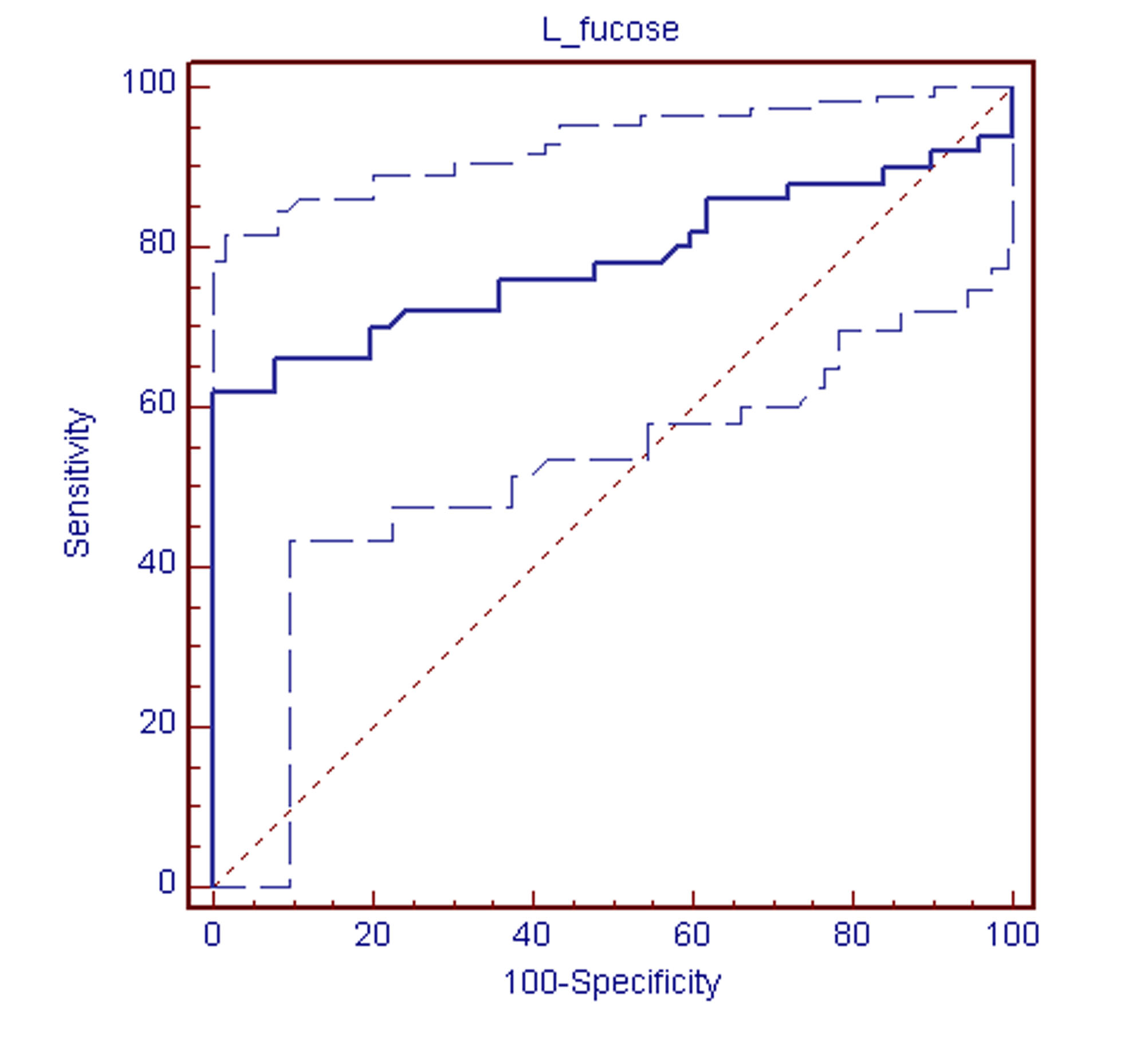
Receiver Operating Characteristic (ROC) curve analysis [12] was performed to find the significance in association of L-fucose, to assess the diagnostic performance for malignancy [Table/Fig-3]. It showed a cut-off of >7.90, sensitivity of 62%, specificity of 100%, positive predictive value (PPV) of 100, negative predictive value (NPV) of 72.45, accuracy of 81.08 and Area Under Receiver Operating Characteristic (AUROC) of 0.784.
ROC curve analysis for glycoprotein L-fucose to evaluate for diagnostic performance ROC curve analysis shows cut-off >7.90, sensitivity 62%, specificity 100%, positive predictive value (PPV) 100, negative predictive value (NPV) 72.45, accuracy 81.08 and Area Under Receiver Operating Characteristic (AUROC) 0.784
Discussion
Head and neck malignancy is a major form of cancer in India, accounting for a major proportion of all cancers in males and a significant number of cancers in females. HNCs make up 30% of all cancers in males and 11% cancers in females [1]. World’s highest reported incidence of HNCs in women is from India [2]. Generally, several authors have documented a male predisposition, with a common male to female ratio of 3:1 in head and neck malignancies.[13,14]. However, we found a higher sex ratio among males and 80% of HNC cases were men, with a male to female ratio of 5:1. Most of them belonged to 41-50 year age-group (36%), followed by 61-70 year age-group (34%). This finding was consistent with that of a study from Gujarat done by Shah et al., who reported that out of 130 patients of oral carcinoma, 94% were men, with a male to female ratio of 15.7:1 [15]. These high incidences of HNCs in males are mostly attributed to smoking in urban population and tobacco chewing and snuffing in rural India. Tobacco and alcohol consumption play a crucial role in the pathogenesis, which is widespread in a country like India. A major proportion of oral and laryngeal malignancies occur among smokers and tobacco chewers. Smoking (60%), alcohol consumption (56%) were the most common habits found in the current study, along with betel nut chewing (54%) and tobacco chewing (48%). Half of the study group (50%) had habits of both alcohol consumption and smoking. Similar risk factors in head and neck malignancies were reported in various other studies [16–18].
A majority of these neoplasms are preventable and curable if they are detected early. Especially, pre-cancerous conditions like submucus fibrosis and leukoplakia, which usually precede oral cancer and are amenable to treatment [19]. Several cancer biomarkers have been studied extensively to provide non-invasive mode of diagnosis, facilitating early detection of neoplastic or pre-neoplastic conditions. Among them, glycoconjugates are of great importance. As per recommendations of the American Physiological Society and The American Society of Biochemists, glycoproteins are compounds of a protein molecule, with a substance or substances containing a carbohydrate group other than nucleic acid [7]. Glycoproteins constitute vast diversity of molecules expressed on cell membranes in both normal and neoplastic cells [19]. Alterations in cell surface glycoproteins are associated with neoplastic transformation [6,20]. Among all serum glycoproteins, L-fucose is of prime importance in cancer research. It is a six-carbon monosaccharide present frequently as a component of various mammalian glycans and glycolipids. The L-configuration and lack of a hydroxyl group on the 6th carbon (C-6) are two unique structural features which distinguish fucose from other hexose sugars. In physiological conditions, it is present in low concentrations in serum but it is increased in cancer and other diseases. Elevation of L-fucose in serum and body fluids may be attributed to release of preformed glycoproteins from the tissue as a result of tissue destruction, or it may be caused by local synthesis and liberation of glycoproteins by tumour cells [21]. However, several workers support the view that an increase in their serum levels reflects tissue proliferation rather than tissue destruction [22]. Fucose, frequently exists as a terminal modification of surface glycoproteins. Fucosyl transferase is responsible for fucosylation of terminal moieties of glycoproteins. Thirteen fucosyl transferase genes have been identified in the human genome [23]. It was found to have an essential role in cancer biology. Increased fucosyl transferase and fucosylated serum glycoproteins have been implicated in surface modulation, decreased adhesion and uninhibited tumour growth [6,19]. Elevated fucosylated serum glycoproteins have been reported in neoplastic, as well as, non-neoplastic conditions [5]. However, there are inadequate published studies which have evaluated serum glycoprotein L-fucose as a potential diagnostic and prognostic marker in HNCs.
Mogra et al., analyzed serum protein bound fucose levels of 25 healthy subjects, 10 patients with benign neoplastic lesions and 30 patients having malignant lesions in head and neck region [24]. They found that the levels were significantly elevated in the malignant group, as compared to those in other two groups. Bathi et al., carried out biochemical analysis of serum protein bound hexose, serum fucose, serum sialic acid levels of 40 patients with HNCs and of 10 healthy individuals comprising the control group [10]. All the head and neck cancer patients showed elevated levels of the serum glycoproteins as compared to those in the control group. It was further noted that the increased levels of the serum glycoproteins correlated well with the clinical staging of the malignancies. In this current study, the mean value of serum glycoprotein L-fucose in HNC patients was 11.33±7.39 mg%. As compared to the mean value of 4.74±1.55 mg% in healthy controls, it was statistically significant. In general, the mean value of normal serum was 6.84±0.13 mg% and any value exceeding 9 mg% was considered to be usually pathological [5]. The aetiology behind such an abnormal rise was mostly a neoplastic process rather than a benign tumour and non- neoplastic conditions [5]. The mean values in the study and control group in this study were essentially similar to those in various other studies. However, Sawke et al., reported a substantial increase in serum fucose levels in different malignancies [7]. We also assessed glycoprotein L-fucose levels according to the site and stage of tumour [Table/Fig-2]. The mean values of early stage, late stage HNC, as well as, oral cavity, oropharynx, hypopharynx and larynx carcinoma were more than those of control group and p-values showed significant association in all except in early stage HNC (stages I and II). Although there are reports on decrease in serum fucose levels in old age, [5] we could not find any association of its normal serum levels with age, gender and dietary habits. The main limitation of this study was small sample size, which could have affected the findings or which may have resulted in a lack of statistical significance. Further investigations are essential to confirm these observations.
Conclusion
Estimation of serum glycoprotein L-fucose levels is a non-invasive, easy to perform and cost-effective technique. The findings of the current study suggest that serum glycoprotein L-fucose level is strongly correlated with stage and site of HNC and that it is not influenced by gender and dietary habits. We conclude that along with clinical diagnostic procedures, measuring serum glycoprotein L-fucose levels can be used as an effective biochemical indicator. It may also be useful in monitoring recurrences.