Breast carcinomas are one of the leading causes of cancer in women. Fine Needle Aspiration Cytology (FNAC) is one of the important components of ‘triple approach’, which has been widely accepted for the preoperative diagnosis of breast lesions [1]. It is a multi-disciplinary approach that includes analysis of clinical and radiological findings in conjunction with FNAC features, to diagnose the breast lesions and to determine the best management plan for the patient.
To correlate the cytological findings with histopathological examinations for breast lesions.
To determine the accuracy of fine needle aspiration cytology in the diagnosis of breast lesions.
Material and Methods
This Fine Needle Aspiration Cytology study was carried out at Smt. N.H.L. Municipal Medical College, Ahmedabad, on 222 patients with breast lesions; among which cyto-histopathological correlations were obtained in 91 cases.
FNAC was done by standard procedure. Palpable axillary lymph nodes were aspirated to exclude metastases. The slides were stained with Haematoxylin and Eosin (H and E) stain. Correlation with imaging studies, including mammography, was done. Statistical analysis of results was performed.
Results
The age of the patients in the present study varied from 16 to 80 years.
Out of 222 cases, female & male patients were 217 and 5 respectively [Table/Fig-1].
Age distribution of patients with breast lesions
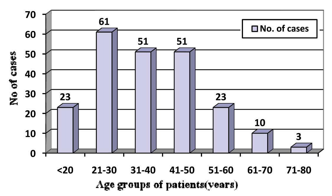
This study documented the fact that benign breast lesions were the most common lesions in young females, among which the Fibroadenoma was the commonest one. The malignant lesions were common in fourth and fifth decades of life, among which infiltrating ductal carcinoma was the most common lesion.
As has been shown in [Table/Fig-2], one case which was cytologically diagnosed as a benign cystic lesion was diagnosed as a malignant phyllodes tumour by doing a histopathological examination (false negative result). Rest of all cases showed good correlations between FNAC and histopathology.
Cytological diagnosis of breast lesions by FNAC (n=222)
| Category | Cytological Diagnosis | No. of Cases | Percentage (%) |
|---|
| Inflammatory Lesions (50 Cases-22.52%) | Acute mastitis/Abscess | 34 | 15.32% |
| Granulomatous mastitis | 12 | 5.40% |
| Tuberculous Mastitis | 01 | 0.45% |
| Fat necrosis | 02 | 0.90% |
| Duct ectasia | 01 | 0.45% |
| Benign Breast Lesions (94 Cases-42.34%) | Fibroadenoma | 67 | 30.18% |
| Fibrocystic disease | 11 | 4.96% |
| Simple cyst | 05 | 2.25% |
| Epithelial hyperplasia | 01 | 0.45% |
| Galactocele | 05 | 2.25% |
| Lactational changes | 02 | 0.90% |
| Gynecomastia | 03 | 1.35% |
| Lesion Not Recognizesd As Benign or Malignant (06 Cases-2.70%) | Phyllodes tumor | 04 | 1.80% |
| Papillary lesion | 02 | 0.90% |
| Atypical/Indeterminate-Probably Benign (01case- 0.45%) | Epithelial hyperplasia with atypia | 01 | 0.45% |
| Suspicious of Malignancy (02cases-0.90%) | Atypical cells suspicious of malignancy | 02 | 0.90% |
| Malignancy (69cases*- 31.08%) | Ductal carcinoma | 65 | 29.28% |
| Lobular carcinoma | 01 | 0.45% |
| Mucinous carcinoma | 01 | 0.45% |
| Stromal sarcoma | 02 | 0.45% |
| Unsatisfactory | | 00 | 00% |
| Total | | 222 | 100% |
*Six cases in which lymphnodes were palpable revealed evidence of metastasis
The two cases which were categorized as “suspicious for malignancy” by cytology turned out to be malignant lesions on histopathology and they were diagnosed as ductal carcinoma in situ with foci of invasion and mucinous carcinoma.
The statistical analysis showed high sensitivity (97.82%) and specificity (100%) of FNAC in breast lesions, with Positive Predictive Value (PPV) and the Negative Predictive Value (NPV) being 100% and 97.82% respectively. The diagnostic accuracy was found to be 98.90% [Table/Fig-3,4].
Cyto-Histopathological Correlation (n=91)
| FNAC ↓ | Histopathological Diangnosis → |
|---|
| Inflammatory Lesion | Fibroadenoma | Fibrocystic disease | Phyllodes tumor | Lactational hyperplasia | Breast carcinoma |
|---|
| Inflammatory Lesion | 13 (14.29%) | | | | | |
| Fibroadenoma | | 23 (25.27%) | | | | |
| Fibrocystic disease | | | 05 (5.49%) | | | |
| Benign cystic lesion | | | | 01 (1.10%) | | |
| Phyllodes tumor | | | | 03 (3.30%) | | |
| Lactational changes | | | | | 01 (1.10%) | |
| Breast carcinoma* | | | | | | 45 (49.45%) |
*Two cases from the category of “suspicious lesion for malignancy” by FNAC are included in malignant lesion; as they were confirmed to be malignant by histopathological examination
Cyto-histopathological correlation & statistical evaluation of breast lesions
| Studies | No. of benign lesion | Histological diagnosis | No. of malignant lesion | Histological diagnosis | No. of suspicious cases | Histological diagnosis | Sensitivity | Specificity |
|---|
| Benign | Malignant | Malignant | Benign | Malignant | Benign |
|---|
| Tiwari M [7] | 16 | 15 (93.75%) | 01 (6.25%) | 05 | 05 (100%) | 00 (00%) | – | – | – | 83.3% | 100% |
| O’Neil S et al., [8] | 166 | 153 (92.17%) | 13 (7.83%) | 401 | 398 (99.25%) | 03 (0.75%) | 125 | 84 (67.20%) | 41 (32.80%) | 97% | 78% |
| Zhang Qin et al., [9] | 215 | 213 (99.07%) | 02 (0.93%) | 73 | 73 (100%) | 00 (0%) | 28 | 26 (92.86%) | 02 (7.14%) | 97.1% | 97.3% |
| A.Z. Mohammed et al., [l0] | 61 | 58 (95.08%) | 03 (4.92%) | 27 | 27 (100%) | 00 (00%) | 02 (2.15%) | 02 (100%) | 00 (00%) | 90.6% | 100% |
| Present study | 46 | 45 (97.83%) | 01 (2.17%) | 43 | 43 (100%) | 00 (00%) | 02 (2.20%) | 02 (100%) | 00 (00%) | 97.82% | 100% |
Discussion
FNAC of breast lumps is an accepted and established method for determining the natures of breast lumps with a high degree of accuracy [4,5]. Application of Fine Needle Aspiration (FNA) for the diagnosis of palpable breast masses was first introduced by Martin and Ellis in 1930 and since then, it has been established as an important tool in the evaluation of breast lesions.
Most of the patients with breast lumps are in a state of anxiety. So, in reducing anxiety and unnecessary surgical procedures as well as in minimization of delay in the diagnosis, FNAC proves very fruitful. FNA procedure is a safe method with only a few reported complications. It has been reported in the literature that the incidence of tumour transplantation along the needle track by FNA procedure is only about 0.0045%, and even much lower in superficially located tumours [6].
In our study, out of 46 cytologically diagnosed benign cases, 45 cases were confirmed histopathologically as benign breast lesions. However, one case which was misinterpreted as a benign cystic lesion by FNAC, was later on diagnosed as a malignant phyllodes tumour on doing a histopathological examination (False negative rate-2.17%). This might be due to inadequate sampling, because of the cystic nature of lesion. So, in case of cystic lesions, it is better to re-aspirate the lesion from the solid area after evacuation of cyst or image guided FNA should be performed to locate solid area. It is always necessary to correlate the FNAC findings with clinical diagnoses and mammograms and to go for core biopsies whenever they are needed, to avoid misdiagnoses. The false negative rate varies from 1-8% in different studies [7–10].
In the present study, all the 43 cytologically diagnosed malignant cases were confirmed as malignant on subsequent histopathological examinations. So, in our study, a 100% cyto-histopathological correlation was observed for malignant lesions. Zhang Qin et al., [9], AZ Mohammed et al., [10], Tiwari M [7] had also observed the same results in their studies.
In the present study, 2 cases which were cytologically diagnosed as lesions “suspicious for malignancy” were confirmed as malignant lesions on doing histopathological studies. Other studies also noted an increase in rate of malignancy on histopathology in lesions which were previously diagnosed under the category of “suspicious lesions for malignancy”.
A difference was noted in the incidences of benign and malignant breast lesions amongst various studies, which may be explained on the basis of variables like the duration of study period, number of cases studied, age group of patients, etc.
In this study, sensitivity and specificity of breast FNAC were 97.82% and 100% respectively, which were quite comparable with the findings of other studies. Diagnostic accuracy in our study was reported to be 98.90%. Accuracy rates of 84-99.5% have been reported in various series [11].
Conclusion
Fine needle aspiration cytology is a simple, cost- effective, highly accurate, quick and relatively less painful procedure which can be used for the diagnosis of breast lumps. Some false negative results are inevitable. Sampling errors and interpretation errors are responsible for false negative results.
Therefore, correlation between clinical examination and histopathology holds high significance in diagnosis of breast cancer.
*Six cases in which lymphnodes were palpable revealed evidence of metastasis
*Two cases from the category of “suspicious lesion for malignancy” by FNAC are included in malignant lesion; as they were confirmed to be malignant by histopathological examination