Most methods currently used to manipulate tissues for microscopic examination were developed in the early 1900s. These methods evolved largely after the establishment of formalin as a fixative that could rapidly and permanently preserve fairly large tissue specimens [1].
Fixation is the first step in tissue preservation for pathological diagnosis. Fixation arrests autolysis and putrefaction, coagulates soluble and structural proteins, fortifies tissues against the deleterious effects of subsequent processing, and facilitates staining. Fixation of tissues can be accomplished by formalin to form cross-links in tissues. However, formalin fixation has three disadvantages:
Changing the standard technique for tissue fixation and preparation from the currently used overnight processing to same day microwave tissue preparation could substantially reduce turnaround times, permitting same-day diagnosis that would facilitate patient diagnosis and management on a one day basis. This improvement in turnaround time could reduce costs associated with diagnosis and patient dissatisfaction. It increases the rapidity with which neoplastic diseases are diagnosed and therapy is initiated [3,4].
‘Microwaves’ are defined as electromagnetic waves with a frequency between 300MHz and 300GHz. The advantage of microwave use is decreased fixation time but disadvantages include tissue shrinkage and breakdown of RBCs, however, these artefacts are very minor if the technique is carried out carefully so that no appreciable differences with routinely processed material is evident [5].
The microwave method shortened the total time of fixation and dehydration required by the conventional method [6].
For almost 100 years, the steps followed to prepare tissues for microscopic review have remained practically unchanged. [7] Formaldehyde-Fixed Paraffin-Embedded tissue (FFPE) is the product of a century old histopathology practice. However, tissue processed by this system has limited application beyond routine histology and immunohistochemistry [8]. Substantial shortcomings associated with this practice include at least a one day delay in providing the diagnosis, [9,10] reagent toxicity, and degradation of nucleic acids [11].
In present study routine and rapid methods of tissue fixation were compared in terms of quality of histological sections and turnaround times.
Material and Methods
This was a prospective study conducted over a period of two years (August 2010-July 2012) on the tissue specimens received from surgical departments in Subharti Medical College and associated chhatrapati Shivaji Hospital, Meerut, India.
A pilot study was done on 10 randomly selected tissue specimens using a domestic microwave oven. Alterations in the time for fixation, power (watt) and Variation in the pH of buffer was done. Tissues fixed at low power for less time showed suboptimal fixation whereas at high power and more time tissue destruction was noted. Change in pH of the buffer however caused no significant difference. Since an accurate temperature could not be reached, results of fixation were unsatisfactory.
Hence, an Enzyme retriever microwave, equipped with temperature probe accurate to ± 1oC was used thereafter and in the present study only cases processed by latter technique were analysed.
The material for the study constituted 140 paired tissue specimens taken randomly (including both neoplastic and non-neoplastic tissues). Very tiny endoscopic biopsies were excluded eg: skin biopsies or mucosal biopsies. Tissues were received in 10% Formalin as a transport or collection medium. Two slices not larger than 5 mm in thickness were selectively taken from each gross specimen received. One was kept overnight in formalin and processed conventionally.
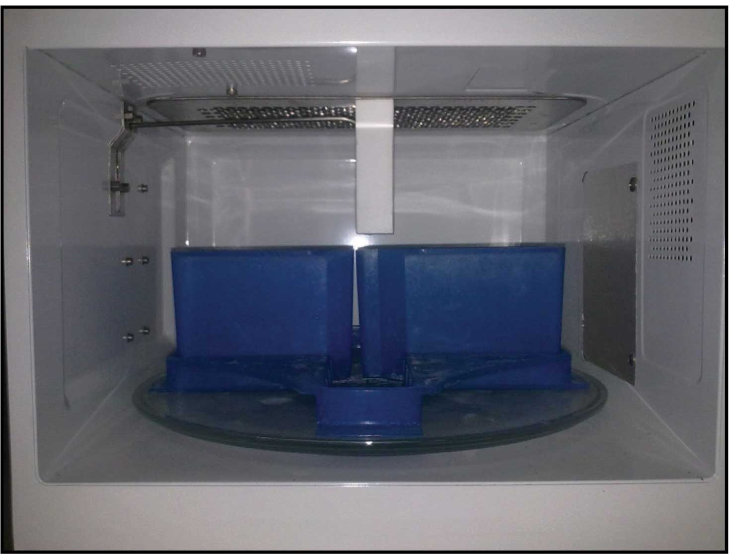
The other slices were placed in plastic cassettes. Four to five cassettes were then immersed in approximately 500ml PBS 0.1M pH 7.4 in a microwave-safe container and microwaved at 68°C for 5 minutes in enzyme retreiver V.2.2 microwave equipped with a temperature probe accurate to +/- 1°C [Table/Fig-1, 2]. Conventional histoprocessing and staining were done. Automated tissue processor Shandon Citadel 2000 was used for histoprocessing. After staining the slides, each slide (of both tissues fixed in formalin and by microwave irradiation) was examined and rated for the adequacy of fixation by two pathologists in a blinded fashion, applying a scale of 1 to 5 (1=poor, 2-fair, 3=good, 4=excellent, 5=outstanding) [12]. Seven parameters were used: Cellular outline, cytoplasmic detail, nuclear detail, erythrocyte integrity, lymphocyte appearance, overall morphology and overall staining. Overall grading was done by adding the scores of each parameter:- 7-13 –>Poor, 14-20–>Fair, 21-27–>Good, 28-34–>Excellent, >34–>Outstanding.
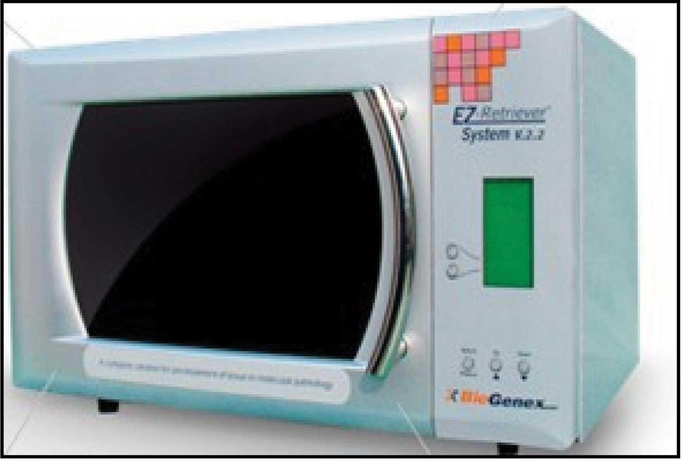
Microwave- Enzyme retriever V.2.2 system
Data management and Statistical analysis was performed using SPSS software version 20.0. Chi-square test was used. The p-value < 0.05 was considered statistically significant.
Results
A total of 140 paired tissue samples were fixed by microwave irradiation and conventional method and comparison of the quality of histologic section and turnaround time was determined [Table/Fig-3].
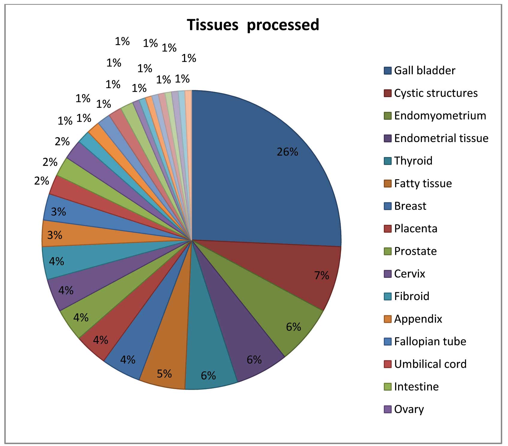
Different tissues processed by both microwave and conventional methods
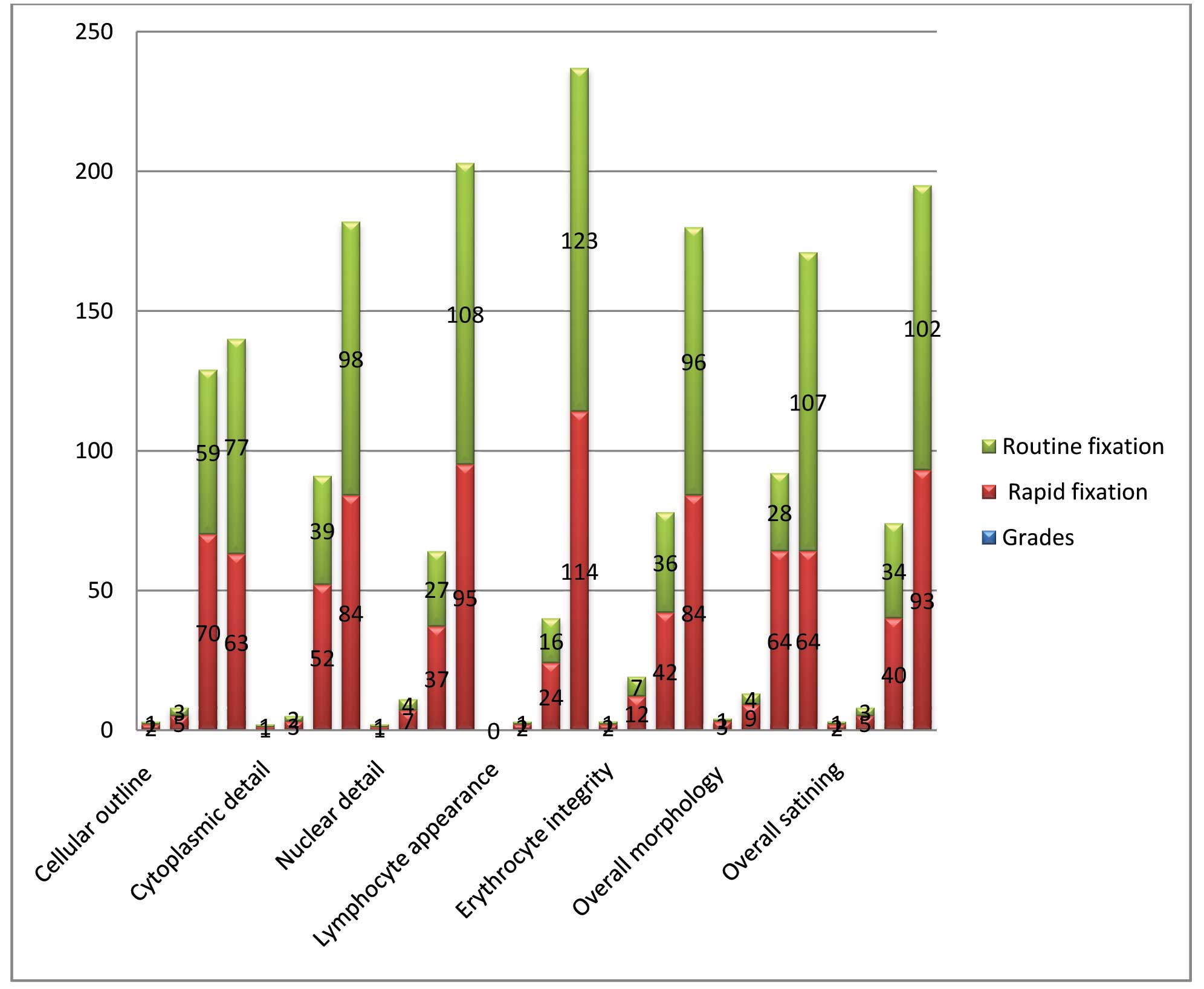
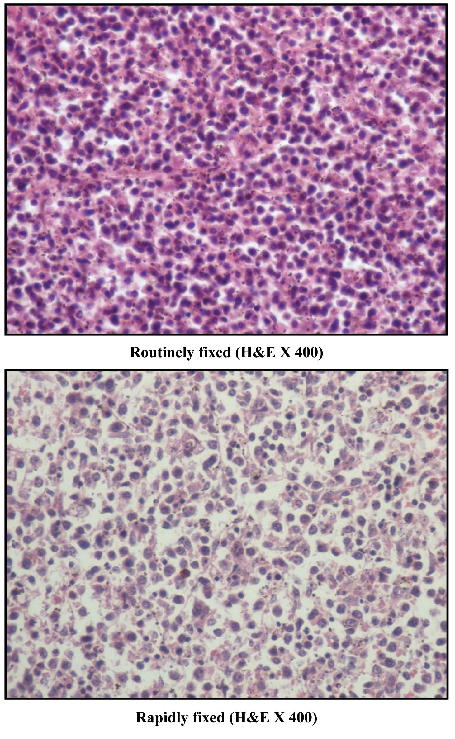
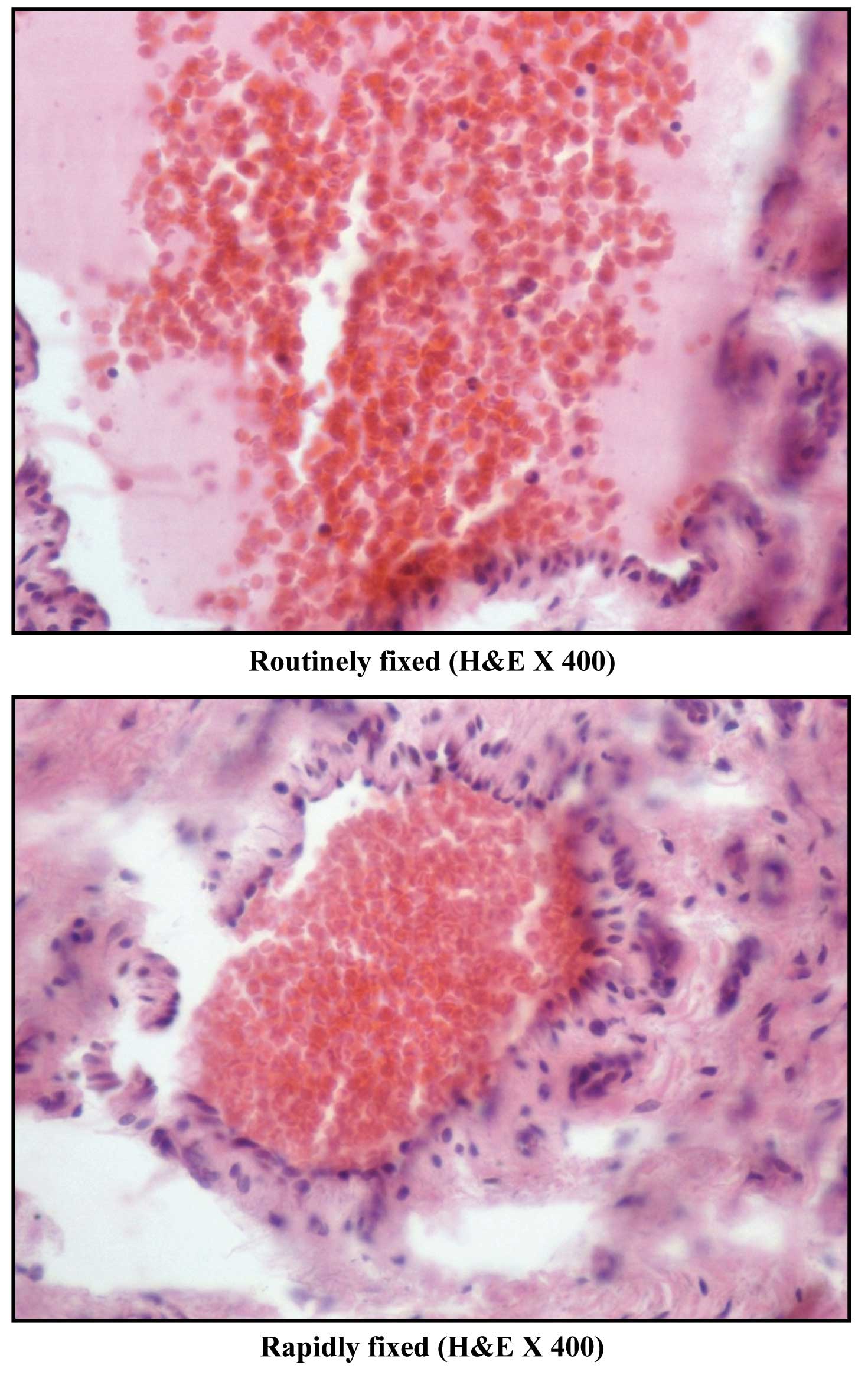
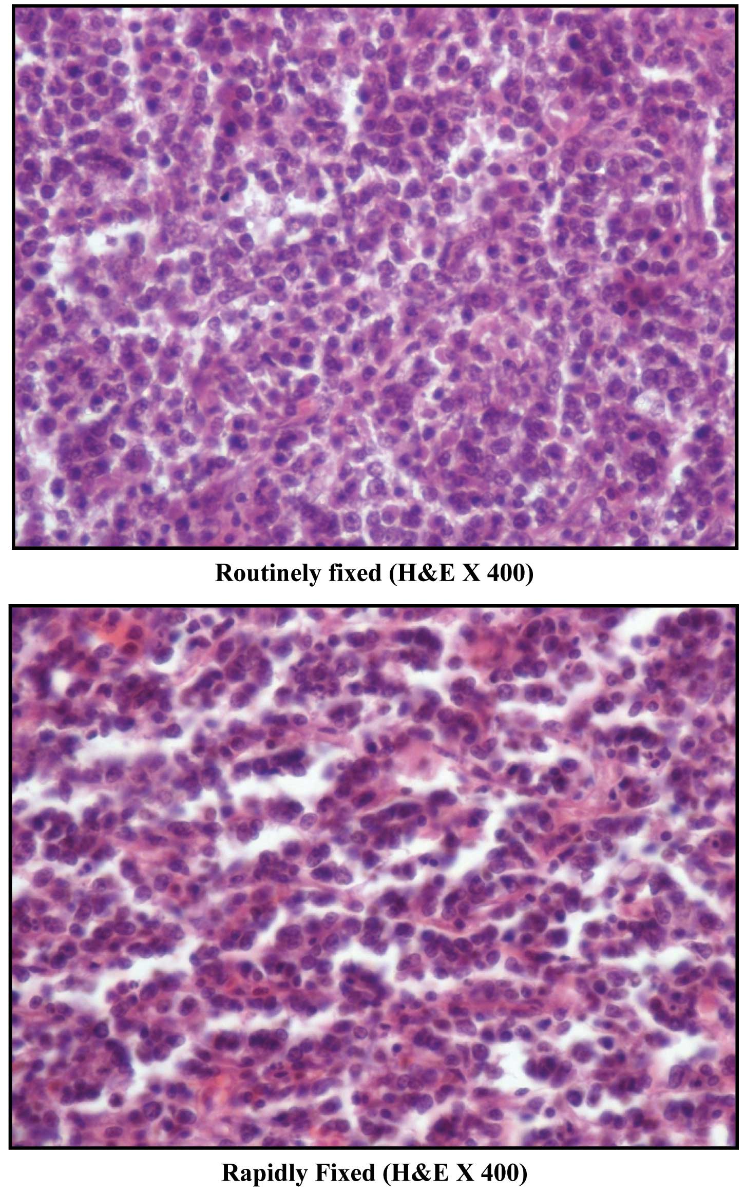
After fixation by microwave irradiation, the tissue sections were firm, shrunken and pale. On microscopic examination by two pathologists, the inter-observer variation was found to be minimal. The cellular outline, cytoplasmic detail, nuclear detail, erythrocyte integrity, lymphocyte appearance and staining as seen after rapid fixation were comparable to that of routine fixation. The p-values were not significant [Table/Fig-4, 5a,B,C,D,E,F,g].
Graph showing grading of each parameter between routinely fixed and rapidly fixed tissue
Comparison of cellular outline of lipoma Interpretation: Cellular membranes are sharper in rapidly fixed section

Comparison of cytoplasmic details of metastatic adenocarcinoma brain Interpretation: Cytoplasmic details are comparable in both sections
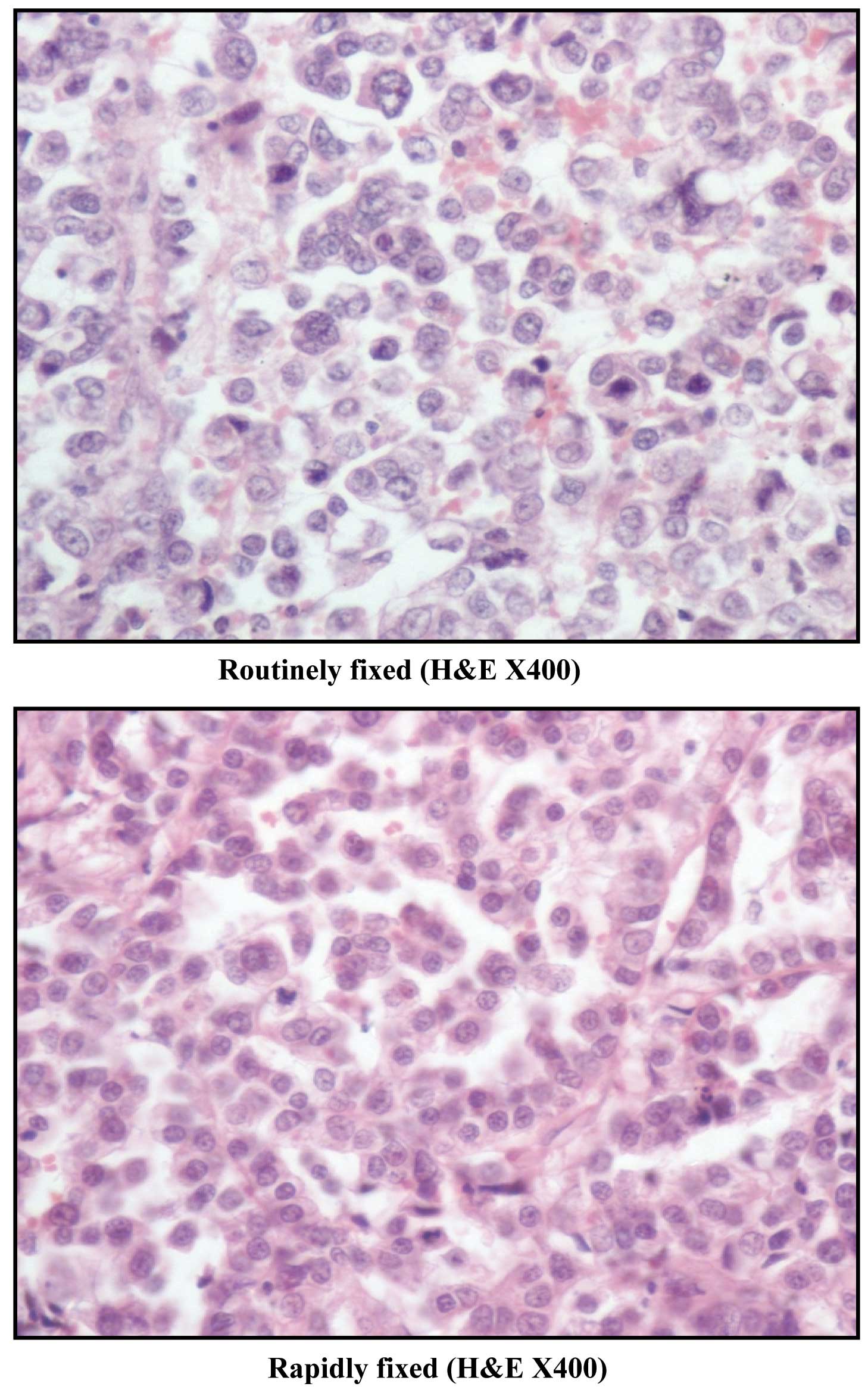
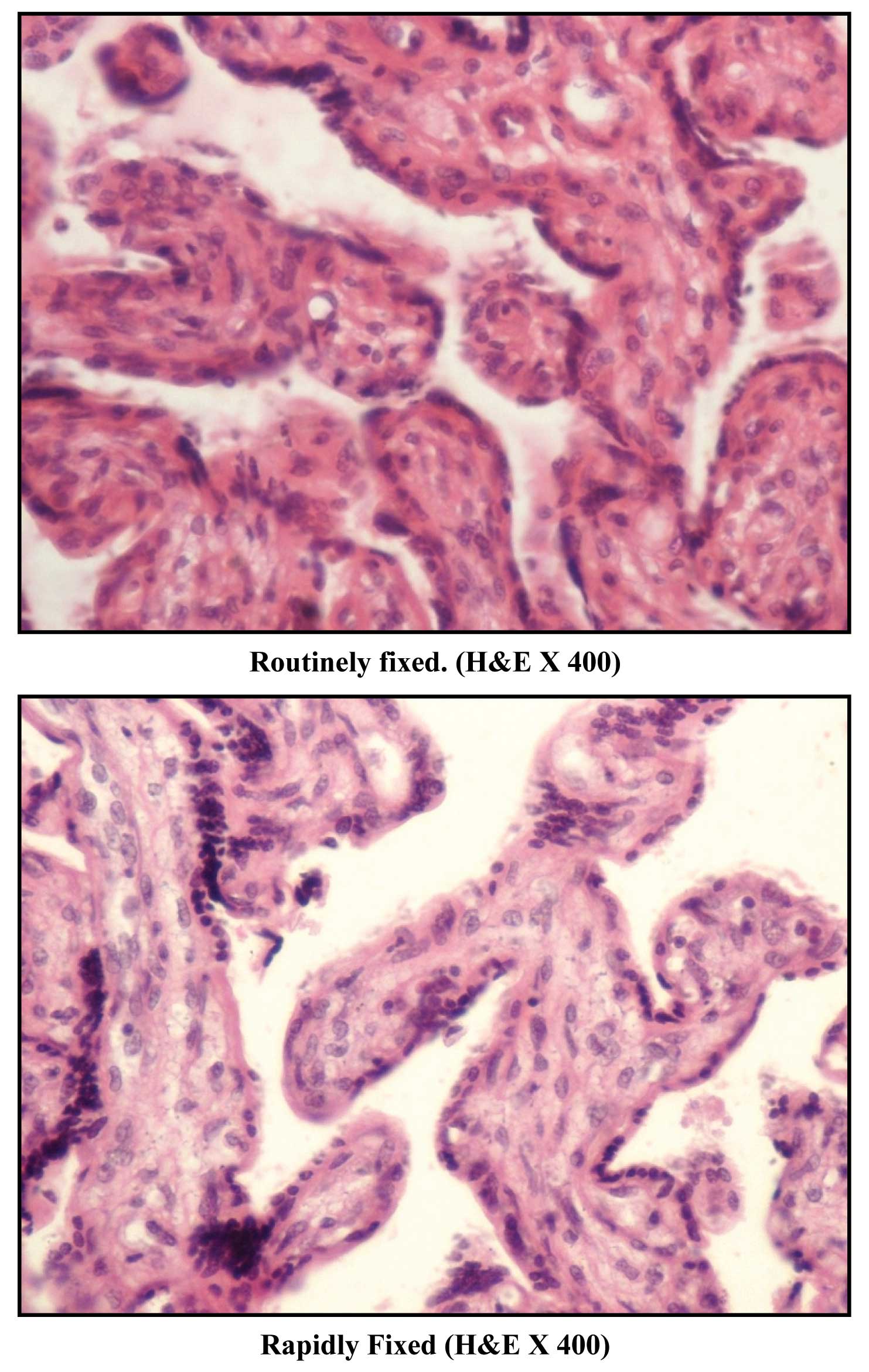
Comparison of nuclear details in spleen Interpretation: Nuclear details are better appreciated in rapidly fixed section
Comparison of erythrocyte integrity Interpretations: Intact RBCs in the blood vessel are seen in both sections
Comparison of lymphocyte appearance in lymph node Interpretation: Lymphocyte morphology was comparable
Comparison of overall morphology of placental tissue Interpretation: Overall morphology depicting cytotrophoblasts and syncytiotropho blast is sharper in rapidly fixed section
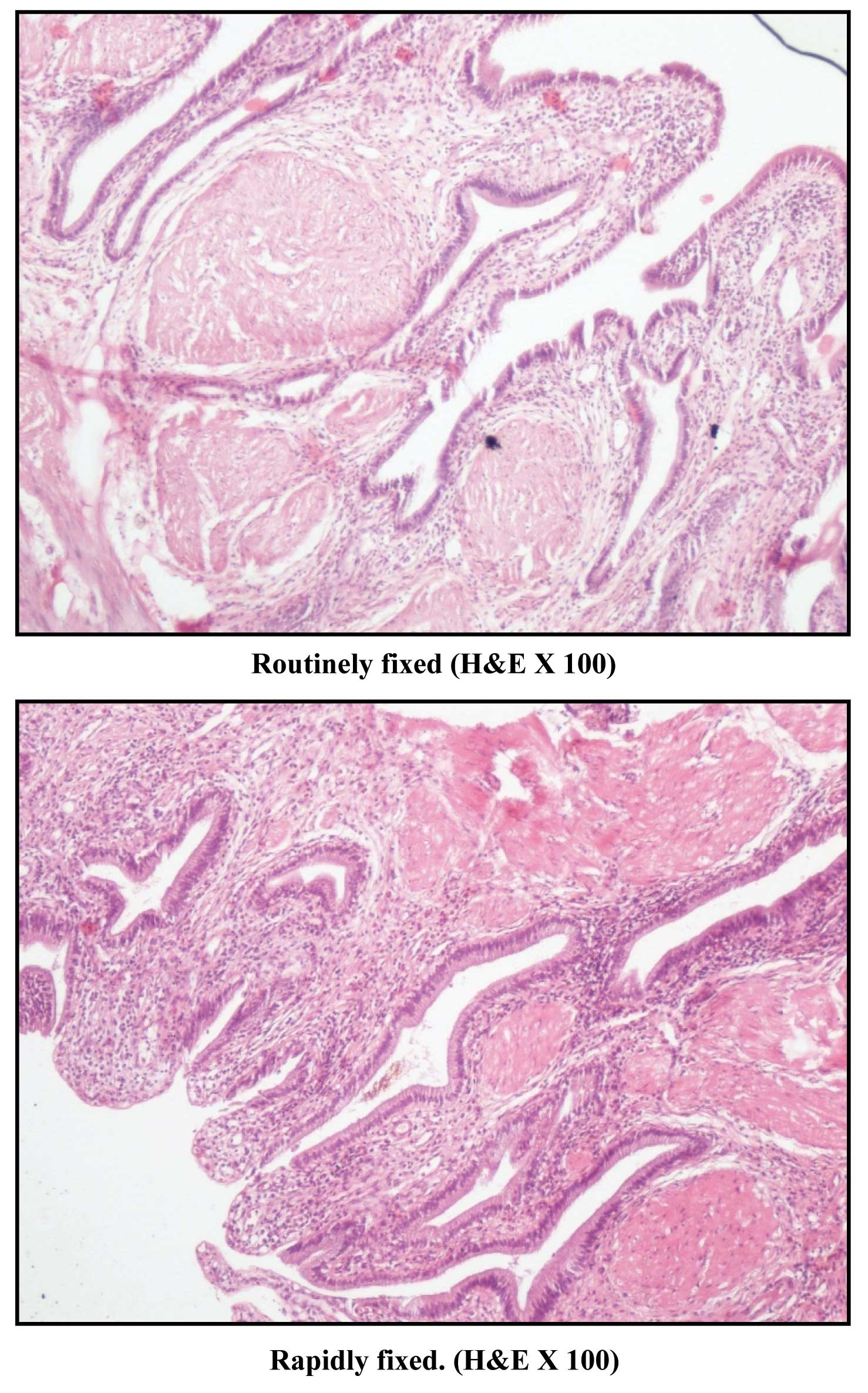
Comparison of overall staining of section of gall bladder Interpretation: Staining appears sharper and brighter in rapidly fixed section
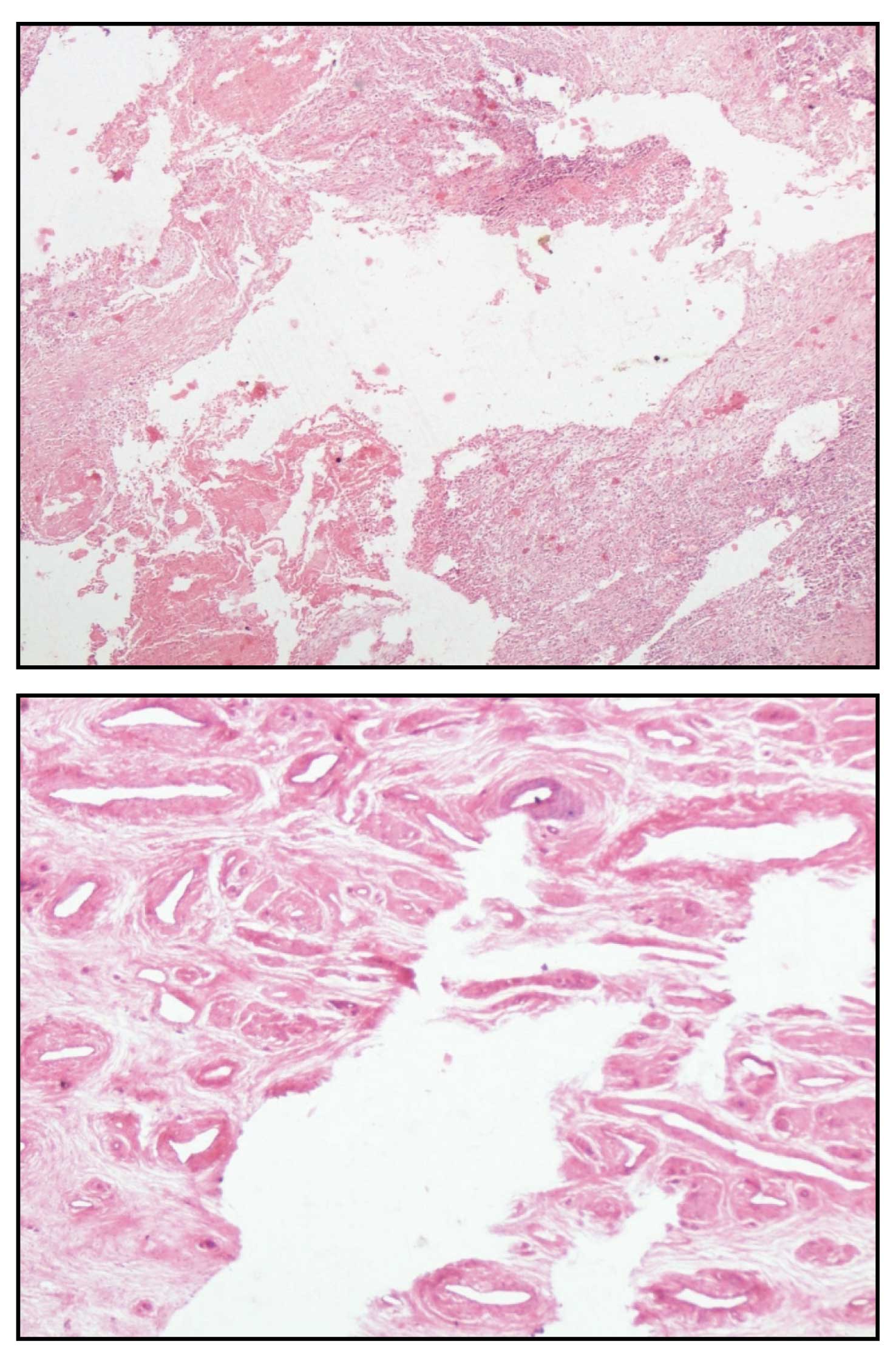
There was partial loss of tissues in rapidly fixed tissues owing to effacement of tissue architecture hence the overall morphology was graded slightly inferior to that of routinely fixed tissues. p value was 0.0001 which is statistically significant [Table/Fig-6].
(A) Rapidly fixed section of breast carcinoma depicting loss of tissue (H&E X 100) (B) Rapidly fixed section of cervix depicting loss of tissue (H&E X 100)
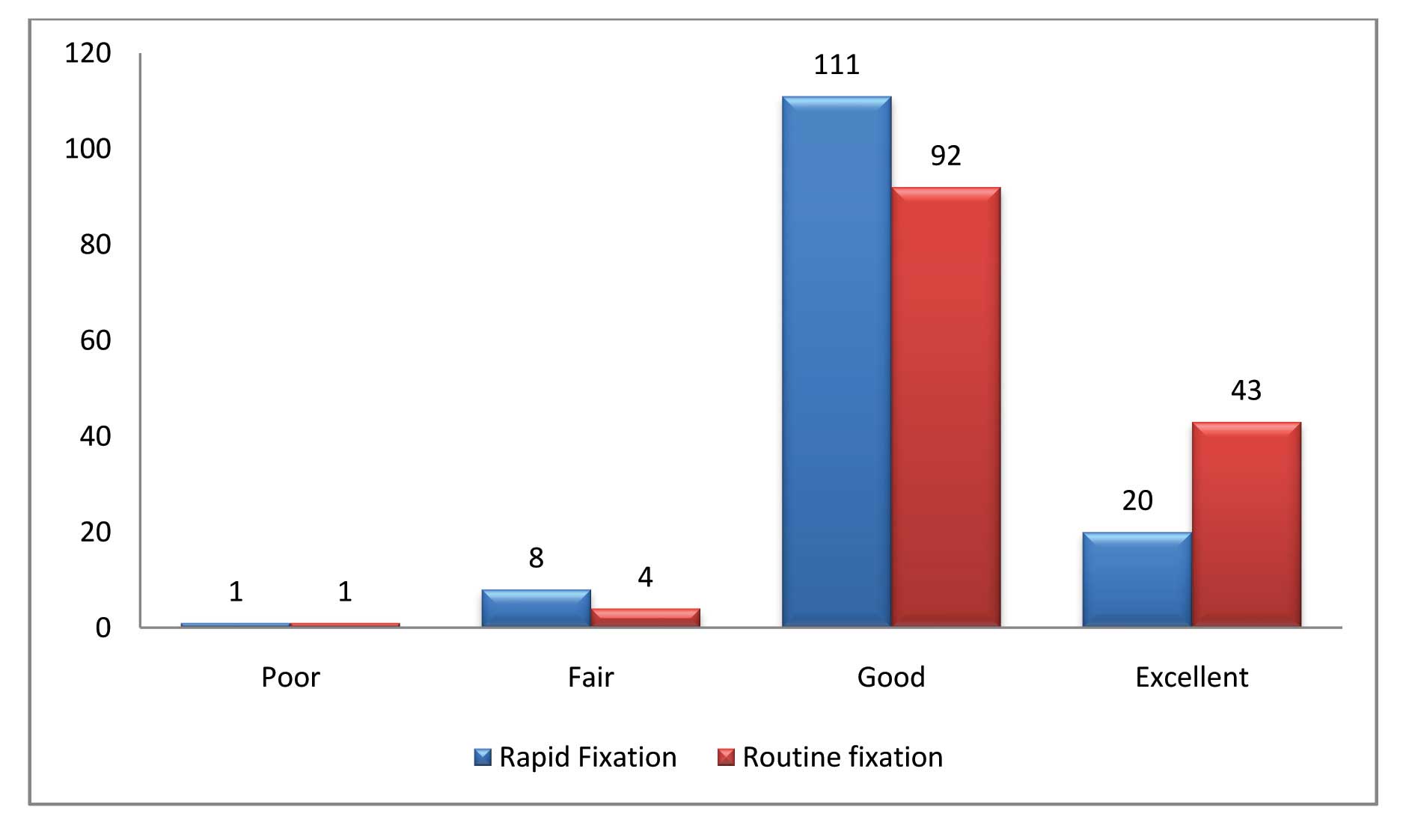
Out of the 140 paired tissues processed, 20 tissues fixed by rapid method were graded as ‘EXCELLENT’ in comparison to 43 of routine fixation. One hundred and eleven cases fixed by rapid method were graded as ‘GOOD’ in comparison to 92 fixed by conventional formalin fixation. 8 rapidly fixed tissues were graded ‘FAIR’ while only 4 were graded so by conventional method. One tissue each was graded as ‘POOR’ on fixation by both rapid and routine method. The p-value of 0.0002 was significant [Table/Fig-7].
Histological Quality of Routine fixation Vs Rapid Fixation
Better morphological details in microwave fixed tissues were noted in all cases of adipose tissue, placental tissue and small specimens like endometrial biopsy and prostate chips. Staining was also sharper in these tissues when compared with formalin fixed tissues.
Tissues were rapidly fixed in 5 mins by microwave irradiation whereas formalin fixation took almost 12 hours. The slides of the tissues fixed by rapid microwave irradiation were received within 24 hours of receiving the specimen when processed by the automated tissue processor whereas the turnaround time was a minimum of two days for conventionally fixed tissues
Discussion
Microwaves are non ionizing radiations that produce alternating electromagnetic fields that result in rotation of dipolar molecules such as water and the polar side chains of proteins through 1800 at the rate of 2.45 billion cycles/second. The molecular kinetics so induced results in the generation of energy flux and continues till radiation ceases [12].
In the present study microwave irradiation has been used only for fixation of tissues like Mayer CP [13] and Pitol DL et al.,[14].
In this study phosphate buffered saline was used as a medium for microwave irradiation and tissues were irradiated at 680C. Login GR et al., [15] found that tissues irradiated to 50± 50C were superior to those at low temperature. Many other studies showed that use of normal saline gave better fixation results than using formalin in microwave irradiation eliminating the noxious and potentially toxic fumes [14,16,17]. Choudhary K et al., [18] demonstrated that only 5 out of 16 tissue (30%) were stabilized in normal saline whereas 22 out of 34 (80%) fixed in formalin with microwave exposure showed satisfactory results.
After fixation by microwave the tissues became firm and pale in colour as also noted by Mayer CP [13] and Leong ASY et al., [17] on macroscopic examination.
On microscopic examination tissue was partially lost in 39.28% cases, predominantly in tissues like gall bladder, lymphoid tissues, cervix and tumour tissues. However, Morales et al., [19] found no loss of tissue architecture between conventionally and microwave processed tissue.
Ragazzini T et al., [20] observed that the loss of tissue in domestic microwave was due to the fact that a constant temperature cannot be reached or maintained in a specific time frame which has now been overcome by computerized systems. In other studies this parameter has not been used as a part of their analysis.
The loss of tissue in this study was seen maximum in cases of lymphoid tissue. All lymphoid tissues were received in fresh state and were not partially fixed. Tissues kept in formalin for 1-2 hours before microwave irradiation gave the best results without any loss of architectural details. In another study, it was found that fixation in neutral buffered formalin followed by microwave irradiation gave better results when compared to microwave irradiation alone [21]. Similar finding was obtained on microwave fixation of tissues previously soaked for four hours in formaldehyde [22].
The cellular outline and cytoplasmic details of the microwave fixed tissue was comparable with that of conventional formalin fixed tissues. This is consistent with other studies [12,23]. However, in our study the nuclear details were slightly less clear in microwave fixed tissues. This finding is supported by the study of Panja P et al., [24] who found that there was loss of nuclear details under high power when complete histoprocessing was done using the microwave oven whereas Kennedy A and Foulis AK [25] and Reed W et al., [26] found that the cytological details of the nuclei was much clearer using the microwave technique.
In the microwave irradiated tissues there was no loss of erythrocytes which is in contrast to the studies of Mayer CP [13] and Hopwood D et al., [27] who demonstrated erythrocytolysis. Boon ME et al., [22] performed the microwave irradiation of brain and found that the erythrocytes were intact. In both methods of fixation inflammatory cells like plasma cells and lymphoctyes were distinguishable from one another as is also demonstrated in this study.
The overall morphology was slightly inferior to that of conventionally formalin fixed tissues owing to the partial loss of tissue which is not in agreement with other studies [1,13,28–30].
Staining of the rapid microwave fixed tissues was sharper and brighter in most of the tissues than the tissues obtained after conventional fixation. This is consistent with the findings of Panja P et al., [24], Barrett C et al., [31] and Ragazzini T et al., [20] who showed that the microwave processed tissue showed a brighter staining with eosin and a stronger reaction with haematoxylin as compared to routinely processed tissue.
Neoplastic tissues in the present study were equally well preserved as the non-neoplastic tissues in the process of microwave fixation. This was similar to the findings of Hopwood D et al., [27] and Emerson LL et al., [32] who showed that malignant cells were equally well preserved by both conventional and microwave fixed tissues. Right from precancerous conditions and lesions to malignancy, reactive lesions to benign tumours, microwave technique has shown a remarkable difference without losing the architecture and morphology of the cells [23].
In this study adipose tissue showed the best morphology and staining characters post microwave irradiation. This is in agreement with Babu TM et al., [4] Other observers like Suri V et al., [33] found that tissues containing fat responded poorly to microwave irradiation. Steri V et al., [34] also found that adipose tissue showed artifacts due to incomplete fixation.
Small specimens like endometrial currettings and prostate chips were morphologically better when fixed in buffered saline in microwave oven. This is in agreement with the findings of Hafajee ZAM et al., [35] and Rohr LF et al., [3].
The overall time for fixation was reduced from 12 hours to 5 minutes when using microwave irradiation. This was found in many studies where fixation time was decreased from hours to a few minutes [17,18,27,30,36,37].
Conclusion
This study demonstrates that microwave irradiation under controlled conditions can reduce fixation time to about 5 minutes and thus reduce the overall processing time in surgical pathology without significant loss of specimen quality. This allowed same-day tissue processing and diagnosis of specimens which is essential when urgent reporting is required. It eliminates the use of formalin which is a potential hazard for health.
Adoption of microwave fixation in the regular histopathology laboratory on a regular basis can only be done after standardizing the time and temperature for each specimen as different tissues respond differently to microwave irradiation. Thus drawbacks like partial loss of tissues could be minimized.