Periodontitis is one of the most common infections that affect the gingiva and the bone supporting the teeth. The main underlying pathogenic mechanism involves the generation of host inflammatory response, which leads to tissue damage. Toll-like receptors play an important role in triggering this inflammatory response [1].
Toll-like receptors function as key pattern recognition receptors of the innate immune system [2]. They recognise and distinguish highly conserved structures on different microorganisms, known as pathogen associated molecular patterns such as bacterial lipopolysaccharide, peptidoglycan lipoprotein, bacterial DNA and double stranded RNA and trigger immune responses to clear them [3]. Ten Toll-like receptors have been described in humans and they have been classified according to the types of ligands that they recognise; for e.g. TLR-2 and TLR-4 recognize lipid based structures [4]. On interaction with their complementary ligands, the TLRs transfer this information through intracellular signaling pathways, resulting in activation of innate immune response: this interaction is also important for the activation of the adaptive immune system. Therefore, TLRs also play a role in linking the innate and adaptive immune responses in microbial infections [5].
These Toll-like receptors are known to be expressed in cells of the immune system, but they are also increasingly expressed in other cells [4]. Several types of cells from oral cavity also express these receptors. The gingival epithelium protects the underlining periodontal tissue from microorganisms and other harmful agents entering the oral cavity. Gingival epithelial cells are multilayered and they constitutively express TLR-2, 3, 4, 5, 6 and 9 [6]. Besides the epithelial cells, the connective tissue and the fibroblasts also express variable amounts of TLR-2, 4 and 9 [7–9].
In general, TLR-2 recognises signals of the gram-positive bacterial ligands, such as peptidoglycans and lipoproteins. Several studies have been performed in the recent past for detection of TLR-2 receptors in the cells of oral cavity by immunohistochemistry (IHC). However, there are not many reports on TLR-2 detection using immunofluorescent techniques (IFTs). Hence, in the present study, we have made an attempt to evaluate the presence of TLR-2 receptors in epithelial cells and fibroblasts of the periodontal tissue by using indirect IFTs. The data have been presented below.
Material and Methods
Subjects and Samples
The present study was carried out in the Department of Microbiology, Maratha Mandal’s N.G. H. Institute of Dental Sciences and Research Center, Belgaum, Karnataka. Fifty subjects were selected from the population which was referred to dental clinics in Belgaum for the study. Individuals recruited included 25 healthy subjects without periodontal disease and 25 patients with periodontal disease. In patients with chronic periodontitis, gingival samples were collected during routine periodontal operations, which included scaling and root planning. Samples from the 25 healthy controls were obtained during tooth-extraction operations performed for fully impacted, retained wisdom teeth. Chronic periodontitis subjects were selected, based on the criteria of the American Academy of Periodontology Classification 1999.
The inclusion criteria for patients with chronic periodontitis included presence of at least 20 natural teeth, a minimum of 6 periodontal pockets of ≥5mm probing depths and clinical attachment loss of ≥3mm around the affected teeth. The age range of patients with periodontitis was 20-60 years, among males or females.
The inclusion criteria for healthy controls comprised the absence of periodontal diseases, having at least 20 natural teeth, age ranging from 20-60 years, among males or females.
The exclusion criteria for both healthy subjects and patients with periodontitis included individuals with any systematic diseases/conditions, pregnant or lactating women and individuals with a history of dental treatment or drug therapy in the past 3 months prior to the study.
Tissue Processing
Gingival samples were directly collected in cold 95% ethanol. The tissues were trimmed to a thickness of 2-4mm and they were left for further incubation for 18-24hrs at 4°C in ethanol. Tissue samples were then processed by dehydrating them in 4 changes of pre cooled absolute alcohol each for 1hr. They were then transferred to 3 changes of xylene each for 1hr at 4°C, embedded in paraffin in 4 consecutive baths each for 2hrs at 56°C. Tissue sections of 5μm thickness were cut on a microtome and they were mounted on APS coated slides [10].
Hematoxylin and Eosin Staining
Initial characterisation of tissues was done by performing hematoxylin and eosin staining of all the specimens.
Immunofluorescence
An indirect immunofluorescence technique was performed to detect TLRs on mounted sections of 5μm thickness. Tissue sections were incubated for 45 minutes at room temperature with TLR-2 primary antibody (Purified Anti-Human, BioLegend); at least two slides per sample were tested. We used mouse monoclonal antibody against human TLR-2, at a 1:50 dilution in sterile Phosphate Buffered Saline (PBS). At the end of incubation, the slides were washed with PBS-T [50 ml PBS + 25μl Tween 20] for 5 minutes 2-3 times. Tissue sections were then incubated for one hour in secondary antibody conjugated with FITC (Goat Anti-Mouse IgG, Imgenex) at a 1:200 dilution, blocked with 5% goat serum in PBS-T for 5-10 minutes and then they were washed with PBS-T for 5minutes 2-3 times and mounted in DPX mountant [11].
Immunofluorescence images were acquired using a fluorescent microscope (Olumpus BX41) with a photographic attachment. At least 3 representative images were captured and analysed per slide.
Ethics
The study was approved by the Local Ethical Committee at the Maratha Mandal’s N.G.H Institute of Dental Sciences and Research Centre, Belgaum. Written informed consents were obtained from all study participants before sample tissues were acquired. The periodontal evaluation was performed by well-trained examiners.
Statistics
The prevalence of TLR in healthy group was 5-10%, assuming maximum prevalence of 10%, to demonstrate prevalence of 50% in period group. Using Type I error of 0.05, Type II error of 0.2 or power of 80% the sample size was calculated using the formula:

Where Zα and Zβ are standard normal constants, p1 – prevalence in healthy samples, p2 - prevalence in periodontitis samples,

q = 100-p, Zα : α = 005 = 1.96, Zβ : β = 0.2 = 0.84. On adding the values to the formula, the sample size was found out to be 25.
Results
A total of fifty gingival tissue specimens were studied, with equal numbers from healthy individuals and patients with chronic periodontitis. Each tissue was studied for histopathological characteristics by using haematoxylin and eosin staining and for expression of TLR-2 in epithelial cells and connective tissue cells by using indirect immunofluorescence. A semi quantitative analysis was carried out, based on the number of cells that took up the specific stain. The positivity was expressed in terms of percentage.
Histopathological Characteristics
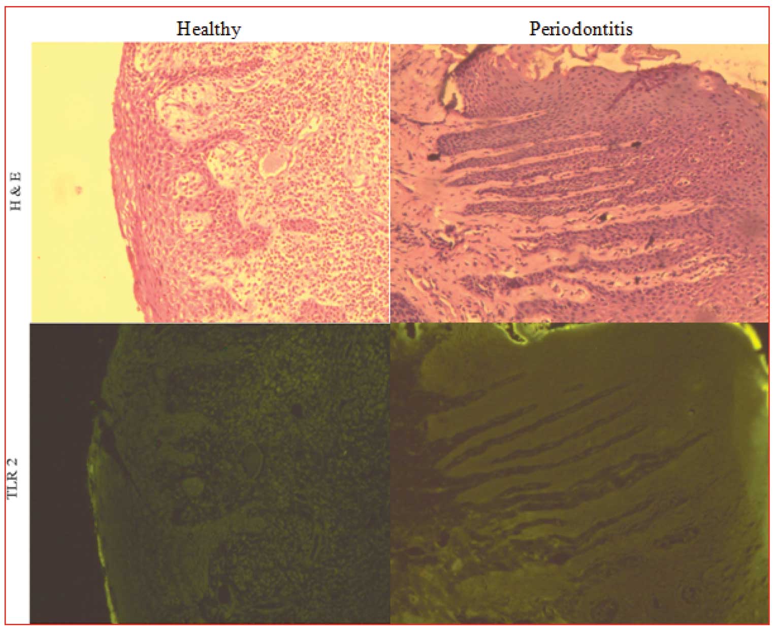
Sections of tissues were stained with haematoxylin and eosin to determine the histological characteristics. Healthy gingival tissues showed a typical keratinized stratified squamous epithelium with minimal inflammatory infiltrates. In patients with chronic periodontitis, keratinized stratified squamous epithelium showed changes such as oedema and exocytosis, mainly caused by the effect of chronic inflammatory infiltrate found predominantly in lymphocytes and plasma cells [Table/Fig-1].
Immunolocalization of toll-like receptor (TLR)–2 in gingival tissue samples from healthy controls and patients with chronic periodontitis. Respective images of Hematoxylin and eosin (H&E)- stained tissue sections are shown in the upper panels
Expression and Localization of TLR-2 in Gingival Tissue
TLR-2 expression in healthy gingival tissues was lower than in the tissues of patients with periodontal disease. In patients with periodontitis, TLR-2 expression was higher in epithelium as compared to its expression in connective tissue [Table/Fig-1].
Analysis of TLR Expression in Gingival Tissue
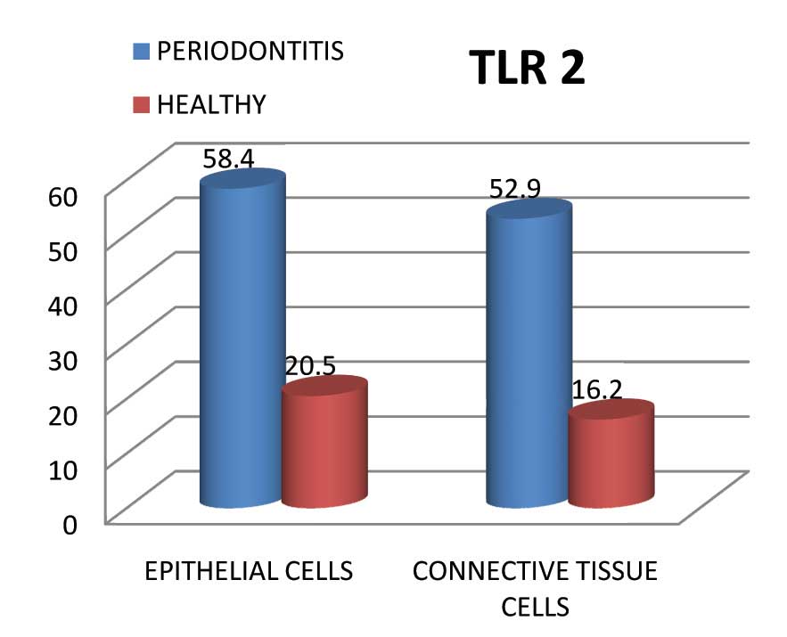
TLR-2 expression in the epithelial cells of periodontitis patients was 58.4 % (+ 19.82) as compared to its expression in epithelial cells of healthy individuals, which was 20.5 % (+3.56). This difference was significant, with a p value of <0.001.
TLR-2 expression in the connective tissue cells of periodontitis patients was significantly higher, with a positivity rate of 52.9% (+ 16.61), as compared to its expression in connective cells of healthy individuals, which was 16.2 % (+3.92) This difference was significant, with a p–value of <0.001 [Table/Fig-2].
Analysis of toll-like receptor (TLR)–2 expression in gingival epithelium and gingival connective tissue of healthy controls and patients with chronic periodontitis
Discussion
Toll like receptors make up an important component of pattern recognition receptors of the vertebrate immune system. They are known to play a crucial role in maintaining the homeostasis in the human body, by attaching to several bacterial cell surface components and by helping in clearing them from various body sites [12]. Amongst the ten TLRs recognised in the human tissues, several of them occur in the cells and tissues of the oral cavity, TLR2 and TLR4 being the dominant ones [4].
TLR2, either alone, or by forming dimmers with TLR1 and TLR6, recognize various surface structures of oral anaerobic pathogens, including LPS and fimbriae [13–15]. TLR-2 is known to be expressed on the surfaces of gingival epithelial cells as well as fibroblasts and these cells are situated at a strategic location, in order to make direct contact with bacterial pathogens, to bring about an immune response [6–8,16].
In the recent years, several workers have attempted to detect and quantify the surface expression of TLR-2 in the oral tissues [10]. Most have used IHC for this purpose and only a few have resorted to IFTs. The method that has been adapted by various researchers to express the levels of TLR-2 are also different: some have considered intensity of staining for quantifying the expression, while others have considered the percentage of cells showing positive results for a given location, for the purpose.
The results of the present study using IFT showed that TLR 2 is expressed in the human periodontal tissues. We found that the expression of TLR 2 in the periodontal tissues of chronic periodontitis patients was 58.4% (±19.82) in epithelial cells and 52.9% (±16.61) in connective tissue cells where as expression of TLR2 in the periodontal tissues of healthy subjects was 20.5% (±3.56) in epithelial cells and 16.2% (± 3.92) in connective tissue cells. These data clearly suggest the involvement of TLR-2 in initiating the inflammatory response.
In our study, we could also see that surface expression of TLR-2 in chronic periodontitis patients was significantly higher as compared to that in healthy subjects. Similar studies have also been done by various workers using IHC [16–21] who have found that the expression of TLR-2 in both epithelium and connective tissue cells was higher in patients with periodontitis than in healthy subjects. Only one study made use of IFTs to evaluate the expression of TLR-2 and it found that the expression of TLR-2 in chronic periodontitis patients was higher than that in healthy individuals [22].
During the course of our study, we realised that quantitative expression of TLR-2 on calculating the percentage of cells showing positive results was a better method than when only intensity of stained cells was taken into consideration, since this could be a variable trait. We did a survey on the work done by various investigators in evaluating the expression of TLR-2 in both the epithelial and connective tissue cells of the human periodontal tissue, the details of which have been shown in [Table/Fig-3].
Showing the comparison of results from various studies and the present study
| S.No | Author | Year | Method | Measured | TLR-2 expression | Reference |
|---|
| Zone 1 | Zone 2 | Zone 3 |
|---|
| 1. | Mori et al.,[14] | 2003 | IHC | Cell percentage | Mild | 0.41± (0.86) | 0.18± (0.48) | 0.47± (1.05) | 16 |
| Moderate | 0.63± (0.88) | 0.45± (0.85) | 0.66± (1.20) |
| Severe | 1.68± (2.24) | 1.04± (1.84) | 0.5 ± (0.82) |
| 2. | Ren et al., [18] | 2005 | IHC | Cell percentage | Detected only week expression of TLR-2 in healthy gingival tissues. | 17 |
| 3. | Sugawara et al., [19] | 2006 | IHC | Intensity | Compared to healthy controls increased expression of TLR2 by inflamed oral epithelium was located at cell borders | 18 |
| 4. | Saraha et al., [20] | 2006 | IHC | Intensity | Showed significantly elevated TLR-2 expression in tissues of patients with gingivitis and chronic periodontitis compared to healthy control. | 19 |
| 5. | Uehara and Takada [21] | 2007 | IHC | Intensity | Clear expression of TLR2 in normal oral epithelium. | 20 |
| 6. | Beklen et al., [22] | 2008 | IHC | Cell percentage | | Superficial cell layer | Spinous cell layer | Basal cell layer | 21 |
| Healthy | 82.7± 6.6 | 74.1± 7.4 | 77.6± 7.6 |
| Periodontitis | 77.1± 6.2 | 89.8± 3.9 | 88.8± 9.1 |
| 7. | Rojo Botello et al., [23] | 2011 | IFT | Fluresence intensity | Epithelial Tissue | Connective Tissue | 22 |
| Control Group (AU) | Chronic periodontitis (AU) | Control Group (AU) | Chronic periodontitis (AU) |
| 17-19 | 30-31 | 11-12 | 15-18 |
| 8. | Present study | 2013 | IFT | Cell percentage | Epithelial Tissue | Connective Tissue | - |
| Control Group | Chronic periodontitis | Control Group | Chronic periodontitis |
| 20.5 % (+3.56) | 58.4 % (+19.82) | 16.2 % (+3.92) | 52.9% (+16.61) |
To conclude, IFT is a better, rapid and more sensitive method than IHC for detection as well as quantification of surface TLR-2 expression. This has been the experience of other workers also, who used IFT for evaluation of the same. However, the set up is comparatively expensive and it needs trained personnel for evaluation of the stained slides [10]. Several such studies need to be taken up on larger sample sizes, for forming a definite opinion. We are presently performing this study with more number of samples and also other TLR markers, to evaluate their presence in oral tissue in periodontal health and diseases.