Superior Articular Facets (SAF) are present on atlas vertebra facing superomedially & they occupy most of the upper surface of the lateral mass and lie obliquely, their anterior ends being always nearer to the midline than the posterior ends. Facets are usually concave, with concavity in both longitudinal and transverse directions. The facets form an atlanto-occipital joint with occipital condyles and this joint is responsible for nodding movements and also for the weight- bearing of the head. The atlanto-occipital joint strains predominantly induce a tension-like headache which is caused as a result of a prolonged and an inappropriate posture which results from a poor ergonomic adaptation. Cervical spine malformations and craniovertebral junction abnormalities which lead to hypermobility of atlanto-occipital joints, give rise to neurological and vascular symptoms. Alterations in the morphometry of superior articular facet will alter the ergonomics of the joint, leading to restricted movements. The description of the SAF of the atlas vertebra, as has been found in the most of the text books of anatomy, makes no mention of its variations. Literature has revealed marked variations in the shape, symmetry, partial or complete separations of the facets and constrictions of SAF of the atlas [1,2,3]. The non-metrical changes which occur in the superior facets of atlas may be a concordant factor for restriction of a cranio-vertebral motion [4,5]. It has been postulated that antero-posterior axial changes are important for assessing the surgical applications. The metric variants, like the surface area of the SAF of the atlas, has been least reported. So, this study was undertaken to study the morphologies and to measure the surface areas of the SAFs.
The objectives of the present study were to report the analyzed morphological features and metrical values of superior articular facets of the atlas. The morphological features which were included in this study were variability in shapes, constrictions and partial or complete separation of facets. The metrical measure included the surface area measurement of the facets.
Material and Methods
This study was carried out on 50 (100 sides) adult, dry, atlas vertebrae of unknown ages and sexes over a period of 6 months, in the Department of Anatomy, St. John’s Medical College, Bangalore, India. Damaged and pathologically abnormal bones were excluded from the study. The surface areas of the superior articular facets were measured by an online application tool that could be utilized to measure various metric measurements, Image J (Image processing and analysis in JAVA) tool. These images were obtained by tracing the articular facets on a graph sheet, which were then scanned. The scanned images were transferred to the computer and analyzed. Simultaneously, other morphological parameters like shapes, constrictions and partial or complete separations of facets were also noted. This was an observational study and an ethical clearance was not required, as it was done on dry bones.
Results
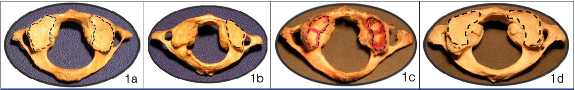
The mean surface area of the SAF of the atlas appeared to be more on the left side 158.5 ± 24.85 (as measured in mm2) as compared that on the right side 157.3 ± 29.02. In our study, various shapes of SAF were observed, like irregular, oval, kidney, etc [Tables/Fig-1a-1d]. The irregular appearance of the facet was most commonly observed, which was succeeded by oval shape and figure of eight shape. Kidney shape was the least observed shape. Asymmetry was the commonest feature of the entire 50 vertebrae.
1a, 1b, 1c & 1d Different shapes of SAFs of Atlas and their percentage of occurrence
1a: Irregular Shape 39% (39 Sides)
1b: Oval Shape 33% (33 Sides)
1c: Eight Shape 18% (18 Sides)
1d: Kidney Shape 10% (10 Sides)
A bilateral, complete separation was observed in 30% (15) vertebrae and a bilateral incomplete separation was observed in 18% (9) vertebrae. A unilateral, complete separation of the facets was observed, with predominance on the right side 6% (3) [Table/Fig-2].
Separation of superior articular facets of atlas with percentages
| Observation | n | % |
|---|
| 1. | No separation of the facets on both sides | 21 (42 sides) | 42 |
| 2. | Complete separation on both sides | 15 (30 sides) | 30 |
| 3. | Incomplete separation on both the sides | 9 (18 sides) | 18 |
| 4. | Left partial separation with right complete separation | 3 (6 sides) | 6 |
| 5. | Right partial separation with left complete separation | 1 (2 sides) | 2 |
| 6. | On right complete separation, left being normal | 1 side | 1 |
| 7. | On left partial separation, right being normal | 1 side | 1 |
A bilateral constriction of the facets was observed in 58% (29) vertebrae and a unilateral constriction was observed on right in 4 vertebrae and on left in 2 vertebrae [Table/Fig-3].
Constriction of superior articular facets of atlas with percentages
| Sl. No. | Constriction | n | % |
|---|
| 1. | Presence on both the sides | 29 (58 sides) | 58 |
| 2. | Absence on both the sides | 15 (30 sides) | 30 |
| 3. | Present only on right side | 4 sides | 4 |
| 4. | Present only left side | 2 sides | 2 |
Discussion
The stability of any joint in human body depends on morphological organization of the bones which are involved and the surrounding soft tissue. The atlanto-occipital joint is the one which involves an approximate reciprocal configuration of occipital condyles with superior articular facets of atlas. The superior articular facets which assume a horizontal orientation during development, which will assume a concave appearance by 6 years of development, will establish stabilization process of the joint [5]. As the age advances, the physical anthropometry of the joint may variate, that may cause either symptomatic or asymptomatic clinical conditions. It is a debatable task to postulate that variant changes in the articulating surfaces of atlanto-occipital joint are the responsible and reasonable factors for neck strains that involve the joint. There are reported findings in the literature on the physical profile of superior articular facets of atlas, with reference to the samples from northern part of Indian subcontinent. The present study reports the existing findings with reference to south Indian samples, along with an innovative method for calculating the surface area, that could help head and neck and vascular surgeons during different degrees of operative procedures. Specific data from the literature on the metrical measure of atlas was very minimal to say that it had an accurate reciprocal surface with occiput. The only report on dry bone measurement said that there was a tendency to form large superior articular facets, in association with large left occipital condyles [6]. With the new technical innovations, an attempt was made to study the surface area in an unknown sample of 50 dry individual atlas vertebrae. IMAGEJ (Image processing and analysis in JAVA) is an online application tool that can be utilized to measure various metric measurements. Rationale of use of this tool is the accuracy in its measurement. The difference in surface area from left side to right side was 1.2 degree, with more surface area to the left. This degree of difference in surface area was not correlated with phenotype of the facet. Angulations in anteroposterior, medial to lateral directions of superior articular facets of atlas, by using mathematical applications, were reported in 20 fresh spine specimens, with a significant difference between the antero-posterior to transverse dimensions on left side [5]. These observations indicated that the metrical values of superior articular facets were important in certain types of complications like Whiplash distortions of cervical spine.
The morphometric observations of superior articular facets were reported in a significant number in the literature. These reports have mentioned about the asymmetry, constrictions along the margins, shapes and separations of facets. In the present study, similar observations were carried out on samples from Southern India.
The present observations and the reported findings from the literature were discussed. Oval, figure of 8, kidney, trilobed, bilobed, irregular, triangular, V and leaf type shapes were the reported shapes of superior articular facets of atlas [7]. In recent studies [7,8], oval shape was reported to be the dominant shape. In contrast to the reports, irregular shape was observed to be the dominant shape (39%), followed by oval shape (33%). Similarly, a significant portion (10%) of the facets showed kidney shape due to the presence of constriction on medial margin of the facet, in comparison with the reported findings. If a complete constriction was present, then the shape was considered to be a figure of 8, which was observed to be 18% less dominant as compared to the reported findings.
In the present study, we observed that not a single atlas vertebra had symmetrical facets. Similar observations were made by Singh et al., [1] and Gottlieb [9]. The parameters of the present study were comparable to the findings of Singh et al., [1] [Table/Fig-4]. Partial or complete partition of the facet is an important finding, that could be interpreted as a derived characteristic for human species, which could indicate the functional modification of the joint, due to acquisition of erect posture and bipedalism [10]. The partial or complete separation of facet can be observed by the presence of the constriction. In the present study, a bilateral constriction was observed in 58% vertebrae, which was agreeable with reported findings, other than a recent study from Northern India. The significant difference could be interpreted well, if a complete morphometry which included various dimensions were studied in detail. The bilateral presence of a constriction that never separated the facets into two halves was seen in 30% vertebrae and a unilateral separation was observed only in 2% vertebrae, which was in contrast to the reported findings. Similar observations were also noticed for either complete or incomplete separations. When it was complete, the separation of facets was well marked all the times. Georgios Paraskevas et al., [9] reported an increase in the incidence of separation of SAF and that a decrease in constrictions could possibly be the result of a restriction in the atlanto-occipital motion in old age. In recent years, considerable innovations in the internal fixation techniques have created a need for a more detailed quantitative description of the anatomy of this bone. Thus, the anatomy of the cervical vertebrae or the spine is of great clinical importance to surgeons, as a surgical procedure may be done through the anterior or posterior cervical spine, with gratifying results.The observations of the present study played an important role in understanding biomechanics and ergonomics of the atlanto-occipital joint. The roles of these asymmetries were related to the biomechanics of atlanto-occipital joint and encroachment on the spinal cord warrants further investigation.
Percentages of constrictions and separations of SAF present study compared with Singh et al.,
| Parameters | Singh et al., [1] | Present study |
|---|
| No constriction | 27.75% | 36% |
| Constriction | 76.25% | 64% |
| No separation | 21.5% | 42% |
| Incomplete separation | 9.75% | 23% |
| Complete separation | 5.5% | 35% |
Conclusion
The study of SAFs of 50 adult atlas vertebrae showed different shapes. The most commonly observed shape was irregular shape, followed by oval shape and figure of eight shape. However, in contrast to most of the common observations, kidney shape was the least observed shape in the present study. No vertebra showed symmetrical facets. Bilateral constrictions of facets were seen in 58 % of vertebrae. Bilateral, complete separations of facets were seen in 30% of vertebrae. Surface areas of SAFs showed no significant differences. Findings of the present study may be useful for anatomists, in enhancing their teaching skills and for clinicians, especially neuro / vascular / ortho surgeons during performance of any invasive procedures.