Palatal Lipoma: A Case Report
Sreenivasulu Pattipati1, M. Naveen Kumar2, Ramadevi ,3, B. Praveen Kumar4
1Senior Lecturer, Department of Oral Medicine & Radiology,St. Joseph Dental College & Hospital, Duggirala, Eluru, Andhra Pradesh-534004, India.
2Associate Professor, Department of Oral & Maxillofacial Pathology,Kamineni Institute of Dental Sciences, Sreepuram, Narketpally, Nalgonda-508254, India.
3Associate Professor, Department of Pathology,Santhiram Medical College & Hospital, Nandyal, Kurnool, Andhra Pradesh, India.
4Reader, Department of Oral Medicine & Radiology,St. Joseph Dental College & Hospital, Duggirala, Eluru, Andhra Pradesh-534004, India.
NAME, ADDRESS, E-MAIL ID OF THE CORRESPONDING AUTHOR:Dr. M. Naveen Kumar,Associate Professor, Department of Oral & Maxillofacial Pathology, Kamineni Institute of Dental Sciences,Sreepuram, Narketpally, Nalgonda-508254, India.
Phone: 9885227600,
E-mail:naveen_motupalli@yahoo.com
Intraoral lipomas are benign mesenchymal neoplasms that originate in mature adipose cells with differential diagnosis of other soft tissue lesions. Lipoma, rarely, occurs in the oral cavity, and it corresponds to less than 4.4%, of all benign oral soft tissue tumors. Here, we are reporting a case of lipoma that occurred in the hard palate, which is extremely rare.
Adipocytes, benign tumor, hard palate, lipoma
Case Report
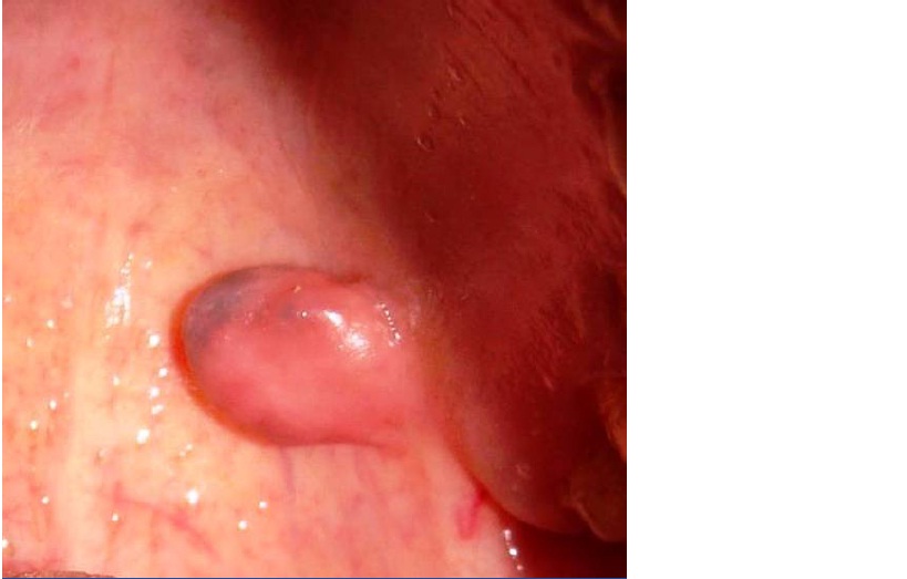
A 37-years-old male patient reported to the Department of Oral Medicine and Radiology with a chief complaint of growth in the left half of the upper jaw since 5 years [Table/Fig-1]. Patient noticed the growth 5 years back. The growth was gradual in onset, slowly increased in size over a period of 2 years and attained to the present size. On inspection, the growth present in the left posterior hard plate region was located 2cm away from maxillary tuberocity. The growth was asymptomatic, slightly brown at the tip of growth and surrounding mucosa appeared to be normal. On palpation, the growth was pedunculated which measured approximately 2×2 cm in diameter. The growth had a smooth surface, soft in consistency and slippery. No signs of pain and discharge from the growth. Occasionally, the patient complained of discomfort while eating. Patient’s medical, dental, family and personal histories were non-contributory and the review of his systems was normal. Based on history and clinical findings, provisional diagnosis of lipoma was considered. Routine blood investigations were carried out which also showed normal.
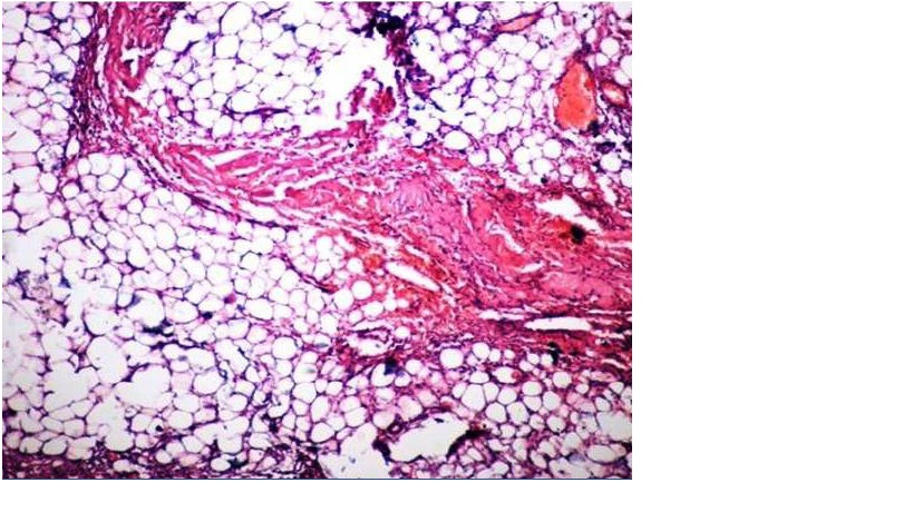
The growth was completely excised under local anesthesia and the specimen was sent for histopathological examination. On microscopic examination of H&E stained slides, mature fat cells arranged in distinct lobular pattern were seen [Table/Fig-2]. The nuclei were fairly uniform and connective tissue richly vascular, which was suggestive of lipoma. Based on clinical and histopathological findings, the growth was finally diagnosed as lipoma of palate.
Lipoma on the posterior palate
H&E section showing Adipocytes ; 40x magnification
Discussion
Lipoma was first described by Roux in 1848, in review of alveolar masses, where he referred it as “yellow epulis” [1]. Lipoma, usually a solitary lesion may be sessile, pedunculated or submerged, ranging in size from 1cm in diameter to a massive tumor of 5cm. The surface is typically smooth and non-ulcerated with deeper lesions varying in contour and shape [2]
Despite the increasing understanding of fatty tissue and fat metabolism, the etiology for lipoma remains enigmatic, since some authors believe that trauma could be a causative factor. Certain factors like heredity, hormones, congenital factors and progressive infection have been implicatedin genesis of lipoma, regardless of site, while others include diabetes mellitus, hypercholesterolemia, obesity and rheumatoid arthritis. Lipomasare seen in patients after the age of 30 years. Although they may be congenital, more than half of cases occur between 4th and 5th decade of life.Oral lipomas have been reported in individuals from 6 weeks of age to 75 years with a mean age of 60 years, rarely seen in children [3]
.
Extraorally, lipoma is one of the commonest benign tumors occurring anywhere in the body, predominantly affecting subcutaneous tissues of the trunk, nape of neck and limbs [4]. Hands, legs and feet are rarely involved [3].
Oral lipomas tend to occur with equal predilection for involvement of men and women. But, Furlong et al., mentioned that oral lipoma is seen more frequently in men (as presented here). Although, it should be mentioned here that Freitas et al., showed oral lipoma has a predilection of occurrence for women [5,6].
Hatziotis et al., [7] had reviewed the literature from 1945 to 1967 and had found 145 cases of intraoral lipomas of which only six cases occurred in the hard palate. ER Fregnani and his associates [8] reviewed 46 cases of lipomas and found none occurring in palate. Review of a few large reported series of intraoral lipoma and its variants seen in the literature did not show any case of oral lipoma occurring in hard palate [9].
Most oral lipomas arise within the superficial connective tissue and exhibit the characteristic yellow color of adipose tissue, which is visible through the thin overlying epithelium. In our case, the lipoma appears like normal mucosa with mild hyperpigmentation.Its consistency varies from soft to firm depending on the quantity of fibrous tissue in it. In some cases, it may be so soft that pseudofluctuancy can be elicted [10]. In the present case, the growth was soft and slippery in consistency.
Histopathologically, most oral lipomas are composed of mature fat cells very similar in appearance to the surrounding normal fat.Individual cells have a clear cytoplasm with a flat nucleus located on the periphery of the cell. Lipoma is usually well-circumscribed and may demonstrate a thin fibrous capsule [5].
According to their histopathologic aspects, benign tumor of adipose tissue (lipoma) can be classified as [2]:
Simple lipoma
Fibrolipoma
Angiolipoma
Intramuscular lipoma
Pleomorphic lipoma
Sialolipoma
Myxiod lipoma
Atypical lipoma
Treatment of lipoma is conservative surgical excision and recurrence is rare except for intramuscular variety which is infiltrative having a high recurrence rate [11]. Malignant degeneration of intraoral lipoma in the form of liposarcoma is extremely rare [12]. After excision of the lesion, we did not notice any signs of recurrence on regular followup for 2 years.
Conclusion
Clinicians should have awareness, that lipoma can occur in the oral cavity and they should be able to identify intraoral lipomas to provide appropriate treatment, thereby, ensuring comfort and quality of life for the patients.
[1]. Raj Mahendra, Ramdoss Tanuja, Anuradha G, Dave Shobhana, Intraoral Lipoma: Review of Literature and Case ReportJournal of Indian Academy of Oral Medicine and Radiology 2012 24(1):36-8. [Google Scholar]
[2]. Raninwala Amena, Kale Hemanth, Modi Tapan, Dave Kajal, Intr-oral lipomacase reportJournal of the International Clinical Dental Research Oraganization 2010 2(3):157-60. [Google Scholar]
[3]. Bagewadi Naik, Aggarwal Keluskar, LIPOMA-SINGLET RING CELL TUMOR REVIEW OF 10 CASES.Int J Dent Case Reports 2011 1(2):1-6. [Google Scholar]
[4]. S Das, A concise textbook of SurgeryComstock Publishing3rd editionDas Publishers:92-94. [Google Scholar]
[5]. MA Freitas, VS Freitas, AA Lima, FB Pereira, dos Santos JN. Intraoral lipomas: A study of 26 cases in a Brazilian population.Quintessence Int. 2009 40:79-85. [Google Scholar]
[6]. Motagi Ahmad, Aminzadeh Atousa, Razavi Seyed M, Large oral lipoma: Case report and literature review in Iran.Dental Research Journal 2012 9(3):350-2. [Google Scholar]
[7]. CH Hatziotis J, , Thassaloniki, , Greeze, Lipoma of the oral cavityJr. of oral surg 1971 31(4):511-21. [Google Scholar]
[8]. ER Fregnani, FR Pites, Falzoni R, Lipomas of the oral cavity, clinical findings and histological classification and proliferative activity of 456 cases.Int J Oral maxillofac Surg. 2003 32:49-53. [Google Scholar]
[9]. A Miles Dale, P Langlains Robert, B Aufdemorate Thomas, Birigit Junfin Glass, Lipomas of the soft palate.Oral Surg Oral Med Oral Path 1984 57:77-80. [Google Scholar]
[10]. A Winnifred Christy, Anitha Bojan, Babu Mathew and S Shanmugam. Lipoma in the Palate: A Rare PresentationJournal of Indian Academy of Oral Medicine and Radiology. 2010 22(4):S51-52. [Google Scholar]
[11]. A Epivatianos, AK Markopoulos, P Papanayotou, J Oral Maxillofac Surg. 2000 58:1113-17. [Google Scholar]
[12]. DJB Ashley, Tumors of adipose tissue, in Evan’s Histological appearance of Tumors (ed4) Edingburgh: Scotland, Churchill-Livingstone 1990 :63-75. [Google Scholar]