Introduction: The cross finger flap is a useful option for coverage of finger defects. The Adipofascial flap, a variant has been a good option being aesthetically more acceptable. Since hand is generally exposed part, we tried to find out the aesthetic value of the adipofascial cross finger flap.
Methods: We operated on 15 patients who had complex defects on the fingers, who needed a flap cover and accepted for the adipofascial variant. We asked for ten independent non-medical literate persons to rate the photos of donor site and recipient site of these patients. We also requested the same ten people to rate the photos of similar, paired defects of the standard flap, which were obtained from the department archive.
Results: All the flaps survived. The mean score for adipofascial flap donor finger was 3.859 and it was 3.6 for the donor finger of the standard cross finger flap. The Cornbach’s alpha of the interpretors was more than 0.7 for donor fingers and more than 0.9 for recipient fingers. The ICC was 0.767 foradipofascial donor finger and more than 0.9 for recipient fingers of both flaps.
Conclusion: Adipofascial cross finger flap may be an aesthetically better flap.
Introduction
The cross finger flaps have been commonly used for coverage of complex finger defects. It has undergone various modifications like the innervated cross finger flap, reversed dermis cross finger flap, de-epithelialised cross finger flap and adipofascial flap etc. The cross finger Adipofascial flap is one of the variant of the cross finger that flap can be widely applied to dorsal finger defects [1]. The Adipofascial flap is yet another option for finger defects [2]. We have tried to improve on the aesthetic value of the reconstruction by using this cross finger flap variant. Further, we have tried to compare the photos of the same with standard cross finger flap with the help of independent non medical reviewers.
The study was conducted from December 2008 to December 2009 at Victoria Hospital, Bangalore, India. During this period, patients with wound over the fingers with critical defects 2.5 cm X 2.5 cm were selected. The critical defects were defined as defects with exposed bone, tendon or joint and those which definitely required a flap cover for the wound. The various options for the management of the wound were explained to the patients. 15 patients, who opted for the cross finger Adipofascial flap were taken for the study. Valid consent was taken for the surgery and they were posted for surgery.
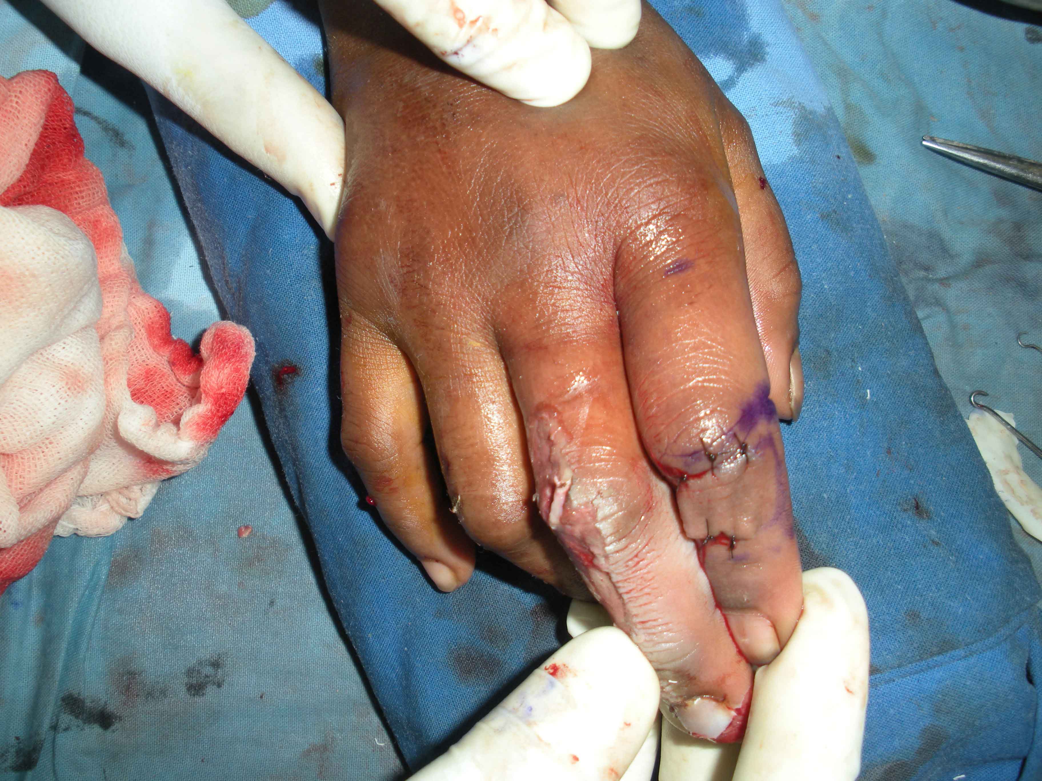
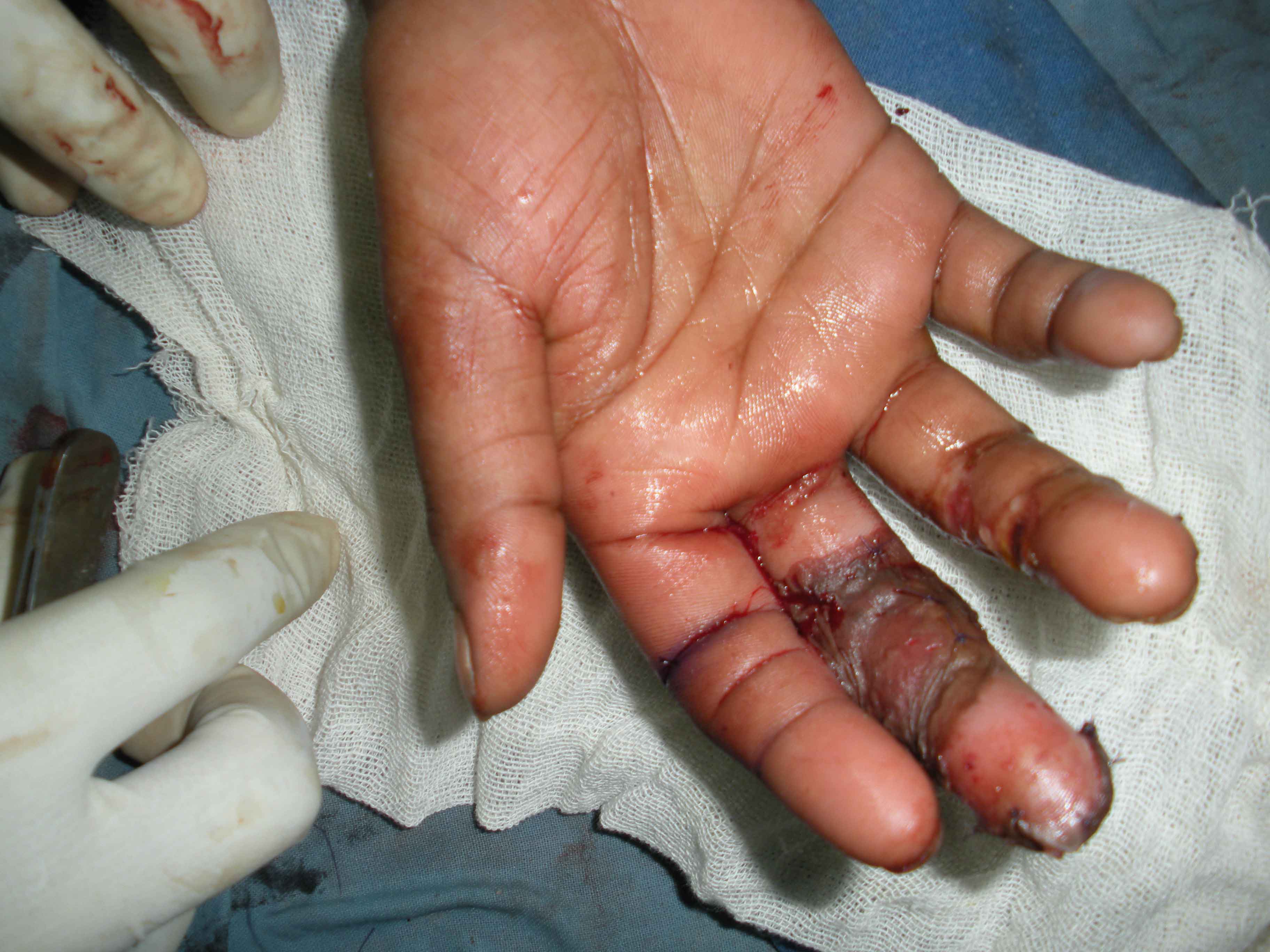
The surgery was performed under wrist block/digital block. Tourniquet was used and the procedure done with loupe magnification. The critical area of the defect was marked. The donor fingers were selected by planning in reverse and considering that finger which would be most comfortable for the patient in the post–operative immobilisation state. Those adjacent fingers which were comfortable, but, injured, were not considered as a donor finger. The skin (epidermis and dermis) were raised, like a page of the book with the base being on the contralateral side of the recipient finger. The skin was elevated, without transgressing the neuro-vascular line of the digits. The adipofascial flap, included the dorsal veins, fat and fascia was raised like a page of a book, with the base on the Ipsilateral side [Table/Fig-1,2]. The flap [Table/Fig-3] was inset to the defect and Split thickness skin graft that was minimally hand meshed was covered over the flap. The Skin graft was harvested from the medical side of arm in all our patients.The fingers were immobilised for 3 weeks. After 3 weeks, flap was divided and fingers mobilized slowly. Adequate physiotherapy was given after the wound healed.These patients were given crepe bandage compression and were advised for regular massage over the donor finger and recipient fingers for six months. Regular follow-up was done for 6 months.
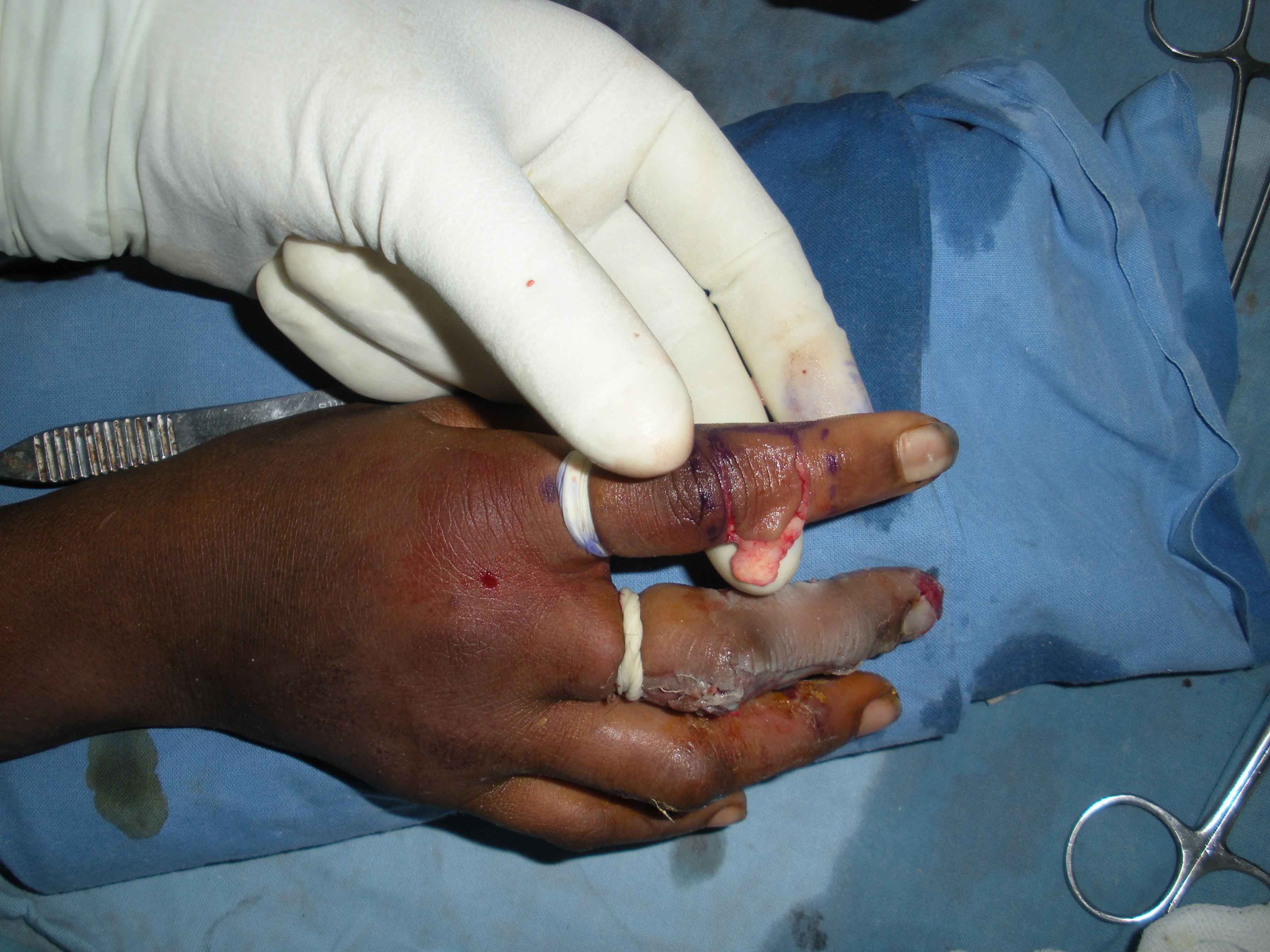
Per-operative photo, Adipofascial flap from the index finger was raised like a page of a book and skin over the dorsum raised like a page of the book. Both of them are based on opposite sides
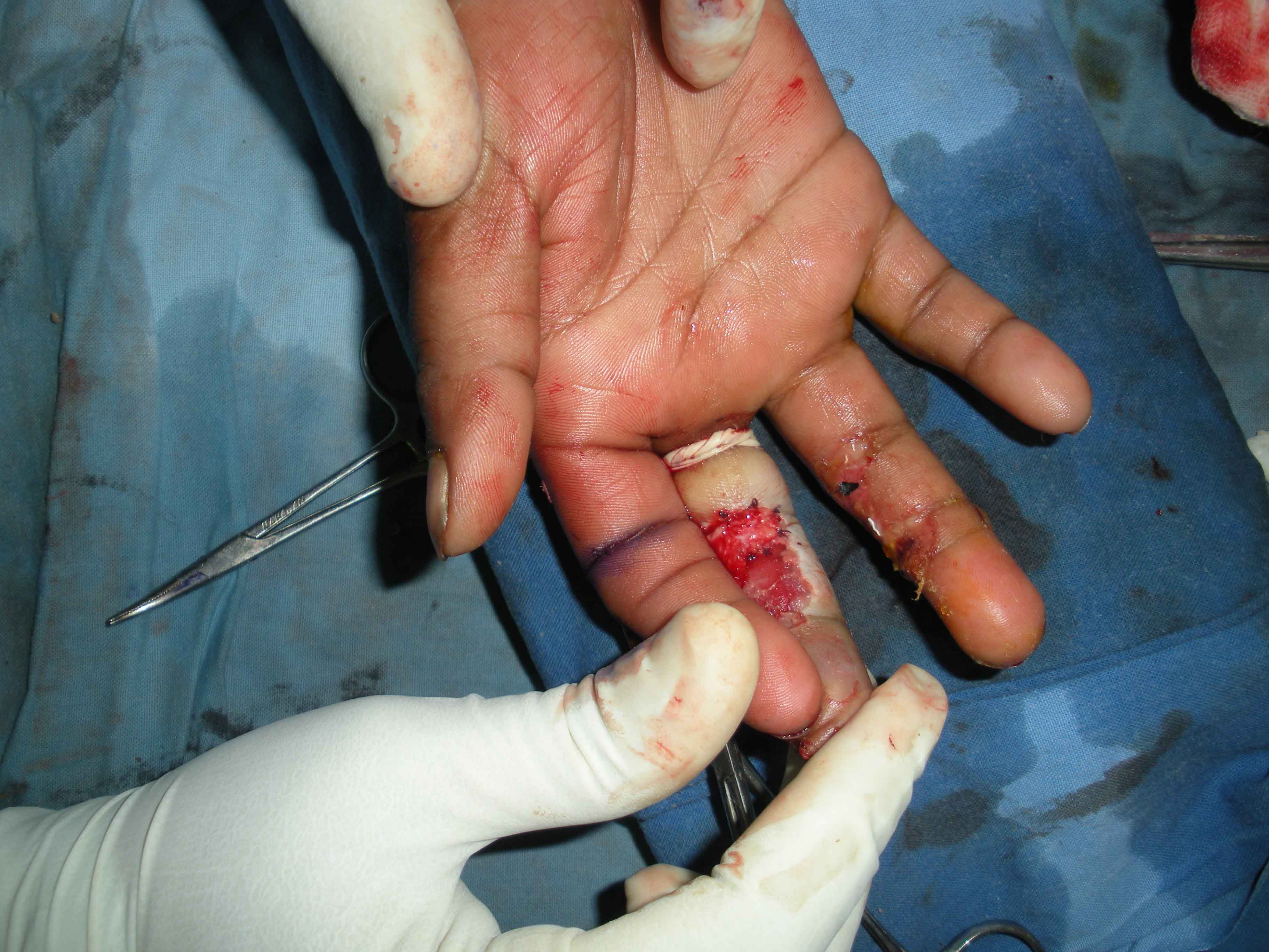
Per- operative photo, Adipofascial flap is inset into a palmar defect
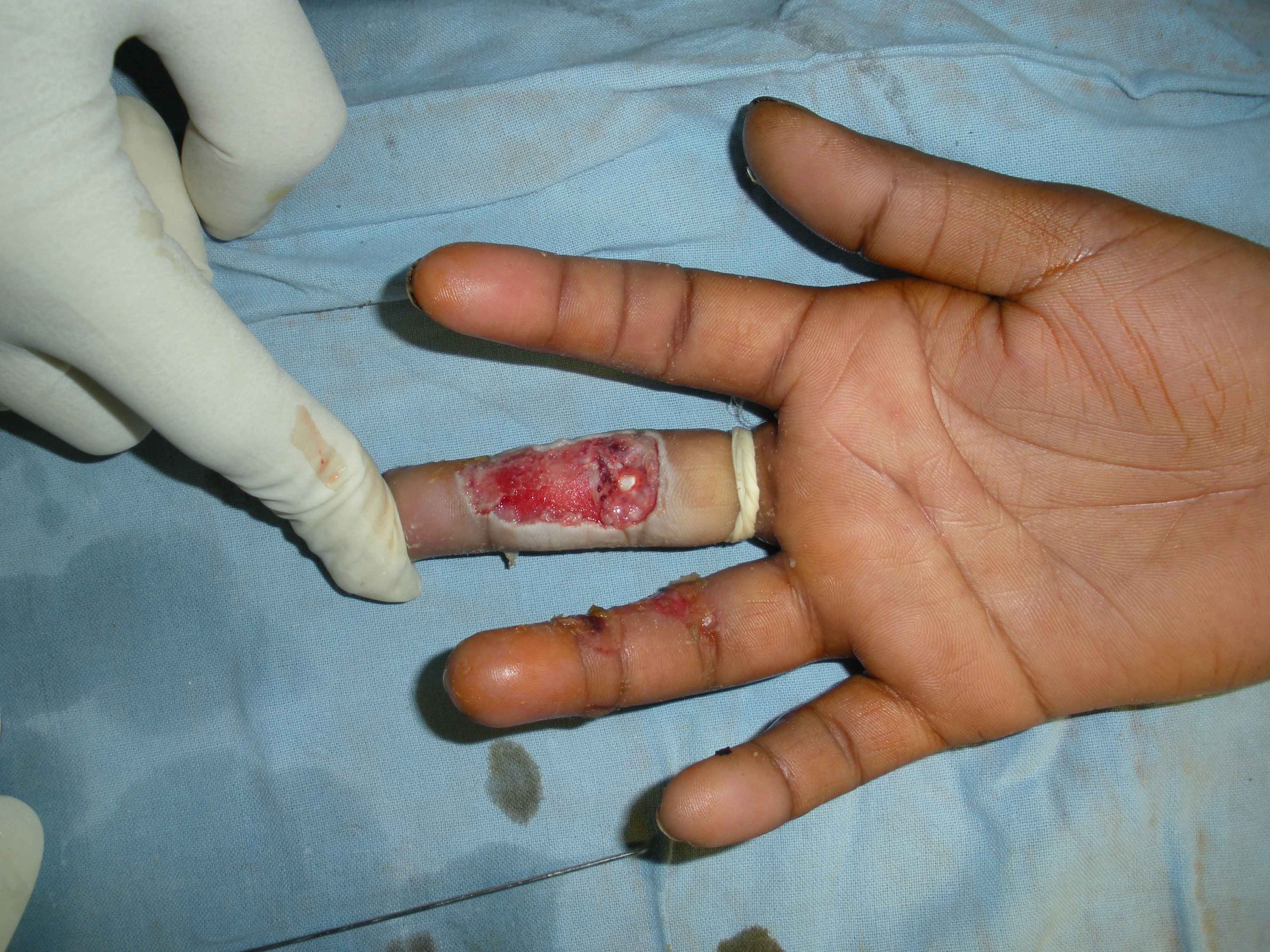
Per-operative picture after covering the recipient site with adipofascial flap and skin graft
Complications
We had complications with respect to the recipient finger. There were minotions in five patients. Minor infections were defined, as those with mild soakage of the dressings and wound swab showing no growth. These patients settled with regular dressings. There was partial loss of SSG, in five patients. We defined partial loss of SSG in those, who had lost, less than 20% of the total skin grafted area. These cases settled with regular dressings [1].
There was complete loss of graft in one case. It was re-grafted, as the flap was viable. There was no problem with respect to the donor finger. We did not encounter any complication with respect to the donor fingers [2]. At the end of six months, assessment was done for sensation, mobility, functional ability, appearance, colour match, patient satisfaction for the donor and recipient finger. They were graded from 1-4 (excellent, good, fair, poor). Mobility and functional assessment were done according to Graham Lister [3].
At the end of six months, these patients were asked to rate about appearance, function, wound healing, independently, without seeing other cases. These patients scored for both the donor and recipient fingers.
Regular standard photographs were taken as per the protocol of the institute.The photos were the pre–operative, peri–operative and late post–operative time.
We selected photos of 15 patients from our department archive; these were photos of the standard cross finger flap and care was taken to pair the photos, so that they have flap, donor site, recipient site at same anatomical position, similar skin colour, no tendency of hyper tropic scarring, similar range of movements.
The photos of the patients of this series, and the paired photos were given to a set of 10 independent, non-medical literate people. These people rated the photos independently with regards to appearance, between 1-4(excellent, good, fair, poor). We did a non randomised comparitive study of the scores given by the reviewers. We calculated the mean of scores given by each reviewer for the photos of donor site and recipient site of adipofascial flap and standard cross finger flap. Further we calculated the reliability, cornbach’s alha, inter rater correlation coefficient to overcome the bias.
Results
The patients were aged between 10-45 years. There were 6 males and 9 females. All patients presented with history of trauma. Off the 15 patients, 13 patients presented within 72 hours of the trauma, 2 patients presented after 7 days of trauma. Fourteen patients presented with defects on the dorsum of the fingers. One patient presented with defect on the palmar side of the finger. Among these patients thumb was involved in two patients, index finger was involved in two patients, middle finger was involved in four patients, ring finger in five patients, and little finger in two patients.
The Index finger was donor site for four fingers, middle finger for seven patients, and ring finger for four fingers. In this series, middle finger was used as donor fingers for two index and five ring finger.The index finger was used for two thumb and two middle fingers. The ring finger was used for two middle fingers and two little fingers. The defect size varied from 2.25 square centimetres [Table/Fig-4] to 6.25 square centimetres with an average of 3.75 square centimetres.
The site, size of the defect, donor site and the complications
| Case No. | Defect | Donor finger | Size of the defect | Complications |
|---|
| 1 | Thumb, distal phalanx, dorsal | Index | 4 sqcm | Nil |
| 2 | Thumb distal phalanx, dorsal | Index | 3 sqcm | Nil |
| 3 | Index, distal phalanx, dorsal | Middle | 2.25 sqcm | Minor infection |
| 4 | Index, middle phalanx, dorsal | Middle | 5 sqcm | Partial SSG* loss |
| 5 | Middle, middle phalanx, palmar side | Index | 2.25 sqcm | Nil |
| 6 | Middle, middle phalanx, dorsal | Ring | 4 sqcm | Partial loss SSG* |
| 7 | Middle, distal phalanx, dorsal | Ring | 4 sqcm | DIP joint arthrosed |
| 8 | Middle, middle phalanx,dorsal | Index | 5 sqcm | Partial loss SSG* |
| 9 | Ring, distal phalanx, dorsal | Middle | 2.25 sqcm | DIParthrosed |
| 10 | Ring, middle phalanx, dorsal | Middle | 2.25 sqcm | Partial loss SSG* |
| 11 | Ring, middle phalanx, dorsal | Middle | 5 sqcm | Partial lossSSG* |
| 12 | Ring, distal phalanx, dorsal | Middle | 6.25 sqcm | Nil |
| 13 | Ring, distal phalanx, dorsal | Middle | 3 sqcm | Nil |
| 14 | Little, distal phalanx, dorsal | Ring | 4 sqcm | Nil |
| 15 | Little, distal phalanx, dorsal | Ring | 4 sqcm | SSG total loss |
SSG*- split thickness skin graft DIP- distal interphalyngeal joint sqcm-squarecentimeter
Mobility of the fingers were of normal range, when compared to the contralateral fingers, in 13 cases. In 2 cases (case no7 and case no 9) we had to arthrodise the DIP joints, the other joint movements were of normal range in comparison to the contralateral finger. Ten patients developed sensation, with a two point discrimination of less than 8 mm similar to that of standard cross finger flap [4]. Five patients developed sensation, with a two point discrimination of more than 8 mm.
Thirteen of the patients were able to use their fingers, respectively with good function [Table/Fig-5, 6]. Two patients had DIP arthrodised of the ring and middle fingers. These two patients were bothered only with functions associated with flat hand. They did not opt for any further procedures. All the patients were satisfied with wound healing, appearance [5], function.
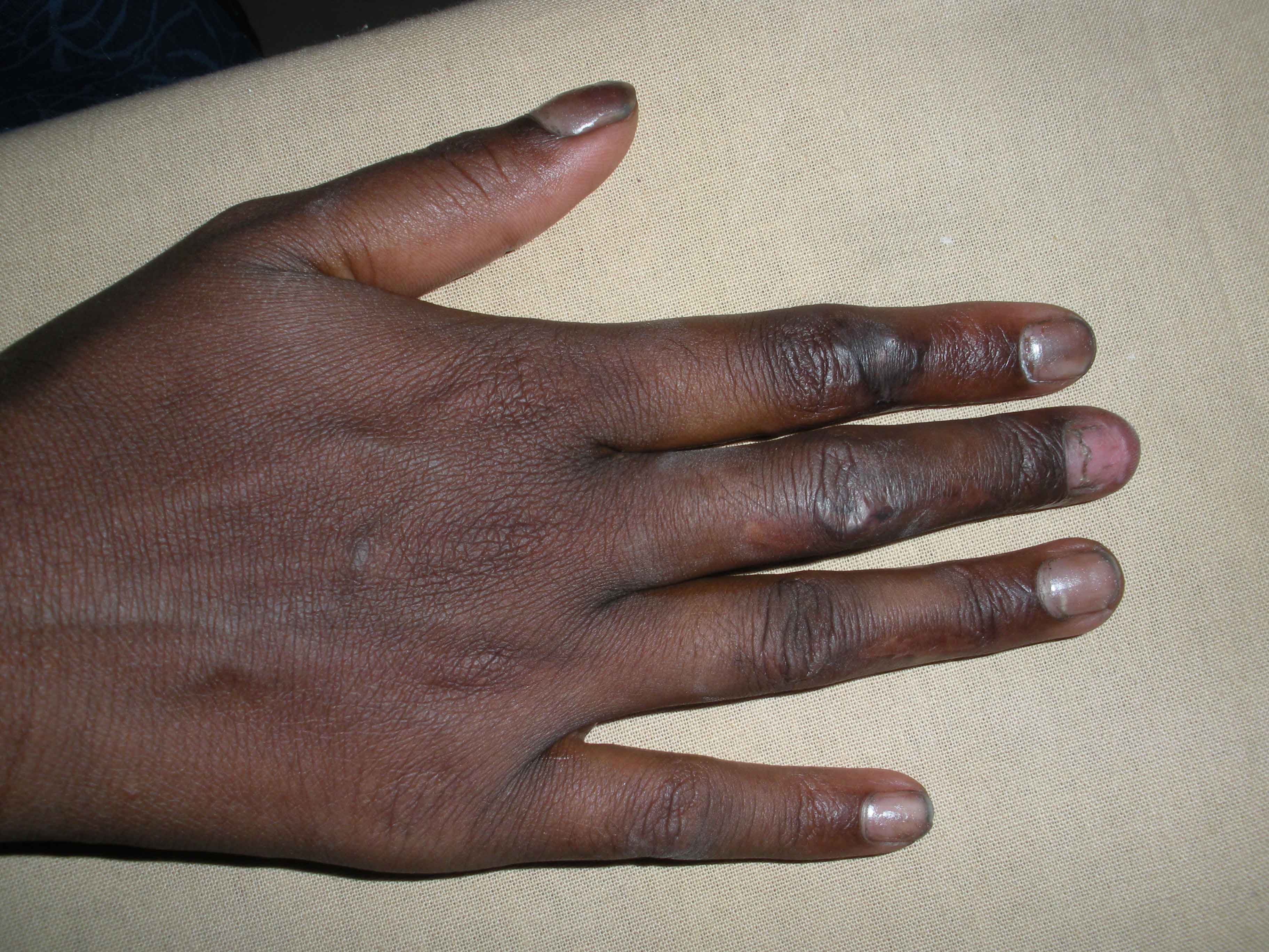
Post-operative photo after 6 months - dorsum of the hand shows well healed scar on the dorsum, which is better than a patch of skin graft
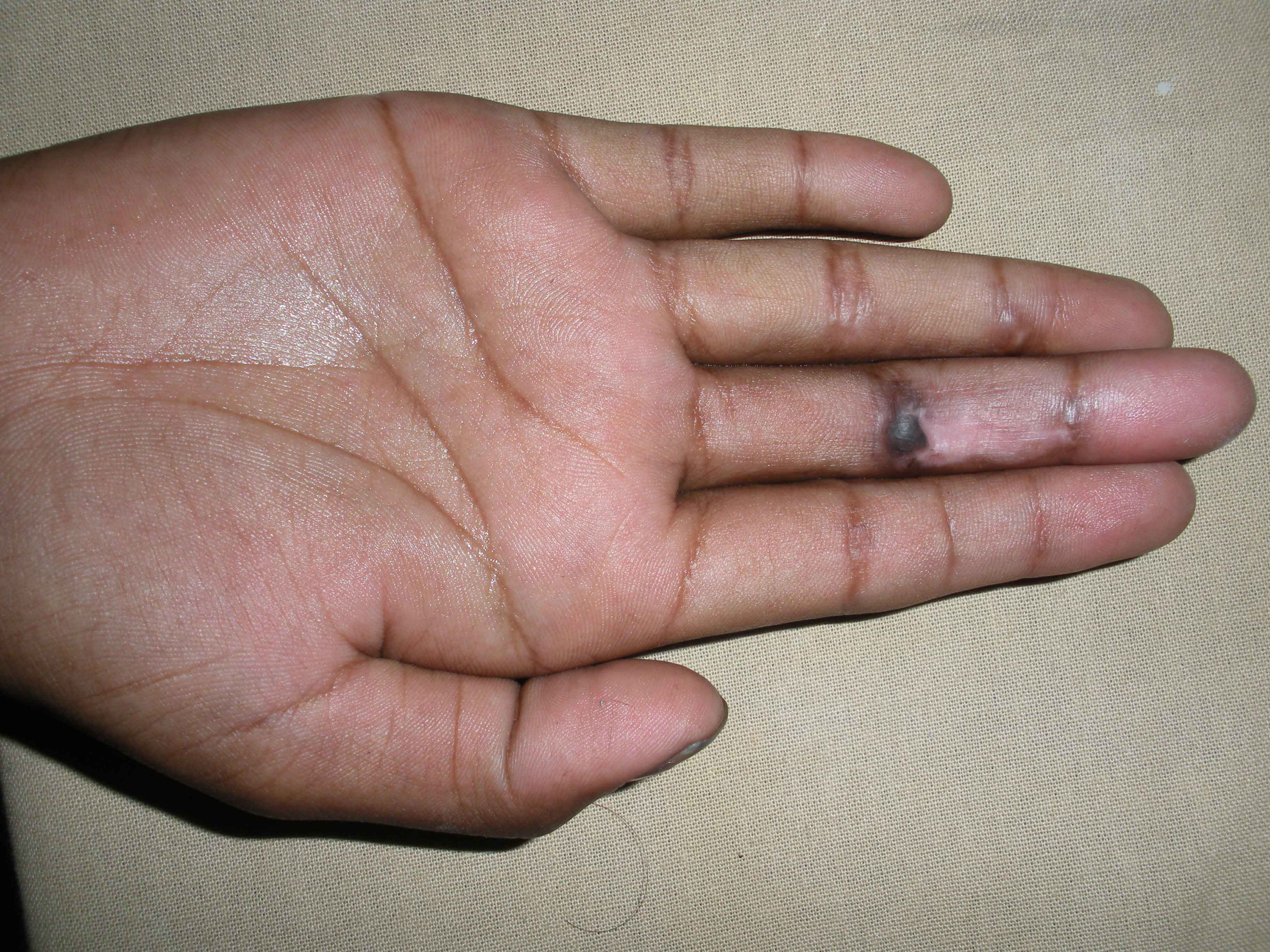
Post operative photo- six months- Recipient site on the middle finger where skin graft has contracted and the scar is small. Further patient has no problem in full extension of the finger
All patients scored their donor fingers 4 out of 4. Nine patients scored their recipient site 3 out of 4 and six patients scored 2 out of 4. The non–medical reviewers scored on the photos of the patients with Adipofascial cross finger flap and the photos of those patients with standard cross finger flap. The mean score that each of the reviewers have rated for the photos are as given in the [Table/Fig-7]. The mean score of all the reviewers for the donor finger in case of Adipofascial flap was 3.859 as compared to the donor finger of the standard crossfinger flap which was 3.6. The mean score of all the reviewers for all the recipient finger of the Adipofascial flap was 2.681, which was more than that of the Standard cross finger flap which was 2.119.
Comparison of mean score of reviewers of photos for appearance score
| Reviwers | A | B | C | D | E | F | G | H | I | J | Mean scores of all the reviewers |
|---|
| Adipofascial cross finger flap |
| Donor finger | 3.87 | 3.93 | 3.80 | 3.93 | 3.87 | 3.73 | 3.80 | 3.93 | 3.80 | 3.93 | 3.859 |
| Recipient finger | 2.67 | 2.67 | 2.80 | 2.80 | 2.80 | 2.53 | 2.67 | 2.67 | 2.53 | 2.67 | 2.681 |
| Standard cross finger flap |
| Donor finger | 3.67 | 3.60 | 3.53 | 3.60 | 3.53 | 3.67 | 3.60 | 3.53 | 3.60 | 3.67 | 3.6 |
| Recipient finger | 2.20 | 2.13 | 2.13 | 2.13 | 2.13 | 2.07 | 2.13 | 2.07 | 2.13 | 2.13 | 2.119 |
What about bias. Inter-observer co-relation? [Table/Fig- 8]
| Numbers | Adipofascial cross finger Flap | Standard cross finger flap |
|---|
| Donor finger | Recipient finger | Donor finger | Recipient finger |
|---|
| Reliability | 0.571 | 0.894 | 0.828 | 0.863 |
| Cronbach’S Alpha | 0.765 | 0.925 | 0.797 | 0.937 |
| ICC | 0.767 | 0.927 | 0.807 | 0.941 |
The reviewers agreed upon that donor finger and recipient finger in case of adipofascial flap were aesthetically better than the standard cross finger flap as the mean score for the adipofascial flap fingers were more than that for the standard cross finger flap.
The consistency/reliability of the interpretors were acceptable for the donor fingers of the adipofascial and standard flap, excellent in case of the recipient fingers of the adipofascial flap and the standard cross finger flap.
The inter–class co-relation coefficient was, strong agreement in case of donor finger adipofascial flap, perfect agreement for the donor finger of the standard cross finger flap and the recipient fingers of adipofascial flap and standard cross finger flap [Table/Fig-9 & 10].
Pre–operative photo, palmar complex defect of the middle finger with flexor tendon exposed
Per-operative photo, finger after closure of the skin on the donor site of the index finger
Discussion
It is as robust a flap as a standard cross finger flap. It is an aesthetically better accepted flap with the native skin draped and absence of SSG on the donor site. The fascia allows the skin graft to contract on the recipient site; hence, it decreases the scar on the recipient site. Even with contracted SSG, we did not find any appreciable problems with the movement. The total area of flap size as seen on the recipient site is less than that of conventional cross finger flap. This is due to contraction of the SSG. It has no complications like inclusion cysts like in the de-epithelised cross finger flap [6]. The mobility may be better with the adipofascial cross finger flap since, the protective adipofascial system was over the tendon instead of the lubricant adipofascial system [7].
Any abnormalities over the hand are appreciated by the people, since it is not always covered. Hence, the need for better aesthetic outcomes is necessary. With that intent, we tried to compare the adipofascial flap and the standard cross finger flap from a non medical persons’ view. The reviewers rated the donor finger better in terms of appearance when they compared it with the standard cross finger flap.
With our results we would like to state that the donor and recipient fingers in case of adipofascial flap looked better than the standard cross finger flap. The reliability of interpretors were acceptable for the donor fingers of adipofascial flap and standard cross finger flap, who have given a mean score of 3.859 and 3.6 respectively.
The recipient fingers were definitely better looking in case of the adipofascial flap, as the cornbachs’s alpha is >0.9 and the mean values being 2.6 and 2.1 for the adipofascial flap and the standard cross finger flap.
The inter–class correlation coefficient shows that the interpretors had strong agreement in case of donor finger adipofascial flap, perfect agreement for the donor finger of the standard cross finger flap and the recipient fingers of adipofascial flap and standard cross finger flap.
Our study design was for 15 patients, which may be small to conclude that Adipofascial flap has a better aesthetic outcome. Our study compares photos of only Standard cross finger flap with the Adipofascial cross finger flap.
Through the present study, we recommend that adipofascial cross finger flap is aesthetically better than the standard cross finger flap with respect to appearance from non medical reviewer’s point of view.
Conclusion
We would like to conclude that the Adipofascial flap is a robust flap for finger defects. We recommend that this flap is aesthetically better than the standard cross finger flap.
Limitation
Our study involved only fifteen subjects, we have compared only standard cross finger flap and adipofascial flap, we cannot say categorically that the Adipofascial cross finger flap gives the best aesthetic results.
SSG*- split thickness skin graft DIP- distal interphalyngeal joint sqcm-squarecentimeter
[1]. Atasoy E, Reversed cross finger subcutaneous tissue flap. In Luis o Vasconez, Elizabeth J hall-findley editorGrabb’sencylopedia of flaps 1998 2nd edNew yorkLippincott-Raven:912-15. [Google Scholar]
[2]. Nawfal Fejjal, Redouane Belmir, Samir El Mazouz, Noureddine Gharib, Abdellah Abbassi, Amin Belmahi, ‘Reversed cross finger subcutaneous flap: a rapid way to cover finger defects’Indian Journal of Plastic Surgery41(1):55-57. [Google Scholar]
[3]. Graham Lister, ‘Reconstruction’, in Graham Lister (ed.)The Hand: Diagnosis and Indications3rd editionEdinburghChurchill-Livingstone:158-61. [Google Scholar]
[4]. Kleinert HE, McAlister CG, Macdonald CJ, Kutz JE, ‘A critical evaluation of cross finger flap’Journal of Trauma14(9):756-63. [Google Scholar]
[5]. Al-Qattan MM, ‘The Cross-digital dorsal adipofascial flap’Annals of Plastic Surgery60(2):150-53. [Google Scholar]
[6]. Al-Qattan M.M., ‘De-epithelialized cross-finger flaps versus adipofascial turnover flaps for the reconstruction of small complex dorsal digital defects: a comparative analysis’Journal of Hand Surgery May,2005 30(3):549-57. [Google Scholar]
[7]. Hideo Nakajima, Nobuaki Imanishi, Toshiharu Minabe, Kazuo Kishi, Sadakazu Aiso, ‘Anatomical study of subcutaneous adipofascial tissue: a concept of protective adipofascial system and lubricant adipofascial system’Journal Plastic Surgery and Hand Surgery38(5):261-66. [Google Scholar]