Acute-Onset of Multiple Painful Nodules over Forearms and Back
Praveen Kumar S.1, Sulatha M. Kamath2, A.L. Shyam Prasad3, Vijaya V. Mysorekar4, T.K Sumathy5
1 Assistant Professor, Department of Skin and STD, M.S. Ramaiah Medical College and Hospital, Bangalore, India.
2 Professor, Department of Pathology, M.S. Ramaiah Medical College and Hospital, Bangalore, India.
3 Professor & Head, Department of Skin and STD, M.S. Ramaiah Medical College and Hospital, Bangalore, India.
4 Professor, Department of Pathology, M.S. Ramaiah Medical College, Bangalore, India.
5 Senior Professor, Department of Dermatology, M.S. Ramaiah Medical College and Hospitals, Bangalore, India.
NAME, ADDRESS, E-MAIL ID OF THE CORRESPONDING AUTHOR: Dr. Praveen Kumar S., 1547, Saikrupa, 26th Main, 26th Cross, HSR Layout 2nd Sector, Bangalore-560034, India.
Phone: 09886320760,
E-mail: drpraveen.1982@gmail.com
Angiolipomas are benign encapsulated, well circumscribed tumours, which show excessive degree of vascular proliferation. Clinically, lesions present as sudden onset of multiple painful nodules. Pain usually does not respond to analgesics. We herein, report a case of a young male, presenting with multiple painful nodules over the forearm and back, which on histopathological examination revealed, encapsulated benign tumour, comprising of proliferated small-caliber vascular channels with microthrombi and variable amounts of mature adipose tissue. Pain subsided on treatment with intralesional steroids and the nodules were excised through a narrow-hole extrusion technique.
Angiolipoma, Intralesional steroids, Nodule
Case Report
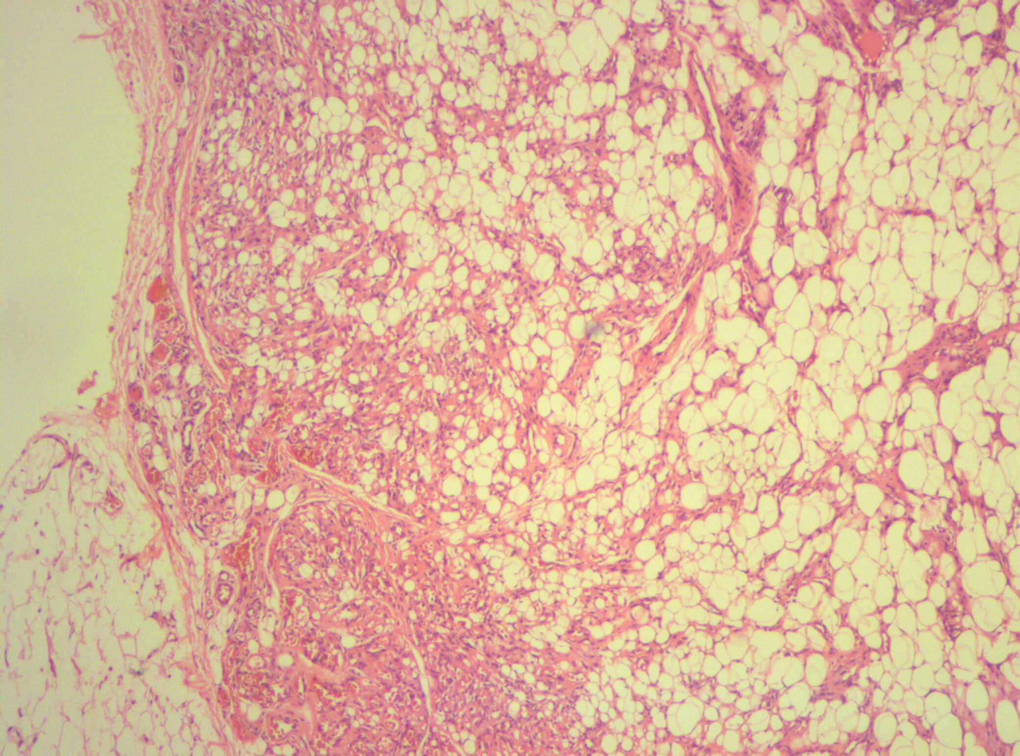
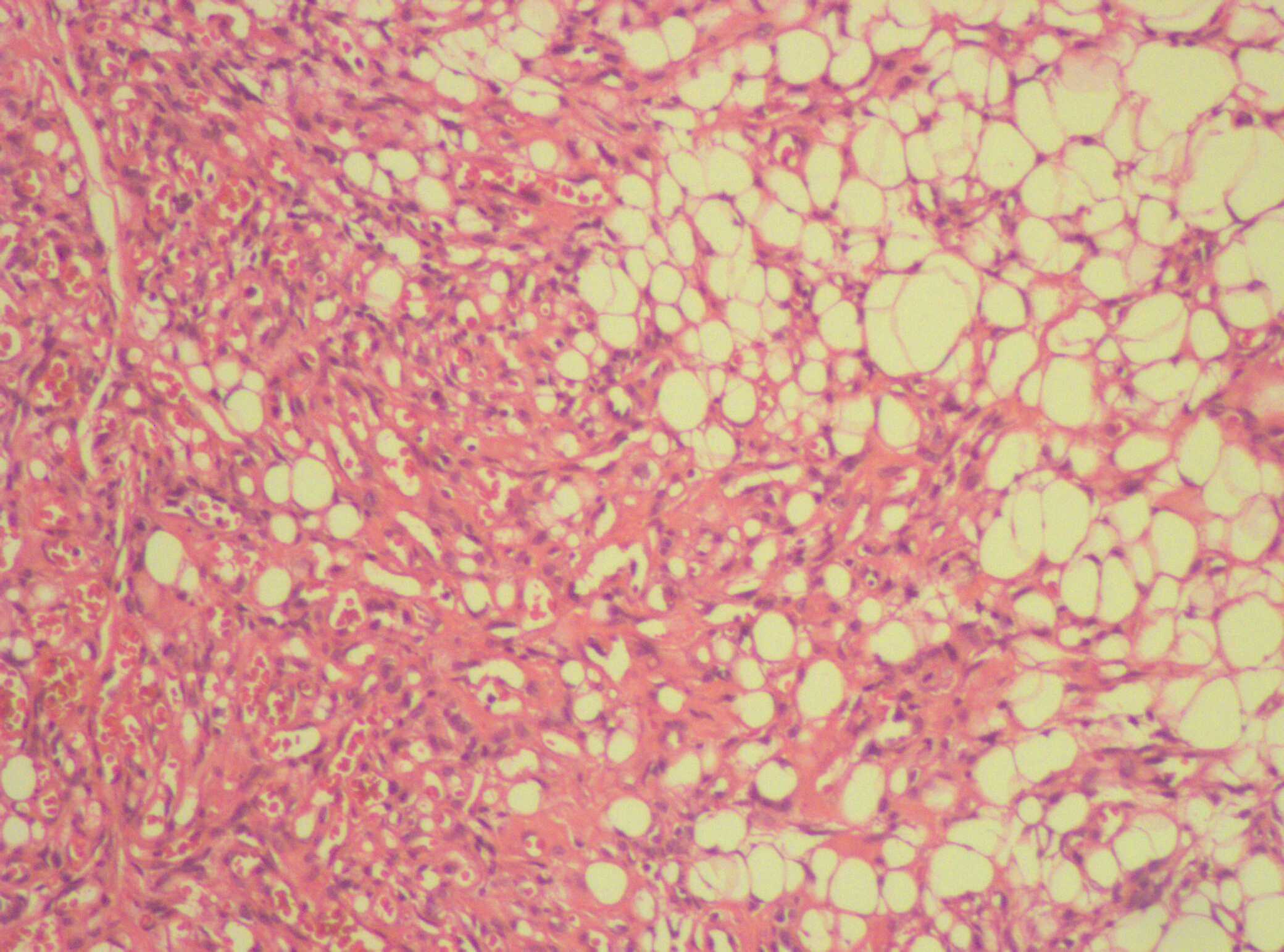
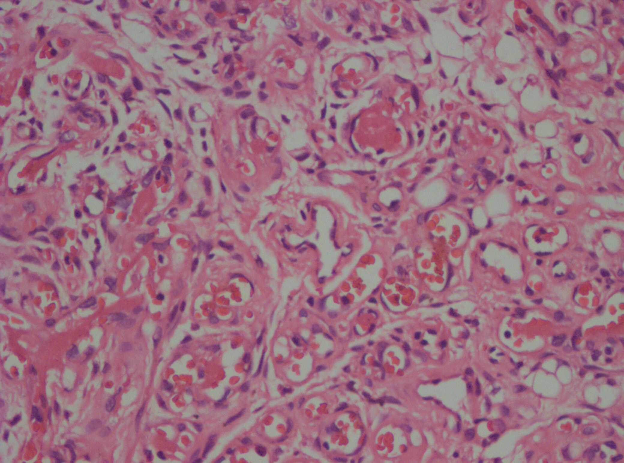
A 15-year-old male presented with 1 month history of multiple painful skin lesions over forearms and back. There was no history of sore-throat, fever, weight loss or joint pains. On examination, multiple, sessile skin-colored nodules around 2 x 2 cms in size with diffuse borders over forearms and lower back were present. On palpation, nodules were firm in consistency with diffuse margins and tenderness was elicited. Slip-sign was positive. Routine investigations were within normal limits. With a provisional diagnosis of erythema nodosum, patient was treated for pain with oral Diclofenac and subsequently with oral Indomethacin (75mg). Patient did not show any improvement. Skin biopsy on histopathological examination revealed well-circumscribed, encapsulated, lobulated benign tumor, composed of numerous proliferated capillaries, engorged with erythrocytes and variable amounts of mature adipose tissue [Table/Fig-1] in the subcutaneous fat. The capillaries were well-formed, dilated and showed no endothelial atypia [Table/Fig-2]. Fibrin thrombi in vessel wall [Table/Fig-3] with scant perivascular fibrosis was seen. The pain subsided with intralesional triamcinolone acetonide injections and the other nodules were excised through a narrow-hole extrusion technique.
Sub-cutaneous encapsulated nodule showing proliferated capillaries and mature adipocytes (H & E, X 100)
Close-up view of the proliferated capillaries and mature adipocytes. (H & E, X 400)
Fibrin thrombi in the vessel lumina (H & E, X 400)
Discussion
Angiolipomas are benign encapsulated lobulated tumors, which show an excessive degree of vascular proliferation. As a rule, they arise in young adults. Approximately less than 10% of lipomas examined pathologically are angiolipomas [1]. Clinically, lesions present as painful skin colored bluish nodules 0.5 cm to 5 cm in diameter, resembling lipomas. The most common site involved is forearm and multiple lesions are common.
Histopathology of angiolipoma is diagnostic. Inapparent at the gross level, angiolipomas microscopically show sharp encapsulation and small-caliber vascular channels with small microthrombi and variable amounts of mature adipose tissue [2]. Small angiomatous foci to dense vascular and stromal tissue may be seen. Perivascular fibrosis may be prominent. Non-infiltrating lipomas are typically multiple, painful nodules [3]. Cellular angiolipoma can be confused with a vascular tumors, especially immature capillary hemangioma and Kaposiform hemangioendothelioma. In both these lesions, mature adipose tissue is absent. In addition, Kaposiform hamangioendotheliomas [4] are infiltrative lesions with fascicles of bland endothelial cells. Spindle cell lipomas lack encapsulation, have slender uniform spindle cells in a mucinous matrix [2,5]. In our case, the lesion was in the subcutaneous adipose tissue with all the histological features as described above. This did not infiltrate the muscular or osseous tissue.
Angiolipomas are exceptionally known to be associated with diabetes mellitus or as a complication of antiretroviral therapy [6]. In our case, the patient was neither a diabetic nor immunocompromised.
Multiple lesions are treated with atenolol and I.V. lignocaine [1]. Patients usually do not respond to analgesics. Solitary infiltrating angiolipomas need wide surgical excision [7]. In our case, lesions were treated with intralesional steroids and pain subsided with one session of treatment. Other nodules were surgically excised with narrow-hole extrusion technique. Patient is currently under follow-up.
Conclusion
This case is presented to highlight the differential diagnosis of angiolipomas as a possibility, with sudden onset of multiple painful nodules. The case specifically highlights the histopathological aspects of angiolipoma as a vital clue to the diagnosis. The response of pain to the intralesional steroids is highlighted.
[1]. McGibbon DH, Subcutaneous Fat. In: Burns T, Breathnach S, Cox N, Griffiths C, EdsRooks textbook of Dermatology 2010 Eigth editionWiley-Blackwell publications:46.44-46.45. [Google Scholar]
[2]. Ragsdale BD, Tumors with Fatty, Muscular, Osseous, and/or Cartilaginous Differentiation. In: Elder D.E.,edsLippincott Lever’s Histopathology Of The Skin 2009 Tenth editionWilliams & Wilkins publications:1065-66. [Google Scholar]
[3]. Ghosh S, Haldar B, Biswas A, Multiple AngiolipomasIndian J Dermatol Venerol Leprol 1990 56:143-4.2007; (2): 1531 [Google Scholar]
[4]. Fletcher CDM, Soft-tissue tumoursIn: Diagnostic Histopathology of Tumours, Fletcher CDM 2007 (2)Churchill LivingstoneElsevier publications:1531 [Google Scholar]
[5]. Bartley GB, Yeatts RP, Garrity JA, Farrow GM, Campbell RJ, Spindle cell lipoma of the orbitAm J Ophthalmol 1985 Oct 15 100(4):605-9. [Google Scholar]
[6]. Dank JP, Colven R, Protease inhibitor-associated angiolipomatosisJ Am Acad Dermatol 2000 42:129-31. [Google Scholar]
[7]. Tighe C, Lynn JA, Angiolipomas of the footJ Am Pediatr Med Assoc 1994 84:85-9. [Google Scholar]