Case One
A 19–year-old-man, an Iranian, a professional athlete, (weight, 65 kg; height, 170cm), a candidate for varicocele surgery under general anaesthesia. He was without any history of previous anaesthetic exposure, illness, use of drug and history of allergy. Chest X–ray, ECG, laboratory tests and physical examination were within normal limits. The patient was accepted for anaesthesia, as he had ASA I physical status.
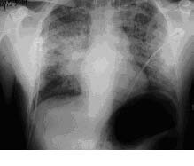
In the operation room, after placing standard monitors, the patient was premedicated with injection fentanyl 100 μgm intravenous and general anaesthesia was induced with Thiopental Sodium 5 mg/kg, Atracurium 0.5 mg/ kg . He was intubated without problems using a 7.5 mm oral endotracheal tube that was fixed 22 cm from the mouth. Over the operative period (1 h), 1000 mL of lactated Ringer’s solution was given. Anaesthesia was maintained by Halotan 0.8%, N 2o: O2 (50 : 50) and Atracurium. The tracheal intubation, anaesthesia, mechanical ventilation, and surgical procedure were uneventful. At the end, oropharyngeal suctioning was done and reversed using injection Neostigmine 2.5 mg IV, and injection Atropine Sulfate 1.0mg IV and subsequently, the trachea was extubated. Directly after extubation, the patient developed laryngospasm and became agitated. Respiratory distress, tachypnoea and cyanosis were observed. Spontaneous ventilation with 100% oxygen could not be assisted by bag and mask ventilation sufficiently and he developed laryngospasm with severe respiratory distress. Subsequently, arterial oxygen saturation decreased to a low 70. The patient was reintubated after administration of Succinylcholine (1mg.kg) and Dexamethasone 8mg. Suctioning of the tube revealed copious amounts of pink, frothy sputum. Chest radiograph showed a small right lower lobe infiltrate. In operation room, he was placed on mechanical ventilation and treated with positive end- expiratory pressure (PEEP) of 10 cm H2O, high fractional inspiratory oxygen concentration (FIO2), intravenous furosemide and morphin initially. Arterial blood gas levels were within normal limits. His extubation was successfully carried out 4 hours after his initial reintubation. He had an uneventful recovery and was transferred to the surgical ward. In surgical ward, remarkable note was derangement in coagulation profile, prothrombin time (PT) and partial thromboplastin time (PTT) (PT=24sec, PTT=54sec). The patient recovered completely and was discharged home on the postoperative day.
Case One – Chest X-ray showing pulmonary edema
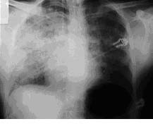
Case Two – Chest X-ray showing pulmonary edema
Case Two
Our second case, brother of the case one, underwent general anaesthesia for septoplasty 3 years later in our hospital. A 17 year- old-man, an Iranian,and a professional athlete, (weight, 82 kg; height, 187 cm). He was also without history of previous anaesthetic exposure, illness, drug use and history of allergy. Chest X–ray ,ECG, laboratory tests and physical examination were within normal limits. The patient was accepted for anaesthesia, as he had ASA I physical status. In the operation room, after placing standard monitors, the patient was premedicated with injection fentanyl 100 μgm intravenous and general anaesthesia was induced with Thiopental sodium 5 mg/kg , atracurium 0.5 mg/ kg. He was intubated without problems using a 8 mm oral endotracheal tube that was fixed 22 cm from the mouth. Over the operative period (2 h), 2000 mL of lactated Ringer’s solution was given. Maintenance of anaesthesia was done by Halotan 0.8%, N2o : O2 (50 : 50) and Atracurium. The tracheal intubation, anaesthesia, mechanical ventilation, and surgical procedure were uneventful. At the end, oropharyngeal suctioning was done and reversed using injection Neostigmine 2.5 mg IV, and injection Atropine Sulfate 1.0mg IV and subsequently, the trachea was extubated. Immediately after extubation, the patient developed laryngospasm and became agitated. Respiratory distress, tachypnoea and cyanosis were observed. As oxygen saturation of > 90% with bag and mask ventilation was difficult to be maintained, the patient was reintubated after administration of succinylcholine (1mg.kg). Suctioning of the tube showed copious amounts of pink, frothy sputum. The chest radiograph showed diffuse alveolar and interstitial infiltration, consistent with pulmonary oedema.
In operation room, he was placed on mechanical ventilation and treated with positive end- expiratory pressure (PEEP) of 10 cm H2O, high fractional inspiratory oxygen concentration (FIO2) , intravenous furosemide and morphin initially. Arterial blood gas levels were within normal limits. Four hours later, in operation room, the patient with endotracheal tube was transferred to the medical intensive care unit (ICU) for further management. In the ICU, he was ventilated (SIMV mode) with oxygen 100%, PEEP= 10 cm H2O, PR=12, TV=750, PS=10, FIO2=10%. Intravenous furosemide and morphin were given. All laboratory blood investigations were found to be within normal limits, but PT, PTT and Normalized ratio (INR) were deranged (PT=20 sec, PTT= 32sec, INR=2.5 Ratio). For patient management, 4 units of FFP were given. His extubation was successfully carried out 10 hours after his initial reintubation. A day later, he had an uneventful recovery and was transferred to the surgical ward. Two days later, his chest radiograph showed a complete resolution of the pulmonary infiltrate and was discharged home [Table/Fig-1,Table/Fig-2].
Discussion
Our current report had several important points. Firstly, both of them were brothers. Secondly, both of them were athletes and thirdly, their PT, PTT and INR were disturbed. NPPE majority before surgery was determined by anaesthetists as laryngospasm issue [1–4]. Young athlete males who undergo head and neck surgeries or painful procedures are more prepared for NPPE than normal patients, because their muscular chest walls could create very high inspiratory pressures [5–7]. NPPE incidence is reported to range from 0.05 to 1%, which is related to all anaesthesia usages [8]. Mortality is more than 40% if NPPE is not detected [9].
Risk factors for NPPE include hanging, strangulation, upper airway tumours, foreign bodies, epiglottitis, croup, choking, hiccups, migration of Foley’s catheter balloon, used to tamponade the nose in epistaxis, near drowning, ETT obstruction, goitre , mononucleosis, post tonsillectomy/ adenoidectomy, postremoval of upper airway tumour, difficult intubation, Ludwig’s angina, choanal stenosis, hypertrophic redundant uvula. NPPP is categorized as type I and type II. Type I is established immediately after acute occlusion of air vessel and Type II is created after rescuing from chronic occlusion of top air vessel [3].
The pathogenesis of NPPE includes changes in starling forces, haemodynamic changes, secondary to markedly increased negative intrathoracic pressure ,alveolar hypoxia, increased catecholamine levels diverting the systemic blood to pulmonary system and failure of lymphatics [10]. Although symptoms usually appear in one hour from the event, in the beginning, the symptoms are reported as well [3]. In clinical presentation, symptoms include tacypnoea, tachycardia, rales, decreased oxygen saturation with pink frothy sputum and chest X-ray abnormalities [2].
Treatment includes supplemental oxygen and supportive care, but PEEP and mechanic ventilation could be needed for a long time period. Not all the patients need PEEP and mechanical ventilation, although continuous positive airway pressure (CPAP) in 9 to 18% of cases and controlled mechanical ventilation in 34 to 43% of patients are needed [11–12]. Diuretics help in reversing the osmotic gradient across the alveolar membrane [6]. Use of hydrocortisone has been recommended in cases where laryngeal oedema is suspected. With adequate management, almost all patients improve within 24-48 hours and have a normal chest X-ray. Immediate diagnosis and treatment are needed for a perfect recovery and they reduce the mortality. Prevention is always better than cure and therefore, a smooth extubation, good oral and nasal suction and good judgment of size of endotracheal tube always helps.
Conclusion
NPPE is effective in some young athlete patients. Anaesthetists must have this standpoint, that every patient who is healthy in other conditions is potentially vulnerable to NPPE. On reviewing medical literature, NPPE caused by coagulation factors and inheritance was found to have been reported to be even rarer in patients. Further research is required, to object association between NPPE, coagulation profile and inheritance.
[1]. Vandse R, Kothari DS, Tripathi RS, Lopez L, Stawicki SPA, Papadimos TJ, Negative pressure pulmonary edema with laryngeal mask airway use : recognition, pathophysiology and treatmentIJCIIS 2012 2(2):98-103. [Google Scholar]
[2]. Gupta A K, Ommid M, Waqar Ul Nisa, Shagufta Qazi S, Mehta A, Negative pressure pulmonary edema after emergency appendicectomyPravara Med Rev 2010 2(1):26-9. [Google Scholar]
[3]. Bhaskar B, Fraser JF, Negative pressure pulmonary edema revisited: pathophysiology and review of managementSaudi J A 2011 5(3):308-13. [Google Scholar]
[4]. Alb M, Tsagogiorgas C, Meinhardt JP, Negative pressure pulmonary edemaAnasthesiol Intensivmed Notfallmed Schmerzther 2006 4:64-78. [Google Scholar]
[5]. Davidson S, Guinnc C, Gacharna D, Diagnosis and treatment of Negative pressure pulmonary edema in a pediatric patient: a case reportAANA Journal 2004 72(5):337-8. [Google Scholar]
[6]. Petrou A, Valmas K, Svarna E, Chortaria G, Giamarelou A, Papadopoulos G, Negative pressure pulmonary edema in a patient ventilated with laryngeal maskJournal of Perioperative Medicine 2003 1:69-73. [Google Scholar]
[7]. Raiger LK, Naithani U, Vijay BS, Gupta P, Bhargara V, Non- cardiogenic pulmonary oedema after neostigmine given for reversal: a report of two caseIJA 2010 54(4):338-41. [Google Scholar]
[8]. McConkey PP, Post–obstructive pulmonary oedema : a case series and reviewAnaesth Intensive care 2000 28:72-6. [Google Scholar]
[9]. Goldenberg JD, Portugal LG, Wenig BL, Weingarten RT, Negative pressure pulmonary edema in the otolaryngology patientOtolaryngol Head Neck Surg 1997 117:62-6. [Google Scholar]
[10]. DeSio JM, Bacon DR, Complete airway obstruction caused by a pseudomembranous cast with subsequent negative pressure pulmonary edemaAnesth Analg 1993 76:1142-3. [Google Scholar]
[11]. Van Kooy MA, Gargiulo RF, Postobstructive pulmonary edemaAm Fam Physician 2000 62:401-4. [Google Scholar]
[12]. Bhattarai B, Shrestha S, Negative pressure pulmonary edema – Case series and review of literatureKathmandu Univ Med J 2011 36(4):310-4. [Google Scholar]