Benign Mass in Tonsil- Cavernous Hemangioma
Sumitha Joseph1, M. Prakash2, Hafida K Mohammed3, Aberna Govar4
1 Asistant Professor, Department of Ear, Nose and Throat, Sree Balaji Medical College and Hospital, Chromepet, Chennai, India.
2 Asistant Professor, Department of Ear, Nose and Throat, Sree Balaji Medical College and Hospital, Chromepet, Chennai, India.
3 Senior PG Resident, Department of Ear, Nose and Throat, Sree Balaji Medical College and Hospital, Chromepet, Chennai, India.
4 Junior PG Resident, Department of Ear, Nose and Throat, Sree Balaji Medical College and Hospital, Chromepet, Chennai, India.
NAME, ADDRESS, E-MAIL ID OF THE CORRESPONDING AUTHOR: Dr. Sumitha Joseph, Asistant Professor, Department of Ear, Nose and Throat, 1A, 8th Block, Kences enclave, No. 1, Ramakrishna Street, T. Nagar, Chennai–600017, India.
Phone: 98840 20003,
E-mail: drsjsbmch@gmail.com
Cavernous hemangioma is also called as ‘ANGIOMA CAVERNOSUM’ or ‘CAVERNOMA’ as benign lesion of blood vessels. They are similar to strawberry hemangioma but deeply situated. Although most often associated with skin it is also sometimes found in mucous membrane, brain and the viscera. The diagnosis of hemangiomas is mainly based on clinical evaluation . Isolated hemangiomas in the tonsillar tissue is a rare occurance. In this we report had a case of adult tonsillar hemangioma of left side associated with recurrent tonsillitis . He was effectively managed surgically without any complications.
Tonsillar tissue, Hemangioma, Benign Lesions
Case Report
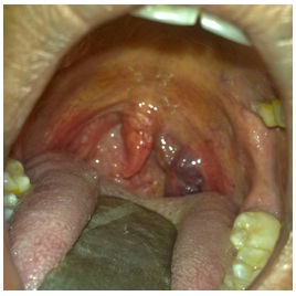
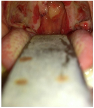
A 28–year–old man presented to Out Patients Department in march 2012 with complaints of recurrent throat pain and fever for the past 5 months. He also gave history of constant foreign body irritation on left tonsillar region. On Examination soft purplish mass was seen filling the left tonsillar region [Table/Fig-1]. On palpation, mass was soft, non-tender, not bleeding on touch. Neck examination revealed non – tender jugulodigastric lymphnode on left side. All other local and systemic examinations were normal. Clinical diagnosis of chronic tonsillitis with left tonsillar hemangioma was made. Since patient was symptomatic tonsillectomy together with excision of hemangiomatous tissue was done under general anesthesia by snare method. The Histopathological report confirmed cavernous hemangioma of left tonsil. Patient had no per operative complication including hemorrhage and is on regular follow up till now [Table/Fig-2].
Soft purplish mass in left tonsillar fossa
Discussion
The most common tumour of head and neck in children are hemangiomas. Tongue and floor of the mouth are the commonest site of the lesions in oral cavity [1]. They are present at birth and gradually increase in size and spontaneously resolve by five years of age. No active surgical intervention is needed in most of the cases. They are histologically benign malformations composed of capillary tangles. Although it is adhered to surrounding parenchyma there is no involvement of parenchyma in hemangioma itself. They do not contain tissue of the organ in which they are situated. Most of the cavernous. Earlier concept that cavernous hemangiomas are congenital but now it is considered to be genetically determined, transmitted in an autosomal dominant pattern. In our case the hemangioma was localized to the tonsillar tissue on one side with no cutaneous lesions.
Benign tumour of tonsil occur infrequently. The first case report of benign neoplasm of tonsil was published by Robert in 1827. The most commonly reported benign tumors are papillomas, angioma, fibroma, adenoma, lymphangioma, teratoma, myxoma, lipoma, chondroma, inclusion cyst and teratogenous cyst. Histological confirmation is very essential for a confirmative diagnosis of these rare benign lesions. Our case was a rare diagnosis of hemangioma of tonsil. Most hemangiomas are developmental in origin and have both hemangiomatous and lymphangiomatous components [1]. Harris and Jakobiec found a 7:3 occurrence ratio of women to men, while Henderson reported an almost equal ratio, 8:7 in women and men. Most cavernous hemangiomas of the head and neck region have recently been renamed as venous vascular malformations [2]. The major distinction between hemangiomas and the venous vascular malformations is that the latter do not involute and may actually grow with time, hormonal influences, infection, thromboses, or trauma Hemangiomas [2] are histologicallly irregular blood filled spaces lined by a single layer of endothelial cells and surrounded by connective tissue. Grossly hemangiomas appear like a well circumscribed red to purple raspberry like lesions. Microscopically it has dilated thin walled vascular caverns lined by a single layer of endothelium and a variable layer of fibrous adventitia. Vascular spaces lack elastic fibres, There may be associated areas of thrombosis and haemorrhage within the vascular spaces. Certain hemangiomas can be associated with venous malformations. Seventy percent of cavernous hemangiomas resolve on their own by the time of adolescence and 50% have an association with the skin hemangioma [2]. They are classified into capillary hemangioma and cavernous hemangioma depending upon the size of the blood filled spaces. A third variant- a cellular(juvenile hemangioma) has also been reported [3]. In our case section show large vascular spaces lined by single layer of endothelial cells confirming the diagnosis of cavernous hemangiomas . In some cases they can be disfiguring without spontaneous regression. They can also cause spontaneous or traumatic bleeding and ulcer. Therapy is indicated when hemangiomas cause uncontrolled hemorrhage, pain, infection, ulceration, airway obstruction and cosmetic deformity. In our case though the patient did not have any complication associated with the mass but he had recurrent exacerbation of chronic Tonsillitis. Localized tumour of tonsil and/or with chronic tonsillitis is an indication of tonsillectomy CT scan usually detects hemangiomas as sharply marginated, homogenous lesion and does not give a definite diagnosis. Ultrasound study can find a uniform high-echogenicity on A-scan. Doppler flow study may reveal subdued blood flow within the angioma [4]. Ultrasound shows smoothly circumscribed lesions with moderate to high internal ecogenicity. No flow can be detected in Doppler [5]. CT scan also shows well circumscribed lesions with attenuation less compared to muscles. Hemangiomas gradually fill up with administration of contrast material [6]. In cases of sclerosing hemangiomas, calcifications can be seen [7]. In general radiological investigations like CT scan and MRI can help in pre–op–diagnosis but are not totally reliable in differentiating cavernous hemangiomas from vascular malformations [7].
Until recently, systemic corticosteroids were the first-line medical therapy for most complicated hemangiomas. In which mechanism of action is poorly understood. The standard treatment regimen is 2 to 4 mg/kg of oral prednisone or prednisolone daily. Alternatives to systemic corticosteroid treatment of aggressive and steroid-unresponsive hemangiomas are interferon-α and vincristine. The mechanism of action of propranolol is that, as a β-adenergic antagonist it induces vasoconstriction, resulting in colour change and palpable softening of the hemangioma, even within 24 hours of treatment. Propranolol might also cause down-regulation of growth factors, such as vascular endothelial growth factor, and up-regulation of cellular apoptosis [8].
If medical treatment fails surgical excision is the treatment of choice for cavernous hemangiomas, there are other options that include sclerosing agents, cryo surgery irradiation and carbon – di- oxide laser excision. Stereotactic radio surgery is the minimally invasive surgical mode of treatment for cavernous hemangiomas. High speed computers are used to target specific areas of the body and high energy gamma frequency radiations are focused on a single spot. Single beam is very weak to have any effect but when the beam “cross” a large amount of energy is concentrated at the particular point. In our case tonsillectomy with cold knife instruments required meticulous dissection and precise control of bleeding points to achieve perfect hemostasis. In this technique, all surgeons should realize that adequate hemostasis is essential during surgical dissection and helps in prompt control of any bleeding vessels [2].
Conclusion
Tonsillar hemangioma is a rare clinical entity which all ENT surgeons should be aware of, to clinch a proper clinical diagnosis and plan an effective surgical management. Most of the oropharyngeal hemangiomas are developmental in nature and localized to soft tissues. Though the commonest sites are tongue base and tonsil we had a case of tonsillar hemangioma which has been rarely reported in literature. Surgical removal of the mass along with tonsillar tissue is done as the patient was symptomatic. Patient is on regular follow till date with no evidence of complication or recurrence.
[1]. Hibbert J, Oral cavity. In: Hibbert J, editor. Scott-Brown’s otolaryngologyLaryngology and head and neck surgery 1997 Vol. 56th edOxfordButterworth- Heinemann:1-32.Chap. 3 [Google Scholar]
[2]. Unusual Presentation of Cavernous Hemangioma in the Palatine Tonsil, Fatican ÖZTÜRK, Erol EGELI, Murat ALPER, Ugur HARPUTLUOGLU h OGHAN, ÖzDüzce Tıp Fakültesi Dergisi 2004 2:34-36. [Google Scholar]
[3]. Waal I, Snow GB, Benign tumors and tumor-like lesions. In: Schuller DE, editorOtolaryngology head and neck surgery 1998 Vol 23th ed:1407-17.Part 6. Chap. 76 [Google Scholar]
[4]. Ko F, Dibernardo CW, Oak J, Miller NR, Subramanian PS, Confirmation of and differentiation among primary vascular lesions using ultrasonographyOphthal Plast Reconstr Surg Nov 2011 27(6):431-5.[Medline] [Google Scholar]
[5]. Byrne SF, Green RL, Ultrasound of the eye and orbit 2002 Mosby IncISBN:0323012078 [Google Scholar]
[6]. Som PM, Curtin HD, Head and neck imaging 2003 Mosby IncISBN:0323009425 [Google Scholar]
[7]. Apestrand F, Kolbenstved A, Vascular mass and hypervascular tumor in the head and neck: characteristic at CT, MR imaging and angiographyActa Radiol 1995 36:136-42. [Google Scholar]
[8]. Zimmermann AP, Wiegand S, Werner JA, Eivazi B, Propranolol therapy of infantile haemangiomas: review of the literatureInt J Pediatr Otorhinolaryngol 2010 74(4):338-42.Epub 2010 Feb 1 [Google Scholar]