Haemorrhagic effusions are common findings and are characterized by sanguineous inflammatory exudates [1]. They may be pathological (malignant, tubercular, etc), traumatic or iatrogenic. They are usually associated with malignancy, are mostly metastatic and rarely primary [2]. The diagnostic efficacy of cytology can suffer if large numbers of Red Blood Cells (RBCs) are present in the sample. Haemorrhagic fluids are processed by a variety of techniques in cytology laboratories. Common goal of each technique is selection and concentration of an adequate number of tumour cells with intact cell morphology, without losing them during processing [3]. Some of the commonly used reagents and methods are Glacial Acetic Acid (GAA), Carnoy’s fixative (CF), Saponin method, Normal Saline Rehydration technique (NSRT), Cellular fixation and Concentration methods [3,4]. The present study was undertaken to improve the quality of haemorrhagic samples by using and comparing three different haemolysing agents, namely CF, NSRT, GAA. Background clarity, retention of cells and cytomorphological details were observed to find out which was most effective technique.
Material and Methods
The present prospective study was conducted from September 2009 to August 2011. A total of 51 haemorrhagic fluid samples (pleural, peritoneal and synovial fluids, ovarian cystic fluid and urine) were received in the Cytology laboratory of Department of Pathology from various wards and OPDs were included in the study. Haemorrhagic fluid less than 5 ml, yellow coloured fluid with no RBC button formation and haemorrhagic urine samples with no clinical suspicion of malignancy were excluded. Detailed clinical history and relevant findings were noted. Gross examination of fluid was done in terms of volume, colour and presence or absence of coagulum or cobweb. Each fluid was divided into two parts. One part was centrifuged at 2000 rpm for 10 minutes. Total of 9 smears were prepared from that part. Three smears in which there was no addition of haemolysing agent were used as controls. In remaining 6 smears (3 smears for each), haemolysing agents, namely CF and NSRT, were applied. Second part of fluid was processed after addition of GAA and was centrifuged at 2000 rpm for 10 minutes. Then, sediment was washed twice with normal saline. Three smears were prepared by using each technique [CF, NSRT and GAA], out of which one was air dried and two were wet fixed. All air dried smears were stained with Leishman’s stain and wet fixed smears were stained with H and E stain, and Pap stain respectively. RBC lysis in smear background, retention of epithelial/mesothelial cells and cytomorphological details were noted in these three types of smears prepared after addition of haemolysing agents [CF, NSRT and GAA] and compared with control smears.
Each smear was scored [1–4] according to modified scoring system provided by NG et al., [5]. Number of RBCs in smear background was scored as score1 [same as in control smear], score 2 [approximately 75% of that control smear], score 3 [approximately 50% of that control smear], score 4 [approximately 25% of that control smear]. Retention of epithelial/ mesothelial cells was scored as score 4 [same as in control smear], score 3 [approximately 75% of that control smear], score 2 [approximately 50% of control smear], score 1 [approximately 25% of control smear]. Cytomorphological details of the smears were scored as score 4 [excellent preservation and sharp nuclear and cytological features], score 3 [optimal with nuclear and cytological features], score 2 [sub-optimal – just acceptable for assessment], score 1 [very poor-unsuitable for assessment]. Values were interpreted statistically using ANOVA (analysis of variance) test. All results were analyzed by considering statistical significance at a level of p=0.05.
Results
Fifty one haemorrhagic fluids were studied, among which majority (11 out of 51 i.e., 21.6%) were in the age group of 60-69 years. Out of 51 patients, 49% were males and 51% were females, with male to female ratio of 1:1.04. In maximum number of cases (24/51), pleural fluid was studied. The effect of haemolysing agents on smear background was studied and was compared with control smear. Best effect was obtained by NSRT among all the techniques on smear background with score 4 [Table/Fig-1] in 37 (72.5%) cases [Table/Fig-2]. p value was 0.939 and it was not significant. The effect of haemolysing agents on retention of epithelial or mesothelial cells was observed. Maximum number of cases i.e. 27/51 [70.5%] with score 4 was seen in NSRT [Table/Fig-3], [Table/Fig-1]. The p-value was 0.131 i.e., more than 0.05 and it was not significant. The cytomorphological details were better observed in CF in 31(60.6%) cases [Table/Fig-4]. The p-value was 0.08.
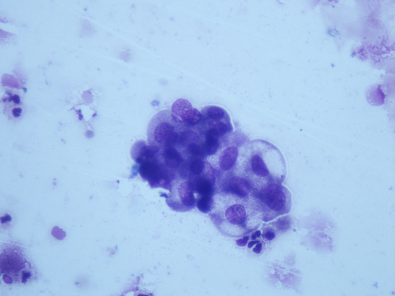
Leishman Stain x400 Pleural fluid cytology showing retention of malignant epithelial cells in clean background - NSRT
Effect of haemolysing agent on smear background
| Processing method | No. of cases | Total cases | Av. score |
|---|
| Score 1 | Score 2 | Score 3 | Score 4 |
|---|
| CF | - | 4 (7.8%) | 16 (31.2%) | 31 (60.8%) | 51 | 3.52 |
| GAA | 14 (27.5%) | 23 (45%) | 17 (33.3%) | 2 (3.9%) | 51 | 2.33 |
| NSRT | 1 (1.9%) | 2 (3.9%) | 11 (21.6%) | 37 (72.5%) | 51 | 3.64 |
Effect of haemolysing agent on retention of epithelial and mesothelial cells
| Processing method | No. of cases | Total cases | Av. score |
|---|
| Score 1 | Score 2 | Score 3 | Score 4 |
|---|
| CF | - | 11 (21.6%) | 11 (21.6%) | 29 (57.8%) | 51 | 3.35 |
| GAA | 3 (5.9%) | 13 (25.5%) | 9 (17.6%) | 26 (50.9%) | 51 | 3.13 |
| NSRT | 2 (3.9%) | 3 (5.9%) | 10 (19.6%) | 36 (70.5%) | 51 | 3.56 |
Effect of haemolysing agents on cytomorphological details
| Processing method | No. of cases | Total cases | Av. score |
|---|
| Score 1 | Score 2 | Score 3 | Score 4 |
|---|
| CF | 1 (1.9%) | 10 (19.6%) | 9 (17.6%) | 31 (60.6%) | 51 | 3.37 |
| GAA | 8 (15.7%) | 7 (13.7%) | 6 (11.8%) | 30 (58.8%) | 51 | 3.13 |
| NSRT | 2 (3.9%) | 6 (11.8%) | 16 (31.4%) | 27 (52.9%) | 51 | 3.33 |
Discussion
Cytological examination of serous fluids is of paramount importance, not only for diagnosis of cancer, but also for its prognosis [4].Haemorrhagic fluids lead to great diagnostic difficulties. The aim of present study was to compare the efficacy of NSRT, GAA and CF in lysing RBCs, in preserving epithelial /mesothelial cells and in retaining the cytomorphological details. The most effective method was also evaluated. Total of 51 haemorrhagic body fluid samples were included in the study, among which maximum number of samples were of pleural fluid (47%), followed by peritoneal fluid (37.3%), urine (7.8%), ovarian cystic fluid (5.9%) and synovial fluid (2%). As in our study, Preeti et al., reported maximum of 50.66% pleural fluid, 46.67 % peritoneal fluid and 2.67% pericardial fluid samples [6]. Malvi and Anthony has also reported maximum of 76.65% pleural fluid, 20% peritoneal fluid and 3.3% pericardial fluid samples [7]. Pericardial fluid samples were not received during our study period. The reason may have been small sample size. With NSRT, almost complete lysis of RBCs was observed in 72.5% of cases as compared to that in controls. Complete lysis of RBCs was noted in 91.33% samples by Preeti et al., and in 93% cases by Malvi and Anthony with NSRT [6,7]. NG et al., who assessed 11 grossly haemorrhagic specimens (2 urine, 4 ascitic fluid and 5 pleural fluid samples) noted complete lysis in all cases [5]. In our study, retention of epithelial / mesothelial cells was seen in 70.5% cases with NSRT as compared to that seen in other studies, where retention was 84.3% and 86.65% respectively [6,7]. Ng et al., observed retention of epithelial or mesothelial cells in 78% of cases with rehydration technique [5]. In the present study, cytomorphological details in NSRT treated smears were excellent in 52.9% of cases, while they were optimal in 31.4% of cases. 11.8% cases were sub-optimal and showed blurring of details and loss of sharpness of nuclear features. 3.9% cases were not suitable for assessment. Preeti et al., observed that 46.67% cases were excellent with NSRT, which was in accordance with our observations. However, in contrast to our observations, they noted that 51.33% cases were optimal and that only 2% cases were suboptimal for assessment [6]. Malvi and Anthony noted no nuclear artifacts with NSRT and morphology was retained in various body cavity fluids [7]. However, in our study, slight blurring of nuclear details was observed in few cases. In our study, on treatment of fluids with GAA, only 3.9% of cases showed complete lysis of RBCs and they showed a clean background. Remaining cases showed incomplete lysis of RBCs and they showed a dirty background. Other studies have reported 56.65% and 53.33% of cases with complete lysis on treatment with GAA [6,7]. Retention of epithelial/mesothelial cells was seen in 50.9% cases and cytomorphological features were excellent in 58.8% cases on treatment with GAA in our study. As in our study, Preeti et al., reported that 56.65% cases showed retention of epithelial/ mesothelial cells as in control smears and excellent cytomorphological features in 68% of cases with GAA, but they reported that 53.33% cases showed complete lysis and a clear background, which were dissimilar to our study findings [6]. Malvi and Anthony also observed the same findings in retention of epithelial/mesothelial cells and preservation of cytomorphological features, but in contrast to our study, only 50% of the smears showed partial lysis of RBCs with a dirty background on treatment with GAA [7]. In the present study, smears treated with CF showed complete lysis with a clear background in 60.8% cases. Retention of epithelial/mesothelial cells was observed in 57.8% cases, as was also observed in control smears. Cytomorphological details were excellent in 60.6% cases; however, few cases showed cellular swelling. Preeti et al., observed complete lysis of RBCs, retention of epithelial/ mesothelial cells and excellent cytomorphological preservation in 82%, 72% and 77.3% cases respectively [6]. Malvi and Anthony reported complete lysis of RBCs in 50% of the cases, along with shrinkage of nuclei of epithelial cells, with subsequent loss of chromatin material [7]. These three techniques were compared with each other and with other studies also [6,7]. For lysis of RBCs in smear background, NSRT was found to be the most effective technique. Our results were in accordance with those of other studies [6,7]. Best result for epithelial/mesothelial cell retention was observed in the present study with NSRT, in which cellular retention was seen in 70.5% cases. Cytomorphological details were best observed with CF by all the three methods. However, cytoplasmic swelling was observed with NSRT and CF, while shrinkage of nuclei of epithelial cells was observed with GAA. These findings were in accordance with those of other studies [6,7].
Conclusion
On the basis of comparative analysis, the most effective method was found to be NSRT for lysis of RBCs in smear background and cellular retention. However, cytomorphological details were best observed with CF. But considering the overall results and procedural simplicity, it was concluded that NSRT was a better technique for processing of haemorrhagic fluid.