Plenty of skeletons were found when excavations were carried out in prehistoric populations of most of the countries. Identification of skeletal remains is of great medicolegal importance. Anatomists and forensic pathologists have been frequently consulted about their opinion on skeletal remains buried in suspicious circumstances and to pronounce an opinion which can form important evidence in the court of law [1].
Establishment of identity of deceased person also assumes a great medicolegal importance. To establish the identity of a person, stature is one of the criteria. To know stature of individual, length of long bones is needed. Use of more than one long bone gives accurate result [2]. Damage to long bones occurs by accidents like plane crash, earthquakes or crush injuries, making them fractured; hence reconstruction of height of body becomes difficult. In such cases, the ratio between fragment of a long bone and length of that bone would help in estimating height of that individual. Therefore, the length of the fragment of the long bone and the total length of that long bone were calculated and the ratio between each fragment and the total length of the bone was found out. Then, by applying the regression formula, the height of the individual can be estimated [3]. This is essential as this method forms one of the big Four parameters in Forensic Pathology such as age, sex, race and stature [4].
The femur is the longest and strongest bone in the human body. It has shaft, proximal end and distal end. The shaft is slightly convex anteriorly [5]. Femur is selected in the present study because it is one of the long bones which helps in assessing the height of the individual more accurately compared to the other long bones. Grossly mutilated skeletal remains are a big challenge for forensic pathologist and physical anthropologist in the identification of the deceased. The application of osteometry is most important in medico legal investigation for estimating the height which is part of achieving the goal of estimating age at the time of death, sex, race, ancestry, ethnicity, stature, body weight and body build. The details of individualizing characteristics i.e. amputation, fractures, ankylosis, deformities and bone pathologies and to some extent the cause of death if reflected in the skeletal remains are also essential in the identification of the individual. The objective is to enable the law enforcement agencies to achieve the ultimate goal of personal identification. Pearson’s derivation of regression formulae for calculation of stature of an individual by the length of long bones was done in case of dead or dry bones [6]. Long bones that make up greatest proportion of stature, i.e. femur and tibia are more accurate than humerus and ulna. The relation between stature and the length of long bone shows a difference amongst the three races like Caucasoid, Negro and Mongoloid. Therefore, there is a need to derive separate, specific formulae for different races [7].
Intact long bones of upper and lower extremities have been used in derivation of regression equations for estimation of stature in different population groups. These bodies are sometimes presented to forensic anthropologists in different states of fragmentation, thereby making derived equations unstable. This has necessitated the need to assess the usefulness of measurement of fragments of long bones [e.g. Femur] in estimation of stature [8]. It is also helpful for the clinicians in the treatment of proximal and distal femur fractures. Femoral neck fractures involve the narrow neck between the round head of the femur and the shaft [9]. Surgeons may treat these types of fracture by replacing the fractured bone with prosthesis. Alternatively, the treatment is to reduce the fracture (manipulate the fragments back into a good position) and fix them in place with three metal screws. In some hip fractures, there is complete removal of the head and neck of the femur, and replacement is done with a prosthetic implant. Intertrochanteric fractures occur between the greater and lesser trochanters. They are usually fixed with a sliding hip screw and plate.
The subtrochanteric fractures of femur are more difficult to treat as compared to intertrochanteric fractures due to the powerful muscle forces acting on the fragments as well as the tremendous stress that is normally placed through this region. In the elderly, they are often low energy injuries involving osteoporotic bone. Fracture may also occur at the site of screw placement for a previous femoral neck fracture if the inferior screw is placed too low (below the lesser trochanter), as this creates a cortical defect and stress riser. Supracondylar femur fractures require anatomically stable internal fixation for best results, which usually necessitates surgical treatment. These fractures usually occur in elderly patients with multiple co-morbidities and osteoporotic bone; thus, a high rate of complication exists [9].
Severely comminuted distal femur fractures are especially difficult fractures to treat properly. Obtaining adequate fixation may be technically challenging, especially when multiple fragments are present. Each device has limitations. Stable fixation depends on the exact placement of the hardware. If comminution and the fracture pattern compromise the use of an implant, the surgeon should be flexible and choose the device that fits best. It is believed that knowledge of the morphometric values of femoral segments is important in forensic, anatomic and archaeological cases in order to identify unknown bodies and stature. It is also helpful for the clinicians in the treatment of proximal and distal femur fractures.
Material and Methods
This cross sectional study was conducted in M.S. Ramaiah Medical College, Bangalore, India. 150 dry and processed femora of both sides were randomly collected from grossly normal and complete adult cadavers, from department of anatomy, MSRMC, Bangalore, and department of anatomy, MSU, GEF, International Medical School, Bangalore, and from the skeletal sets of medical students of both institutes.
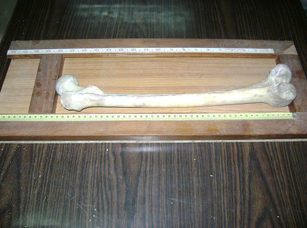
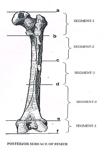
Unossified bones, bones with diseases and injuries were excluded from the study. This estimation was done based on the study, “Estimation of length of femur from its fragments”, considering total length of femur mean± SD= 43.93 ± 2.75. Here we are considering alpha error as 5% and allowable error as 1%. The total length of the femur was noted first by using the osteometric board and then confirmed with the measurements taken by using a thread. These two values were taken because of the forward curvature of the femur. In order to compare all measurements of the segments of femur, it had been divided arbitrarily into five segments by taking following landmarks on the basis of morphological characters of femur from the top of head to distal point on medial condyle, i.e. a-b; b-c; c-d; d-e; e-f. This enabled us to take advantage of numbers of segments and make a study with broad prospective [Table/Fig-1].
Showing the osteometric board

The most proximal point on head.
Lower border of lesser trochanter.
Point where gluteal and spiral lines join to form linea aspera.
Point where linea aspera divides into medial and lateral supracondylar lines.
Most proximal point on the intercondylar fossa.
In this way, with the help of above landmarks, the whole length of femur is divided into five segments, i.e. segment-1= a-b, segment-2= b-c, segment-3= c-d, segment-4=d-e, segment-5= e-f. The lengths of the segments were measured with the help of ‘osteometric scale’ to the nearest millimeter.
Thus the maximum total length of all 150, 72 left and 78 right femur bones were measured with standard procedure, and linear measurements of all segments mentioned above were taken simultaneously to nearest millimeter.
Statistical Analysis
Linear regression analysis was employed to estimate the stature of individual from length of femur.
Descriptive statistics like mean and standard deviation of segement-1, segment-2, segment-3, segment-4 and segment-5 was calculated.
Independent ‘s’ test was used to compare the segment-1, segment-2, segment-3, segment-4 and segment-5 lengths of left and right femur. Data analysis was carried out using Statistical Package for Social Science (SPSS) package.
Method of Statistical Analysis
The following methods of statistical analysis have been used in this study. The results were averaged (mean + standard deviation) for continuous data and number and percentage for dichotomous data are presented in Table and Figure.
1) The student ‘t’ test was used to determine whether there was a statistical difference between two groups in the parameters measured.
Student ‘t’ test is as follows:

2) One way Analysis of Variance (Anova). One way analyses of variance were used to test the difference between groups. Analysis of Variance is a technique by which the total variation is split into two parts, one between groups and the other within the group. If ‘F’ value is significant, there is a significant difference between group means. To find out which of the two groups means is significantly different, post hoc test of Least Square Distance test is used. The formula used:

3) Initially, a simple linear regression was applied. This regression was made considering the right and left, separately for each fragmentary bone measurement. Multiple regressions was used to estimate the total length using fragmentary bone measurement. In this method, the incorporation of variables was made through stepwise regression.
In the entire above test the “p” value of less than 0.05 was accepted as indicating statistical significance. Data analysis was carried out using Statistical Package for Social Science (SPSS) package.
Results
The lengths of 150 femora and the lengths of individual segments of all the 150 femora were noted and subjected to statistical analysis and compared. The following results were obtained:
1] The mean length of femur bone on left side was 43.54±2.78cm while on right side was 43.42±2.43cm with ‘t’ value=0.079 and ‘p’ value= 0.780.
2] The mean length of segments on left side were segment-1= 8.06±0.70, segment-2= 8.25±1.24, segment-3= 10.34±2.21, segment-4= 13.94±1.93 and segment-5= 2.76±0.53, along with ‘F’ value= 540.61 and ‘p’ value= ≤0.001. While mean length of segments on right side were segment-1= 8.08±0.70, segment-2= 8.30±1.34, segment-3= 10.44±1.91, segment- 4= 13.50±1.54 and segment-5= 3.08±0.41, along with ‘F’ value = 664.49 and ‘p’ value = ≤0.001.
3] On left side, mean percentage of segments to total femoral length were segment-1= 18.52±1.20, segment-2= 18.97±2.78, segment-3= 23.70±4.61, segment-4= 32.06±4.18 and segment-5= 6.35±1.20, along with ‘F’ value= 634.292 and ‘p’ value=≤0.001. While on right side, mean percentage of segments to total femoral length were segment-1= 18.63±1.33, segment-2= 19.11±2.85, segment-3= 24.02±4.09, segment-4= 31.10±3.13 and segment-5= 7.12±0.922, along with ‘F’ value= 803.117 and ‘p’ value=≤0.001. These results are depicted in the form of tables [Table/Fig-2,3,4–5].
Showing the mean lengths of five segments of the left and right Femora along with the standard deviation, ‘f’ and ‘p’ values
| Side | Segment | n | Mean | SD | Min. | Max. | ‘F’ Value | ‘p’ value |
|---|
| Left | 1 | 72 | 8.063 | .7098 | 6.6 | 10.3 | 540.611 | <0.001 |
| 2 | 72 | 8.250 | 1.2460 | 4.7 | 10.4 |
| 3 | 72 | 10.349 | 2.2163 | 4.9 | 17.7 |
| 4 | 72 | 13.943 | 1.9318 | 8.0 | 18.6 |
| 5 | 72 | 2.767 | .5389 | 1.5 | 4.6 |
|
| Right | 1 | 78 | 8.088 | .7045 | 6.2 | 9.7 | 664.496 | <0.001 |
| 2 | 78 | 8.303 | 1.3438 | 5.4 | 11.3 |
| 3 | 78 | 10.440 | 1.9156 | 6.8 | 16.4 |
| 4 | 78 | 13.500 | 1.5430 | 9.6 | 17.2 |
| 5 | 78 | 3.088 | .4128 | 1.7 | 4.1 |
Showing length of Femur and the Femoral Segments along with the standard deviation, ‘f’ and ‘p’ values of the segments of femur
| Side | Segment | n | Mean % to Total length | SD | Min. | Max. | ‘f’ Value | ‘p’ value |
|---|
| Left | 1 | 72 | 18.525 | 1.2092 | 15.4 | 21.8 | 634.292 | <0.001 |
| 2 | 72 | 18.974 | 2.7849 | 10.5 | 27.3 |
| 3 | 72 | 23.709 | 4.6165 | 13.5 | 39.3 |
| 4 | 72 | 32.065 | 4.1815 | 17.2 | 40.0 |
| 5 | 72 | 6.356 | 1.2077 | 3.8 | 11.1 |
|
| Right | 1 | 78 | 18.635 | 1.3330 | 14.8 | 22.1 | 803.117 | <0.001 |
| 2 | 78 | 19.116 | 2.8590 | 12.4 | 25.2 |
| 3 | 78 | 24.027 | 4.0967 | 16.9 | 37.0 |
| 4 | 78 | 31.101 | 3.1314 | 23.9 | 38.0 |
| 5 | 78 | 7.120 | .9226 | 4.2 | 9.4 |
Showing the comparison between the mean lengths of corresponding left segments and right segments of Femora along with the standard deviation, ‘t’ and ‘p’ values of the segments of femur bone
| n | Mean | SD | Min. | Max. | ‘t’ value | ‘p’ value |
|---|
| Segment 1 | Left | 72 | 8.06 | 0.710 | 6.60 | 10.30 | 0.050 | 0.823 |
| Right | 78 | 8.09 | 0.705 | 6.20 | 9.70 |
| Total | 150 | 8.08 | 0.705 | 6.20 | 10.30 |
| Segment 2 | Left | 72 | 8.25 | 1.246 | 4.70 | 10.40 | 0.061 | 0.805 |
| Right | 78 | 8.30 | 1.344 | 5.40 | 11.30 |
| Total | 150 | 8.28 | 1.294 | 4.70 | 11.30 |
| Segment 3 | Left | 72 | 10.35 | 2.216 | 4.90 | 17.70 | 0.073 | 0.788 |
| Right | 78 | 10.44 | 1.916 | 6.80 | 16.40 |
| Total | 150 | 10.40 | 2.059 | 4.90 | 17.70 |
| Segment 4 | Left | 72 | 13.94 | 1.932 | 8.00 | 18.60 | 2.426 | 0.121 |
| Right | 78 | 13.50 | 1.543 | 9.60 | 17.20 |
| Total | 150 | 13.71 | 1.749 | 8.00 | 18.60 |
| Segment 5 | Left | 72 | 2.77 | 0.539 | 1.50 | 4.60 | 17.007 | <0.001 |
| Right | 78 | 3.09 | 0.413 | 1.70 | 4.10 |
| Total | 150 | 2.93 | 0.502 | 1.50 | 4.60 |
| Total Length | Left | 72 | 43.54 | 2.785 | 36.30 | 48.80 | 0.079 | 0.780 |
| Right | 78 | 43.42 | 2.434 | 37.70 | 48.60 |
| Total | 150 | 43.48 | 2.600 | 36.30 | 48.80 |
Shows the comparison of present work with other authors
| Authors | Mean total Length of Femur |
|---|
| Mc Kern & Steel10 | 44.90 ± 1.71 cm |
| A G Shroff16 | 42.01 ± 2.75 cm |
| Present study | 43.48 ± 2.6 cm |
Discussion
Plenty of skeletal remains are found either accidently or when exhumation of buried cadavers is carried out. Estimation of stature from skeletal remains has great importance in forensic medicine. Moreover, it has been reported that stature may vary from person to person, throughout the day and in different population [10].
Pan measured the maximum lengths of humerus, radius, ulna, femur, tibia and fibula in 142 male and female east Indians [Hindu], in fresh state with articular cartilages covering the ends [11].
Stevenson studied measurement of cadaver length and dry bone lengths of 48 northern Chinese male skeletons in mongoloid group to find out the ratio between bone length and height of individual [12].
In 1951, Dupertain and Hadden measured the stature of American whites and blacks [100 each sex-race group] from a cadaver population. They suggested that stature can be estimated from a combination of two or more leg bones. Long bones of leg give closer estimates as compared to upper limb long bones [13].
The ratio between the length of the femur and the stature of that individual is significant in Forensic science and anthropology. The measurements of segment-1 and segment-2 of the femur can be used in determining the length of the femur in case only the proximal part of the femur is available [14].
Length of femur and the stature estimated from it of an individual are of forensic and anthropological significance. Length of femur can be determined from the head and neck of femur, only when a fragment of proximal femur available with head and neck of femur [14].
Gehring estimated body height with partial femur measures. However, greater errors were revealed in estimates than the formulae worked out using the total femur lengths [15].
To estimate stature, length of long bones is needed. Damage to these long bones can occur in various circumstances, making them fractured or fragmented. Hence, if such a fragment or segment of bone is found, then it is needed to assess the length of that bone and from it the height of the individual can be calculated. Among all the long bones, the length of the femur helps in estimating the height of that individual [1]. Therefore, present study deals with assessment of ratio between various segments of femur with the length of that bone. Once the length of the femur is obtained, the height of the individual can be calculated using various standard regression formulae. In the present study, femur was divided into five segments using anatomical landmarks which varied from previous works where they divided femur into five segments, but using different anatomical landmarks. This will help in studying the number of fragments of different lengths, which are sent for forensic studies [16].
The previous studies done on femoral fragment measurements were done irrespective of the side of femur bone. In the present study, the mean total length of femur and its segments was calculated separately for right and left sides, and the mean values of total femur length was identified as 43.54 ± 2.78 cm on left side and 43.42± 2.43 on the right side respectively. The right and left femora were studied separately and the ratio of the segments were calculated in the present study and compared with the values of the studies conducted by Steels and Mc Kern in the year 1969 on north eastern Arkansans femora [16], which showed that the present values are lower. In another study conducted by A.G. Shroff 9 in the year 1999, the values of our study were found to be slightly higher than that study. The reason could be due to the height of the individuals in those regions.
Segment-1 [Table/Fig-6] in the present study was taken from highest point on the head to the lowest point of lesser trochanter which is similar to the study conducted by Shroff whereas the points for segment-1, taken by Mc Kern & Steel, was highest point of head and the midpoint of the lesser trochanter & it measured 7.35± 0.61 cm. Therefore, the values of our study (8.08± 0.70 cm) and Shroff’s observations (8.44± 0.96cm) almost coincided, the reason being different landmarks.
Points (a to f) taken to divide five segments – [a] The most proximal point on head [b] Lower border of lesser trochanter [c] Point where gluteal and spiral lines join to form linea aspera. [d] Point where linea aspera divides into medial and lateral supracondylar lines [e] Most proximal point on the intercondylar fossa [f] Most distal point on medial condyle

In a study conducted by Mc Kern and Steel on 72 male and 36 female femora collected from north eastern Arkansas, segment-1 that corresponds to distance between most proximal point on head of femur and mid-point of lesser trochanter [Table/Fig-6] measures 7.35± 0.61 cm. In another study, segment-1 that corresponds to distance between most proximal point on upper end of femur and lower border of lesser trochanter measures 8.44± 0.96 cm. In the present study, segment-1 is different from segment-1 of study by Mc Kern and Steel where the lower point of the first segment was midpoint of lesser trochanter. But in the present study, the segment-1 corresponds more closely to segment of study conducted by A G Shroff. Segment-1 in the present study measures 8.08± 0.70 cm. Therefore, there are some differences in mean value of measurements between our study and the rest because of different landmarks. In the present study, the proportion of segment-1 to total length of femur was calculated. It was 18.58± 0.62% and this varied from previous study done by Mc Kern and Steel and A G Shroff [16]. This can be used for estimation of stature of individual by using Steele’s regression formulae.
In the present study, segment-2 was the distance between lower border of lesser trochanter and point where gluteal and spiral lines join to form linea aspera [Table/Fig-6]. It gave proportion of segment-2 as 19.05± 0.34%. In the study conducted by Steele and Mc Kern, segment-2 was between mid-points of lesser trochanter to most proximal extension of popliteal surface where medial and lateral supracondylar lines become parallel below the linea aspera. It gave proportion of segment-2 as 56.1%. Moreover, segment-2 taken in the present study is different from the segment-2 taken in studies by earlier authors. The 3rd anatomical point for segment-2 was the point where gluteal and spiral lines join to form linea aspera and 4th point at place where linea aspera divides into medial and lateral supracondylar lines. Hence, segment-2 and segment-3 are different obviously from segments being taken in the earlier studies. Combination of segment-2 and segment-3 [Table/Fig-6] in our study corresponds roughly to segment-2 of earlier studies. Proportion of segment-2 was 19.05± 0.34% and of segment 3 is 23.87± 0.41% of the total mean length. These new segments taken in the present study have enough scope to derive a regression equation to find out the length of the femur precisely which will help in determining the height of that individual.
In the present study, segment-4 is between lines where linea aspera divides into medial and lateral supracondylar lines, to the most proximal point on intercondylar fossa [Table/Fig-6] and it measured 13.71± 1.74 cm. This segment-4 corresponds to the segment-3 of the study done by A G Shroff and Mc Kern and is higher than the present values [16]. Proportion of segment-4 to the total length was 31.56± 0.39%. This is because of difference in race, growth pattern as well as nutrition.
In the present study, segment-5 [Table/Fig-6] measured 2.93± 0.50 cm and it corresponds to the Segment-4 of the study by earlier authors and the proportion was 6.75± 0.27% and these values were lesser than the previous studies. This can be also due to difference in rate of growth pattern, racial difference, environmental influence and nutritional issues.
In the earlier studies, segment-5 corresponded to a combination of segment-4 and segment-5 in their study [Table/Fig-6].
Some differences are found in mean values of femoral segments and total length of femur when compared to other studies. This was due to the result of factors such as age, sex, race and also environmental factors affecting bone growth, such as nutrition, physical development and genetic factors. Moreover, these diversities could depend on the differences in the anatomical reference points which are taken as criteria in the measurement of bones.
Hence, the ratio and proportion between the fragmentary length of femur and the total length of femur, for estimating the total length of femur, has been derived in the present [11] studies.
Once the total length of femur has been estimated from a fragment, it was possible to calculate stature of that individual with reasonable accuracy. In the present study, ‘p’ and ‘f’ values indicate significance [Table/Fig-2–4].
Conclusion
In the present study, determination of the total length of femur has been done from various femoral segments. For this purpose, femur has been divided into five segments taking six anatomical points which are different from points being taken in previous studies of some original workers. Hence, it helps us to have fragments of femur which are of varying length from previous studies. In the present study, segment-2 and segment-3 are taken separately and when combined it corresponds to the segment-2 of previous studies. Hence, these extra segment-2 and segment-3 provide further scope to calculate regression formulae in future. The knowledge of the proportion of other segments can be used for estimating the stature for individual by using standard regression formulae. In the present study, mean lengths of femoral fragments and mean total length of femur have been calculated separately for right and left side respectively. This observation has not been done by previous authors.
The knowledge of the morphometric values of femoral segments is important in forensic, anatomic and archaeological cases in order to identify unknown bodies and stature. It is also helpful for the clinician in the treatment of proximal and distal femoral fractures. Therefore, the present study supplies the mean values of different morphometric measurements from the femur. As a result, these measurements may help to indicate the characteristic morphological features of femoral segments in south Indian population and also help the orthopedic surgeon to place various implants in the reconstruction of femoral fragments.In the present study, the sample size is 150, 72 left and 78 right with ‘p’ value of ‹0.001 in all segments. When comparison is done between segments of right and left femur bone, then ‘p’ value of segment-5 is found to be ‹0.001, but when multiple comparison is done between different segments of femur bones, then ‘p’ value of all segments were found to be statistically significant. Therefore, the results are reliable but further study needs to be designed to get more accurate estimates in population considering the age and sex factors apart from race, region and nutritional status.