Surgical and Prosthetic Management of Suction Cup Induced Palatal Perforation: Case Report
Yogesh Rao1, Pankaj Yadav2, Jagjeet Singh3, Divyang Patel4, Amit Aggarwal5
1 Senior Lecturer, Department of Prosthodontics, Maharana Pratap College of Dentistry and Research Centre.
2 Senior Resident, Department of Conservative, Dentistry and Endodontics, Maulana Azad Institute of Dental Sciences.
3 Senior Lecturer, Department of Prosthodontics, Maharaja Ganga Singh Dental College and Hospital.
4 Senior Lecturer, Department of Prosthodontics, Ahmedabad Dental College, Ahmedabad, Gujrat, India.
5 Senior Lecturer, Department of Prosthodontics, New Horizon Dental College and Research Institute, Billaspur, India.
NAME, ADDRESS, E-MAIL ID OF THE CORRESPONDING AUTHOR: Dr. Yogesh Rao, Department of Prosthodontics, Maharana Pratap College of Dentistry and Research Centre, Gwalior, Madhya Pradesh, India.
Phone: 07827774442,
E-mail: dryogesh.yadav@yahoo.com
Construction of complete dentures with adequate retention is a complex procedure. Various techniques have been tried to improve the retention of dentures. Use of suction cup in maxillary denture is one of the techniques to improve retention. Suction cup provides retention and stability by inducing negative pressure on the mucosal surface. Palatal Suction cups which provide high retention are not being recommended because of their pathological effects on the palatal tissues. Here a case report of complete denture with suction cup induced palatal perforation which was surgically treated is presented.
Denture retention, Palatal perforation, Suction cup
Case Report
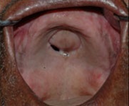
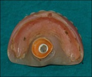
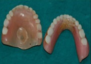
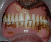
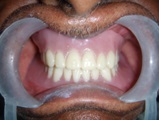
A male patient aged 65 years reported to the department with the complaint of a hole in upper jaw, presence of nasal twang in voice, loose-fitting denture. History revealed that he constructed his denture 20 years back and the defect was present since last one year. Patient was wearing the denture entire day and night and was removed only for cleaning. Examination of the palate revealed a curvilinear perforation of the palatal region approximately 4x3 cm in size [Table/Fig-1]. Inspection of the denture showed a suction cup [Table/Fig-2] and presence of attrited denture teeth [Table/Fig-3]. Also there was presence of crossbite on one side, under extended borders, poor retention and stability of denture and poor denture hygiene [Table/Fig-4]. Rubber suction cup was replaced every month. On palpation margins were non-tender and except for the palatal lesion all surrounding tissues appeared normal. When patient was asked to gargle water it came through his nose.
Maxillary denture with suction cup
Attrited maxillary and mandibular denture
Crossbite on one side and poor denture hygiene
A maxillary impression was made for record. The treatment planned was surgical closure of the perforation and construction of a new complete denture after healing of the area. Another maxillary primary impression was taken for construction of surgical stent. Primary cast was scraped in the perforation area and was blocked with wax. Then a surgical stent was made of clear heat cure.
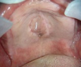
Patients medical history was uneventful and blood investigations were within normal limits. Also serological investigations showed a negative report for venereal diseases. Under local anaesthesia relieving incisions were placed on the alveolar ridge and around the perforation [Table/Fig-5]. The margins of the perforation were excised to remove the epithelial lining. A full thickness palatal flap was raised and rotated over the defect. Then the ends of perforation were sutured using catgut 3-0 suture. Surgical stent was positioned in the palate after surgery. Post-surgical healing was uneventful. When the patient was examined after 1 month for evaluation, there were no signs of inflammation. New maxillary and mandibular denture were constructed 1month after the surgery [Table/Fig-6 & 7]. The perforation had healed completely and all the surrounding tissues appeared normal after one year [Table/Fig-8].
Incision placed on the alveolar ridge and around the perforation

Denture in centric occlusion
Healed palate one year after surgery
Discussion
Complete dentures can induce both hard and soft tissue changes [1–3]. Resorption of the alveolar bone leads to a situation where the denture although well retained by the rubber disc, rocks around the mid palatal area, exerting excessive pressure [4]. This negative pressure leads to the loss of palatal bone as seen in this case. These complications can be more severe if the prosthesis is not constructed properly, just like lesions induced by poor prosthesis fit, improper occlusal relationship [5]. and continuous presence of denture with suction cup. It is known that these dentures have a destructive effect on the oral tissues, which can result in perforation of the palate [6,7]. Leaving the denture out of mouth provides time for the tissues to heal and further prevent the tissues from ill effects.
The management of palatal perforation caused by suction cup begins with educating the patient about the ill effects of continuous use of suction cup denture. The closure of palatal perforation is primarily by surgical means. There are various methods of surgical closure depending on size and destruction of tissues around the perforation. These options are local advancement flaps viz. palatal flaps or tongue flap, distant pedicle flaps like buccal fat pad, temporoparietal fascial flap. In this case a palatal flap was raised and preferred due to the absence of soft tissue thickness around the perforation.
Despite its known ill effects, many clinical practitioners are still using suction cups in the maxillary denture as the retentive aids. So, practioners should be well aware of the older methods of denture retention and render appropriate treatment to stop progression of the existing pathology and provide environment for the healing to take place.
[1]. Zarb GA, Bolender CI, Bouchers prosthodontic treatment for edentulous patients 1985 ed 9St LouisMosby:28-47. [Google Scholar]
[2]. Bergman B, Carlsson GE, A longitudinal two year study of a number of full denture casesActa Odontol Scand 1964 22:3-26. [Google Scholar]
[3]. Stewart JCB, Zarbo RJ, Oral pathologyClinical Pathologic Correlations 1999 ed 3PhiladelphiaSaunders:176-216. [Google Scholar]
[4]. Stafford GD, Perforation of the palate due to a suction disc on a dentureBr Dent J 1974 136:240 [Google Scholar]
[5]. Ordulu M, Emes Y, Ates M, Aktas I, Yelchin S, Oronasal communication caused by a denture with suction cups: A case reportQuintessence Int 2006 37:659-62. [Google Scholar]
[6]. Cayetano O, Boone ME, Suction cups on maxillary dentures: Report of caseJ Am Dent Assoc 1987 115:577 [Google Scholar]
[7]. Shalhoub SY, Palatal perforation related to faulty denture constructionJ Oral Med 1985 40:49-50. [Google Scholar]