Craniofacial Duplication: A Case Report
Pradeep Suryawanshi1, Mandar Deshpande2, Nitin Verma3, Vivek Mahendrakar4, Sandhya Mahendrakar5
1 NICU Incharge & Associate Professor, Division of Neonatology, Department of Paediatrics, Bharati Vidyapeeth University Medical College, Pune, India.
2 Fellow Paediatric Critical Care, Consultant Neonatologist, Amrut Balrugnalaya and Critical Care Center, Aurangabad, India.
3 Clinical Fellow, Nepean Hospital, Penrith, Australia
4 Consultant Paediatrician, Mahendrakar Hospital, Aurangabad, India.
5 Consultant Obstetrician, Mahendrakar Hospital, Aurangabad, India.
NAME, ADDRESS, E-MAIL ID OF THE CORRESPONDING AUTHOR: Dr. Pradeep Suryawanshi, D 404, Treasure Park Sant Nagar Parvati, Pune–411009, India.
Phone: 91-9923540500,
E-mail: drpradeepsuryawanshi@gmail.com
A craniofacial duplication or diprosopus is an unusual variant of conjoined twinning. The reported incidence is one in 180,000-15 million births and 35 cases have been reported till date. The phenotype is wide, with the partial duplication of a few facial structures to complete dicephalus. A complete duplication is associated with a high incidence of anomalies in the central nervous system, cardiovascular system, gastrointestinal system and the respiratory system, whereas no major anomalies are found in the infants with a partial duplication. A term baby with the features of a craniofacial duplication has been described, with the proposed theories on embryogenesis and a brief review of the literature.
Craniofacial duplication, Diprosopus
Introduction
Conjoined twins have been reported in the literature since 1864; however, their existence has been known for centuries. Diprosopus is a rare form of symmetric conjoined twinning with a single neck and body and a wide spectrum of duplication of the craniofacial structures.
The earliest known report on diprosopus has been credited to Ambroise Paré (Of Monsters and Prodigies) of the 16th century [1]. Since 1884, there have been only 35 reports on diprosopus in the world medical literature [2]; however, there is no published case of post natal diprosopus in India.
Case Report
A term male neonate presented at 16 hours of life to our centre for the evaluation of dysmorphic features. The baby was born to a G2P1L1 unregistered mother by a normal vaginal delivery and she had an unremarkable antenatal history (an antenatal scan was not done) in a 2nd degree consanguineous marriage. The previous sibling was normal. The baby is said to be cried immediately after birth, he had dysmorphic features and he developed respiratory distress in the form of tachypnoea after birth.
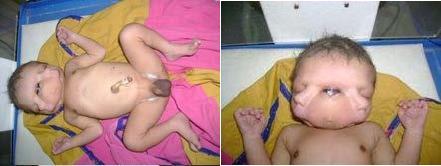
The baby’s features included three eyes, two noses, two mouths, a single large midline orbit, corneal opacity in the third eye, two ears, two tongues and a single neck and trunk. The other features included a duplicated labial frenulum, a duplicated palate without any cleft, duplicated vocal cords and a duplicated mandible and maxilla. The cranium had two frontal and two sets of parietal bones that were medially hypoplastic, one occipital bone, and two temporal bones.
The baby had frequent episodes of de-saturation with cyanosis, which required oxygen support, but the saturation remained below 80%. His chest X-ray showed oligaemic lung fields with no cardiomegaly. A further work up could not be done, as the baby died within 1 hour, due to a sudden cardiac arrest [Table/Fig-1].
Images showing duplicated facial features
Discussion
Diprosopus refers to a partial to a complete division of the facial structures and it is associated with a variety of anomalies. There is a predominance of females over males (2:1) [3]. The duplication can involve as small as the nasal to complete facial structures (diprosopus monocephalus) [4]. A complete duplication or dicephalus is associated with a high incidence of anomalies in the Central Nervous System (CNS), Cardiovascular System (CVS), Gastrointestinal System (GI) and the Respiratory Systems (RS), as well as cleft lip and palate. Partial duplication: the infants have a mandible and a mouth which are most commonly duplicated. The CNS anomalies involve anencephaly, duplication of the brain with two prosencephalons and a single rhombencephalon, two diencephalons (each with a set of thalami and basal ganglia) and two symmetric telencephalons (each with a set of cerebral hemispheres and lobes). Hypoplasia of the medial temporal lobe has also been noted. Multiple spinal abnormalitites with duplication of the cervical spine, with abnormal cervical and thoracic vertebrae, have been seen [5]. The defects in the other organs include diaphragmatic hernia, cardiac defects (VSD, an overriding aorta and a hypoplastic ascending and descending aorta and an aortic arch and dextrocardia), bilateral dysplastic cystic kidneys, hypoplasia of the ureters and the urinary bladder, cleft lip, palate and an imperforate anus [6,7].
A pre-natal diagnosis is possible, with ultrasound findings such as; anencephaly, a partially duplicated CNS, neural tube defects, a wide vertebral column, a bifid cranial vault, polyhydramnios and raised AFP levels.
The embryology of the condition has been a matter of debate. The most accepted theory presently is that conjoined twins result from an embryological disturbance in the separation of the twins during the 2nd week of pregnancy (12–13 days), as a result of the abnormal splitting of the post-implantation blastocyte [8]. Such incomplete, separated, germinal discs lead to this extremely rare foetal anomaly. Although recently, it has been postulated that conjoined twins result from the development of two independent notochords which were initially destined to become separate twins, but which were too close to develop independently [9]. The reported incidence is 1:50,000 to 1:200,000 births and 1% of all the monochorionic twins. They are classified mainly into three groups, terata Catydidymus (iprosopus, Dicephalus, Ischiopagus (6–20%) and pyopagus (10–20%), terata Anadidyma (Dipygus, Syncephalus and Crainopagus (6–12%) and terata Anacatadidyma – Thoracopagus (30–40%), Omphalopagus (25-30%) and Rachipagus.
Diprosopus involves the bifurcation or forking of the notochord, leading to two side by side oriented vertebral axes and the formation of two neural plates and neural crest derivatives [10]. It has been suggested that during the neurulation of the duplicated notochords, each neural tube and crest migrate inwardly and separate from the surface ectoderm. If the distance is narrow between the two, then there would not be enough ectoderm to cover the inner folds, resulting in the failure of the closure of the neural tubes. A normal chromosomal analysis has been reported by various authors [11,12].
The prognosis is poor for the infants with a complete duplication, although the treatment options such as excision of the duplicated parts, which give a normal appearance in partial diprosopus, have been variably successful.
[1]. Walton Michael T, Fineman Robert M, Walton Phyllis J, Historical essay: Of monsters and prodigies: The interpretation of birth defects in the sixteenth centuryAm J Med Genet 1993 Aug 1 47(1):7-13. [Google Scholar]
[2]. Stefan Hähnel, Peter Schramm, Stefan Hassfeld, Craniofacial Duplication (Diprosopus): CT, MR Imaging, and MR Angiography Findings – Case ReportRadiology 2003 226:210-13. [Google Scholar]
[3]. Mason Barr Jr, Facial duplication: Case, review, and embryogenesisTeratology 1982 25(2):153-59. [Google Scholar]
[4]. Amr SS, Hammouri MF, Craniofacial duplication (diprosopus): report of a case with review of literatureEur J Obstet Gynecol Reprod Biol 1995 Jan 58(1):77-80. [Google Scholar]
[5]. Angtuaco TL, Angtuaco EJ, Quirk JG Jr, US case of the day. Complete brain duplication with fusion at the posterior fossa (diprosopus tetrophthalmos)Radio-Graphics 1999 19:260-63. [Google Scholar]
[6]. Chervenak FA, Pinto MM, Heller CI, Norooz H, Obstetric significance of foetal crainofacial duplication. A Case reportJ. Reprod. Med 1985 30:74-76. [Google Scholar]
[7]. Turpin IM, Furnas DW, Amlie RN, Craniofacial duplication (Diprosopus)Plast Reconstruct Surgery 1981 67:139-42. [Google Scholar]
[8]. Strauss S, Tamarkin M, Engelberg S, Ben Ami T, Goodman RM, Prenatal sonographic appearance of diprosopusJ Ultrasound Med 1987 6:93-95. [Google Scholar]
[9]. Spencer R, Conjoined twins: theoretical embryologic basisTeratology 1992 Jun 45(6):591-602. [Google Scholar]
[10]. Machin G, Conjoined twins: implications for blastogenesisBirth Defects Orig Artic Serv 1993 29:141-79.1993 [Google Scholar]
[11]. Sharony R, Diprosopus: A pregastrulation defect involving the head, neural tube, heart and diaphragmBirth defects Orig Serv 1993 29:201-09. [Google Scholar]
[12]. Wu J, Staffenberg DA, Mulliken JB, Shansken AL, Diprosopus: A unique case and review of the LiteratureTeratology 2002 66:282-87. [Google Scholar]