An Accessory Muscle of Pectoral Region: A Case Report
B.M. Bannur1, Nagaraj Mallashetty2, Preetish Endigeri3
1 Professor and HOD, Department of Anatomy, BLDEU’s, Shri BM Patil Medical College, Bijapur, Karnataka, India.
2 PG Student, Department of Anatomy, BLDEU’s, Shri B M Patil Medical College, Bijapur, Karnataka, India.
3 PG Student, Department Orthopaedics, BLDEU’s, Shri BM Patil Medical College, Bijapur, Karnataka, India.
NAME, ADDRESS, E-MAIL ID OF THE CORRESPONDING AUTHOR: Dr. Nagaraj Mallashetty, PG Student, Department of Anatomy, BLDEU’s, Shri B M Patil Medical College, Bijapur, Karnataka, India. Email ID: nagarajsmalashetti@gmail.com; Preetishendigeri_86@yahoo.co.in
Among the variations of pectoral muscles, this case appears to be unique in the literature. This was a case of an accessory pectoral muscle which was located between pectoralis major and pectoralis minor muscles, which was discovered during a routine anatomy dissection. The accessory muscle originated from 6th and 7th ribs at costo-chondral junction, which travelled supero-laterally and inserted by fusing with fibres of pectoralis minor. This unusual muscle holds importance for surgeons while they perform dissectomies, in avoiding complications.
Accessory pectoral muscle, Pectoralis major, Pectoralis minor, Medial pectoral nerve, Costo-chondral junction
Case Report
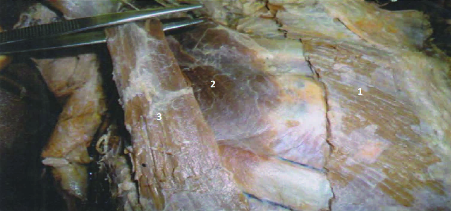
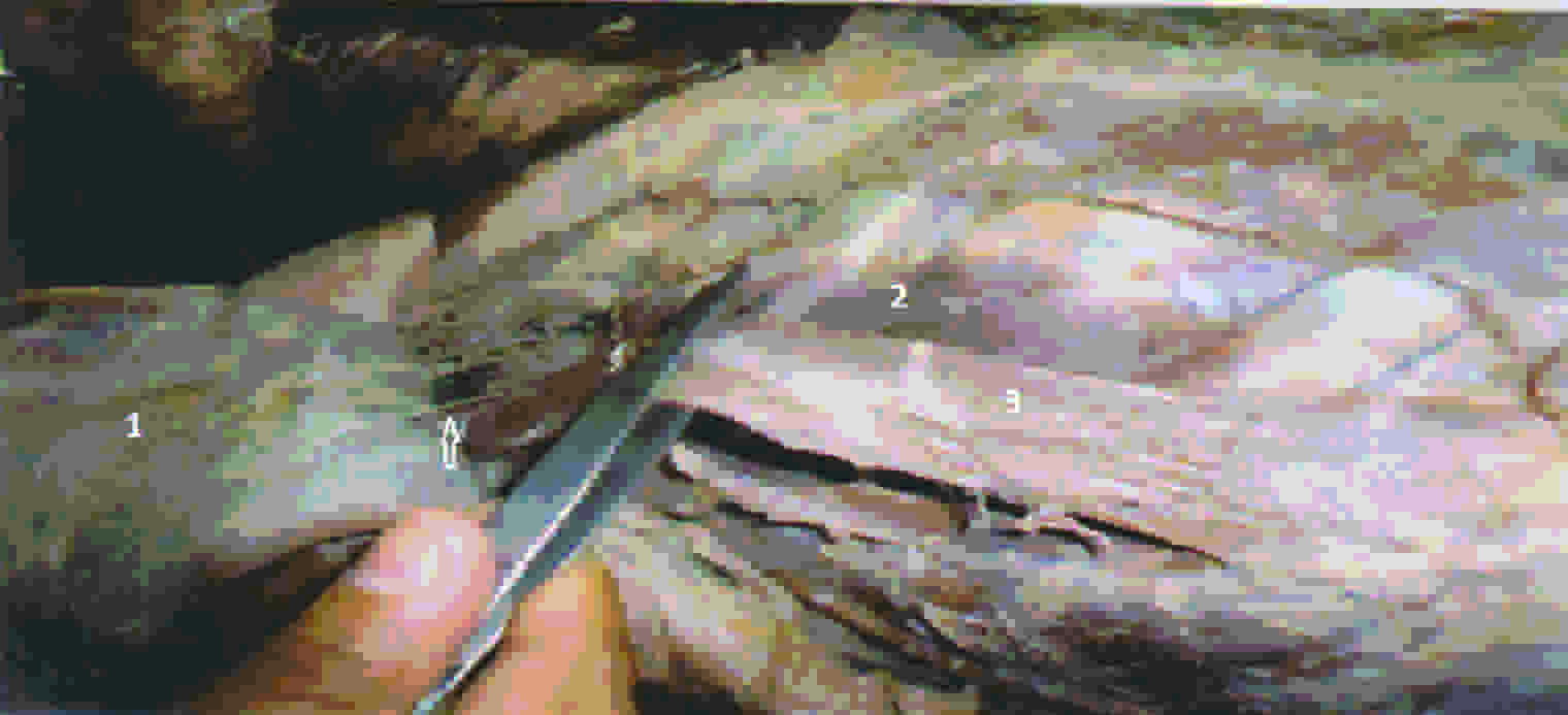
During the routine dissection of a male cadaver in the Department of Anatomy, Shri B M Patil Medical College Bijapur, India, we found a long ribbon like muscle in right pectoral region, which originated from right 5th and 6th ribs at costo-chondral junction, in between the pectoralis major and pectoralis minor muscles [Table/Fig-1]. From its origin, it travelled above and laterally and inserted by fusing with fibres of pectoralis minor. Pectoralis minor was inserted on medial border and upper surface of coracoid process of scapula. Accessory pectoral muscle was 14 cm long and it was 4 cm broad at its origin.The nerve supply to this muscle was through medial pectoral nerve [Table/Fig-2]. No variations were found with regards to origin and insertion of pectoralis major and minor. This accessory muscle was found only on right side and left side pectoral muscles showed no variations.
Pectoralis major (2) Pectoralis minor (3) Accessory pectoral muscle
Pectoralis major (2) Pectoralis minor (3) Accessory pectoral muscle. The arrow indicates the medial pectoral nerve
Discussion
The variants of pectoralis major include: Absence of the abdominal slip of pectoralis major, variations in the number of costal attachments, decussation of right and left muscles across the sternum and ascension of superficial vertical slips from the lower costal cartilages which are called “sternalis”.
Variants of pectoralis minor include: Separation of muscular slips of pectoralis minor and their variations in number and level eg. like one passes from first rib to coracoids process, which is called as pectoralis minimus and costal attachments can be 2nd to 5th ribs, 3rd to 5th ribs or 2nd to 4th ribs [1].
Although variations of pectoral muscles are not uncommon, our case appeared to be unique in that this accessory muscle arose separately from 5th and 6th ribs at costo-chondral junction, on right side and fused with pectoralis minor muscle before its termination.
An embryological explanation for this finding: The pectoralis muscle is derived from dorsal limb bud masses which arise from myoblasts that migrate out of the last five cervical and first thoracic myotomes into the developing limb buds during the fifth week of development [2]. The pectoralis muscles assume their final forms through a combination of migration, fusion and apoptosis of muscle cell precursors [3]. It is possible that accessory pectoral muscle is a result of a failure of designated myoblasts in undergoing apoptosis.
Reported cases of variations of pectoral muscles: Most common anomaly of pectoralis major is its complete deficiency, which is described as Poland syndrome, which is characterised by unilateral absence of pectoralis major and cutaneous syndactyly [4]. Paraskevas et al reported bilateral hypoplasia of clavicular head of pectoralis major, which was associated with a vascular anomaly in an anteriorly placed external jugular vein, which was referred to as “atypical Poland’s syndrome” [5]. Philip A Fabrizio et al., noted that “pectoralis quartus” had originated from 5th costal cartilage and that it had inserted into the fascia which overlay the coracobrachialis muscle. He also noted an accessory muscle, which was found just lateral to the pectoralis minor, which had originated from external abdominal oblique muscle and had inserted into coracobrachialis fascia, deep into insertion of pectoralis quartus during customary dissections [6].
Leonard P Seimon reported that “epicostobrachialis” muscle arose from the lateral edge of the pectoralis major muscle and inserted into the medial epicondyle of the humerus during routine dissection of a male cadaver [7]. M Loukas and G South et al., reported a case of a unilateral occurrence of a right sided accessory head of pectoralis major muscle in a female cadaver, which had originated from the serratus anterior muscle and had inserted into the tendinous fibres of pectoralis major muscle [8]. Katherine Marie Huber B S et al., reported an extra muscle anterior to pectoralis major muscle in a 45 year old female during a left mastectomy and an immediate reconstruction surgery and named the muscle as, “oblique pectoralis anterior” [9]. Voto and Weiner reported the presence of “chondroepitrochlearis” muscle, which had led to restriction of the functional mobility of the limbs [10].
Lama et al., reported a slip which arose from the anterior border of latissimuss dorsi and inserted into the fascia of biceps, which was called “axillary arch of larger”. Another slip which arose from the pectoralis major and inserted into the same biceps fascia was called “chondrohumeralis” in same case, which presented with neurovascular complications as both traverse superficial to axillary neurovascular bundles [11].
Considering the above reported cases, our case appeared to be unique, because the accessory muscle was in between pectoralis major and pectoralis minor and was inserted by fusing with pectoralis minor muscle fibres.
Accessory pectoral muscles have negative and positive implications. Accessory pectoral muscles may be mistaken for masses or tumours during CT or MRI scans. During pectoral flap surgeries, difficulties may be encountered due to presence of these accessory muscles. Pectoralis minor is commonly used as a landmark during surgeries of axillary region. Presence of this muscle may disturb this landmark. Utility of pectoralis major in head and neck reconstructions is immense [12]. Increased size and origin of the pectoralis minor muscle through the supplementation of accessory pectoral muscles may enhance the optimal function of shoulder.
Conclusion
Among the variations of pectoral muscles, this case appears to be distinctive in the literature. The possible clinical importance of the present case should be discussed, as the knowledge on this is important to anatomists as well as surgeons, for preventing complications during surgery and to benefit from the outcomes of surgery. This case report illustrates a need for reporting such cases and discussing about their clinical importance.
[1]. Standring S, Pectoral girdle, shoulder region and axilla. In: Standring S, editorGray’s Anatomy 2008 40th edChurchill LivingstonElsevier:807-8. [Google Scholar]
[2]. Sweeney LJ, Basic concepts of in embryology 1998 McGraw-Hill:834 [Google Scholar]
[3]. Carlson BM, Human embryology and developmental biology 2004 Mosby:224-5. [Google Scholar]
[4]. Clarkson P, Poland’s syndactylyGuys Hosp Rep 1962 111:335-46. [Google Scholar]
[5]. Paraskevas GK, Raikos A, Bilateral pectoral musculature malformations with concomitant vascular anamolyFolia Marphol 2010 69(3):187-91. [Google Scholar]
[6]. Fabrizio PA, Hardy MA, An accessory muscle of thoracic wallIJAV 2009 2:93-5. [Google Scholar]
[7]. Seimon LP, Musculus Costo-epitrochlearisS.A. Medical Journal 1953 :1146-9. [Google Scholar]
[8]. Loukas M, South G, A case of an anomalous pectoralis major muscleFolia Morphol 2006 65:100-3. [Google Scholar]
[9]. Huber KM, Boyd TG, Quillo AR, Wilhelmi BJ, Implications of anomalous pectoralis muscle in reconstructive breast surgery: The oblique pectoralis anteriore Plasty 2012 12:383-9. [Google Scholar]
[10]. Voto SJ, Weiner DS, The chondroepitrochliaris muscleJ Pediatr Orthop 1987 7:213-4. [Google Scholar]
[11]. Lama P, Potu BK, Bhat KM, Chondrohumeralis and axillary arch of Langer a rare combination of variant muscles with unique insertionRom J Morphol Embryol 2010 51(2):395-7. [Google Scholar]
[12]. Dhuria R, Mehta V, Loh H, Unilateral quadripartite composition of pectoral major muscle: Anatomical description and clinical implicationsInt. J. Morphol 2009 27(4):1213-6. [Google Scholar]