Non–Syndromic Bilateral Supplemental Maxillary Lateral Incisors: A Rare Case
Ajay Nagpal1, Manoj Kumar Hans2, Shashit Shetty3, Navpreet Kaur4, Sunil Kumar5
1 Assistant Professor, Department of Conservative Dentistry & Endodontics, Kanti Devi Dental College and Hospital, Mathura, Uttar Pradesh, India.
2 Reader, Department of Conservative Dentistry & Endodontics, Kanti Devi Dental College and Hospital, Mathura, Uttar Pradesh, India.
3 Professor & HOD, Department of Conservative Dentistry & Endodontics, Kanti Devi Dental College and Hospital, Mathura, Uttar Pradesh, India.
4 Assistant Professor, Department of Public Health Dentistry, Kanti Devi Dental College and Hospital, Mathura, Uttar Pradesh, India.
5 Assistant Professor, Department of Conservative Dentistry & Endodontics, Kanti Devi Dental College and Hospital, Mathura, Uttar Pradesh, India
NAME, ADDRESS, E-MAIL ID OF THE CORESPONDING AUTHOR: Dr. Ajay Nagpal, Assistant Professor, Department of Conservative Dentistry & Endodontics Kanti Devi Dental College and Hospital, Mathura, Uttar Pradesh, India.
Phone:91-8979712525,
E-mail: drajayendodontist@gmail.com
Supernumerary teeth or hyperdontia are defined as the teeth that exceeds the normal dental formula, regardless of their location and morphology. A supernumerary tooth is a developmental anomaly and it has been argued to arise from multiple aetiologies. Supernumerary teeth with orthodox shapes and sizes, that resemble the normal dentition are called ‘supplemental teeth’. The supplemental teeth are often overlooked because of their normal shapes and sizes. Supplemental lateral incisors are rare and bilateral cases are even rarer, with only two cases being reported in the literature till date. Therefore, we are describing a rare case of non–syndromic, bilateral, supplemental, maxillary, lateral incisors and we have discussed the management of supernumerary teeth.
Supernumerary, Anomaly, Supplemental
Case Report
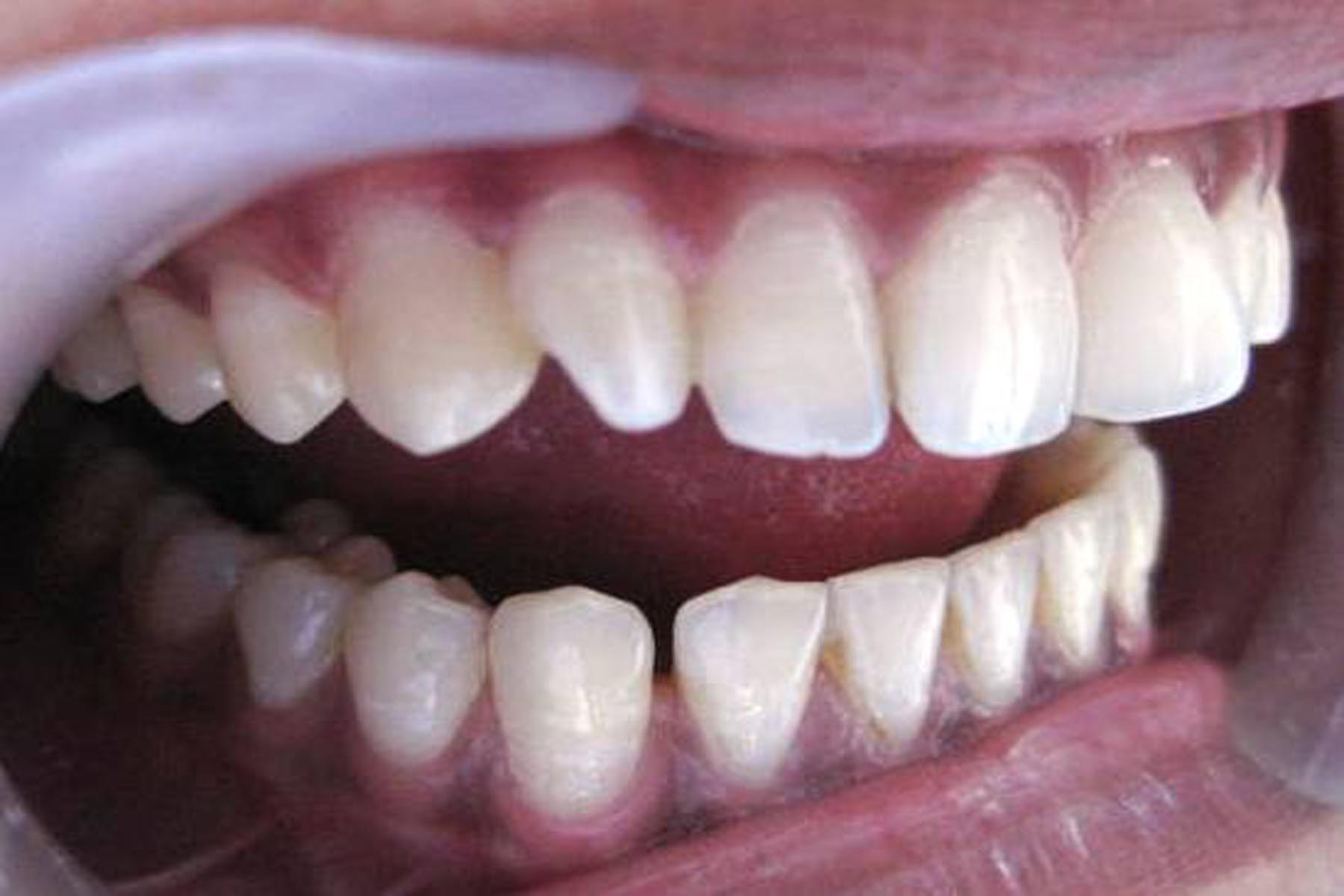
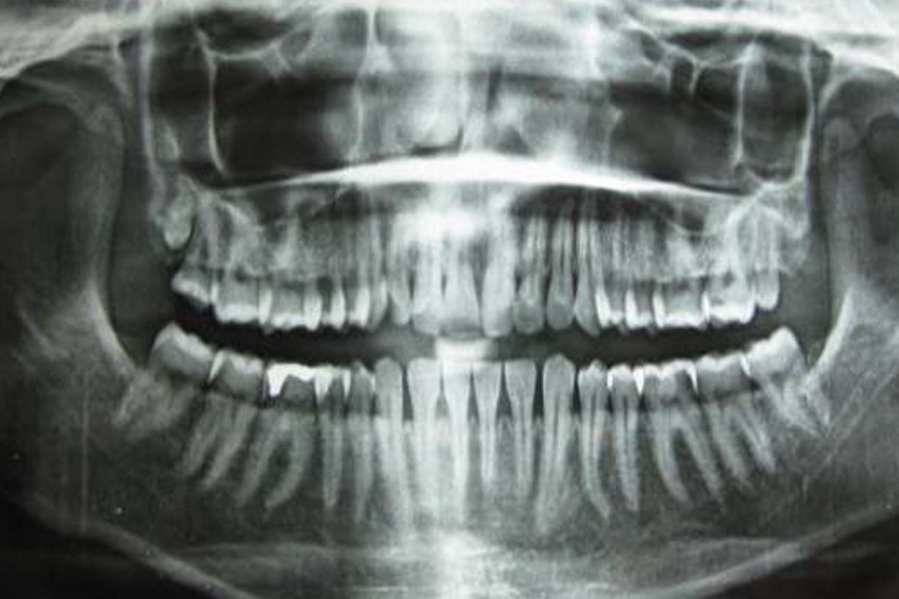
A 21-years-old female patient presented to our outpatients dental department with pain in lower right back tooth region, which had persisted for one month. Her medical and dental histories were non–contributory. On extraoral examination, no abnormalities were detected, and an intra–oral examination revealed a class II silver amalgam restoration with a secondary deep carious lesion in the mandibular right first molar. On further clinical examination, four maxillary lateral incisors were found [Table/Fig-1 and 2]. On the clinical and physical examinations of the patient, no symptoms which were related to any syndrome were revealed. The patient’s parents and siblings were also examined, and no related findings were observed. The lateral incisors which were located towards the midline were termed as supplemental lateral incisors because they were morphologically similar to the adjacent normal lateral incisors. The right supplemental lateral incisor was slightly rotated because of a discrepancy in its arch length [Table/Fig-3]. An Orthopantomogram (OPG) showed four maxillary lateral incisors [Table/Fig-4] and it also showed that the roots of the supplemental lateral incisors were similar to those of the normal lateral incisors. All the four incisors elicited a positive response to thermal and electric pulp testing. The patient was informed of her diagnosis and the potential future complications which were associated with it. The following treatment was advised: extraction of the supplemental teeth, followed by orthodontic treatment for the space closure and realigning of the remaining teeth or cosmetic contouring of the supplemental teeth, but the patient was not willing to undergo any treatment.
A clinical image shows two maxillary right lateral incisors
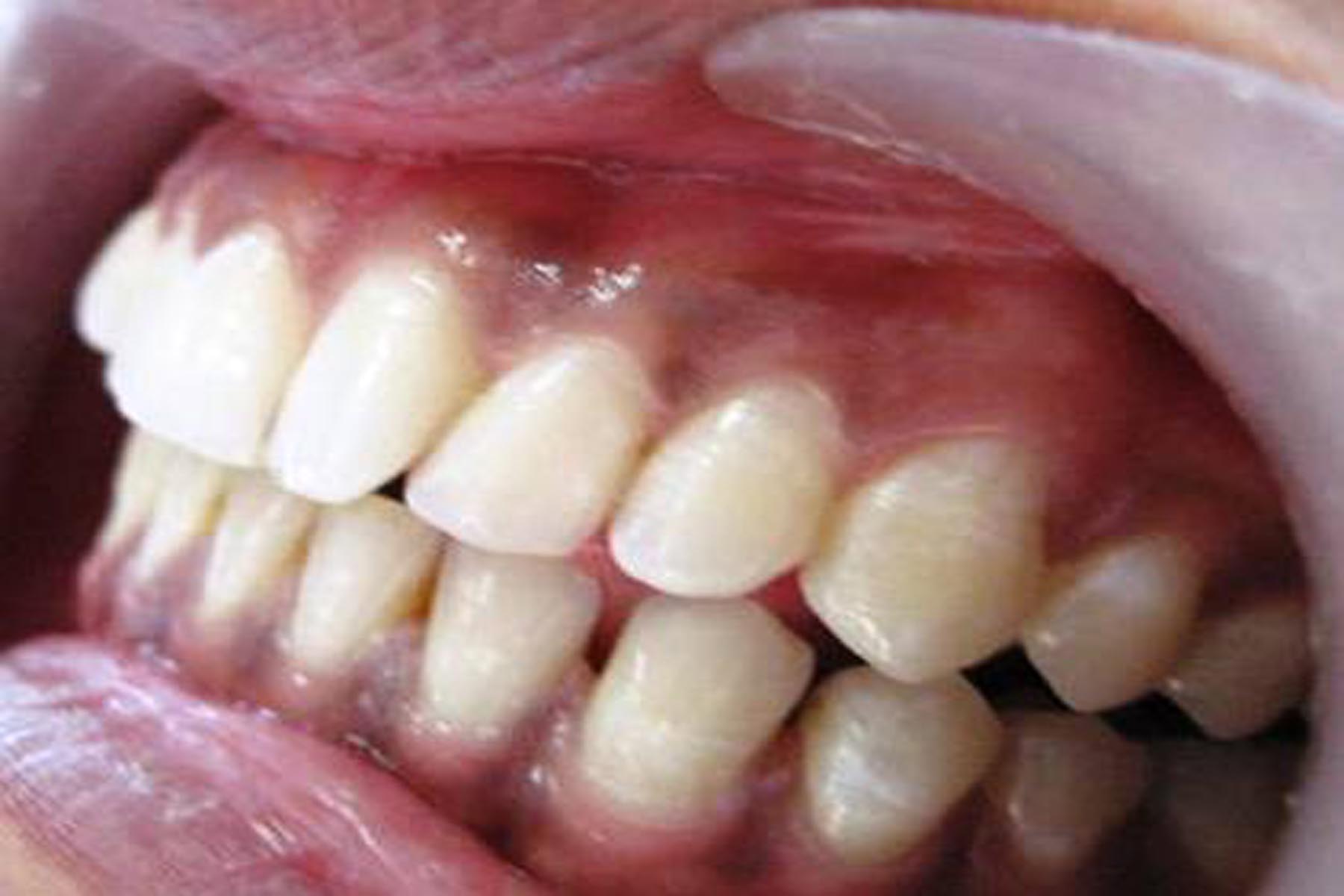
A clinical image shows two maxillary left lateral incisors
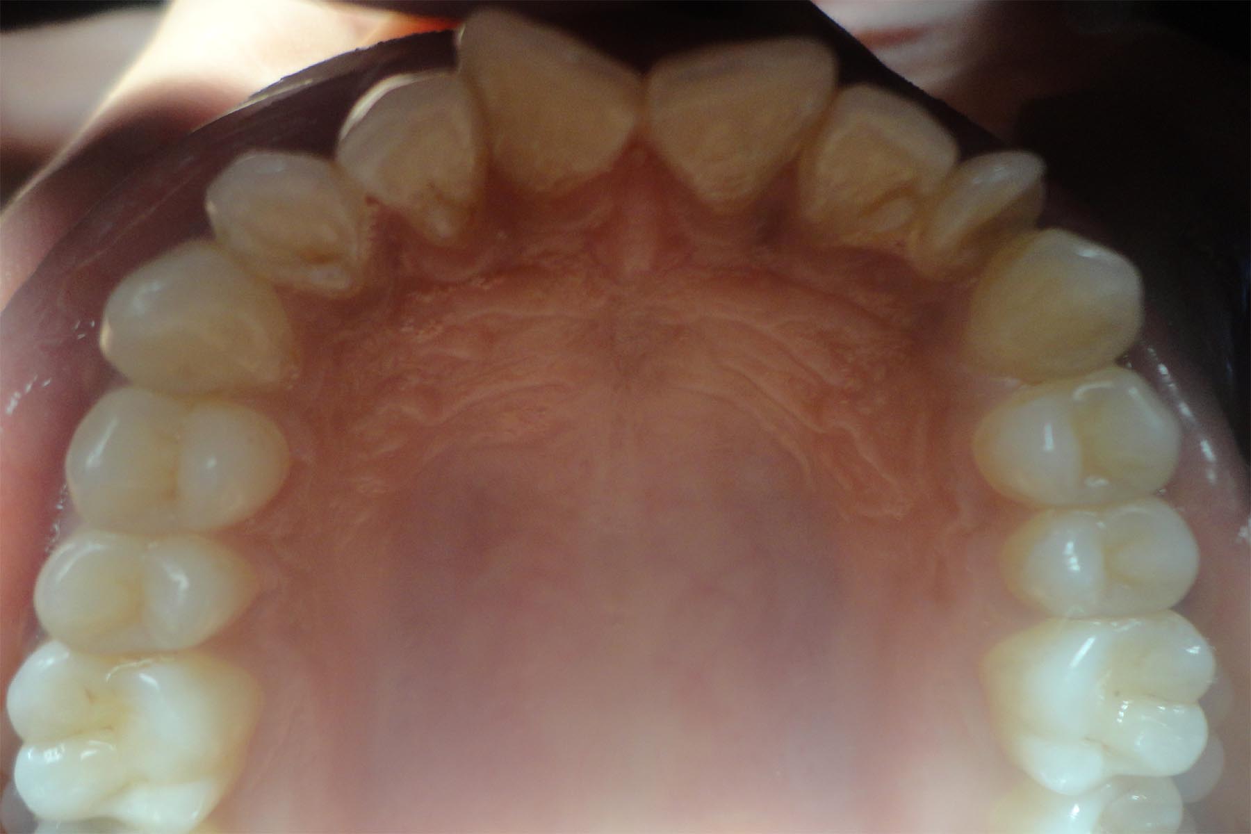
A clinical image shows four maxillary lateral incisors (Palatal view)
An orthopantomogram shows four maxillary lateral incisors.
Discussion
Supernumerary teeth or hyperdontia are defined as the teeth which are in excess of the normal dental formula. Prevalences of the supernumerary teeth of 1%-4% in the permanent dentition and of 0.2%-1.9% in the primary dentition have been reported [1]. The supernumerary teeth can occur as singles or multiples, unilaterally or bilaterally, and in the maxilla or the mandible, or in both. The supernumerary teeth in the maxilla are 8.2 to 10 times more than those in the mandible and they most commonly affect the premaxilla. However, there are variations within the reported relative frequencies of the supernumerary teeth in alternative regions [2]. Primosch classified the supernumerary teeth into two types according to their shapes. The supernumerary teeth which have normal shapes and sizes (eumorphic) are termed as ‘supplemental’, or ‘incisiform’, whereas the teeth which have abnormal shapes and smaller sizes (dysmorphic), are termed as ‘rudimentary’ and they include ‘conical’, ‘tuberculate’ and ‘molariform’ teeth [1].
The aetiology of hyperdontia is still unclear. According to the dichotomy theory, which seems to be the foremost possible cause, the tooth bud splits into two elements of equal orunequal sizes, which end up in two teeth of equal sizes or one normal and one abnormal tooth, respectively. The supernumerary teeth show a strong correlation with developmental disorders such as cleftlip and palate, Cleidocranial dysostosis and the Gardener syndrome and fewer such teeth usually occur with the Ehlers-Danlos syndrome, the Fabry Anderson’s syndrome, Chondroectodermal dysplasia, Incontinentia pigmenti and the Tricho Rhino-Phalangeal syndrome [2]. The erupted supernumerary teeth causes aesthetic issues, particularly when they are located in the anterior jaw region.
Most of the supernumerary teeth are detected as a chance finding during clinical or radiographic examinations, as the supernumerary teeth are mostly asymptomatic. It is essential, not only to enumerate, but also to identify the supernumerary teeth which are present clinically and radiographically, before a definitive diagnosis and a treatment plan can be formulated [3]. This case is unusual, as it demonstdates bilateral supernumerary lateral incisors in the patient, without any associated syndrome. The supplemental incisors are less common than the conical type of supernumerary teeth. The supplemental lateral incisors are rare, and bilateral cases are even rarer. Indeed, only two cases have been reported in the literature till date, to the best of our knowledge [4,5]. The supernumerary teeth can erupt or they can remain impacted and can cause numerous complications. They may be kept under observation without extraction, when a satisfactory eruption of the related teeth has occurred, without any functional or aesthetic disturbances and with no association with any pathology, but if it is associated with any complication, it should be extracted.Most of the supplemental teeth remain unerupted and they have been associated with several pathological conditions, such as widened follicular spaces, dentigerous cyst formation, dental pulp necrosis, pulp canal obliteration, root resorption, and ankylosis [6]. A disturbance in their eruption, a diastema formation, and rotations of the permanent teeth are the common complications [7]. If a supplemental tooth has fully erupted, it may be difficult to determine as to which tooth is supplemental and which is the normal dentition. In such circumstances, if both the teeths are healthy, it is wise to extract the tooth which is most displaced from the arch; so as to avoid crowding. The orthodontic problems and the dental pathology which are associated with the supernumerary teeth can be avoided by an early detection and management, which are the necessary aspects of preventive dentistry. Thus, the early clinical and radiographic assessments of the suspected cases are of utmost importance.
[1]. Primosch RE, Anterior supernumerary teeth-assessment and surgical intervention in childrenPaediatr Dent 1981 3:204-15. [Google Scholar]
[2]. Rajab LD, Hamdan MA, Supernumeraryteeth: review of the literature and a survey of 152 casesInt J Paediatr Dent 2002 12:244-54. [Google Scholar]
[3]. Scheiner MA, Sampson WJ, Supernumerary teeth: a review of the literature and four Case reportsAust Dent J 1997 42:160-65. [Google Scholar]
[4]. Townsend BR, Two cases of duplication of deciduous lateral incisors followed by permanent lateral incisorsBritish Dental Journal 1953 95:47-48. [Google Scholar]
[5]. Yildirim G, Bayrak S, Early diagnosis of bilateral supplemental primary and permanentmaxillary lateral incisors: a case reportEur J Dent 2011 5:215-19. [Google Scholar]
[6]. Stafne EC, Supernumerary upper central incisorsDental Cosmos 1931 73:976-80. [Google Scholar]
[7]. Rock WP, A case of bilateral supplemental maxillary central incisorsInt J Paediatr Dent 1991 1:155-58. [Google Scholar]