Human Immunodefeciency Virus Associated Cryptococcal Meningitis at a Tertiary Care Centre: Diagnostic Tools and Antifungal Susceptibility Testing
Rashmi Munivenkataswamy1, Anjana Gopi2, Shaik Mohammed Usman3, Jagadeesh4
1 Consultant, Department of Microbiologist, Gokula Metro Polis Diagnostics, Bangalore, India.
2 Associate Professor, Department of Microbiology, KIMS Hospital and Research Centre, Bangalore, India.
3 Post Graduate, Department of Microbiology, KIMS Hospital and Research Centre, Bangalore, India.
4 Professor, Department of Microbiology, KIMS Hospital and Research Centre, Bangalore, India.
NAME, ADRES, E-MAIL ID OF THE CORRESPONDING AUTHOR: Dr. Rashmi Munivenkataswamy, Consultant, Department of Microbiologist, Gokula Metropolis Clinical Laboratories, MSRMH, MSRIT Post, New BEL Road, Bangalore-560054, Karnataka, India.
Phone: 9880772903
E-mail: mrashmi22@gmail.com
Context: Cryptococcal meningitis has emerged as a leading cause of the infectious morbidity and mortality in HIV sero-reactive subjects and it is the second most common cause of the opportunistic neuroinfections in it. As this is a indistinguishable from other causes of meningitis, its early diagnosis is the key to the therapeutic success.
Objectives: This study was undertaken to know the incidence of Cryptococcal meningitis in HIV sero–reactive individuals and to assess the role of the microbiological parameters in its specific diagnosis, with a perspective of evaluating the anti–fungal resistance.
Material and Methods: A total of 66 CSF samples from suspected cases of meningoencephalitis were subjected to standard microbiological procedures. The Cryptococcal isolates were identified by microscopy, the cultural characteristics, melanin production on Niger Seed agar, urea hydrolysis, the Nitrate assimilation test and by capsular antigen detection by latex agglutination. The Cryptoccal isolates were further biotyped by using Canavanine–Glycine–Bromothymol blue agar. The Minimum Inhibitory Concentrations (MIC) of Amphotericin B and Fluconazole for the isolates were detected.
Results: The incidence of Cryptococcal meningitis in our study group was 18.2% (12/66). The Cryptococcal antigen was detected in all the 12 cases, whereas microscopy was positive only in 9 cases and Cryptococcus was isolated by culture in 10 cases. All the isolates were sensitive to Amphotericin B and 90% of the isolates were sensitive to Fluconazole. The CD4counts ranged between 22-138 cells /μl.
Conclusion: A high incidence of Cryptococcal meningitis in HIV sero-reactive subjects necessitates the importance of a precise and an early microbiological diagnosis for better management of such subjects. Due to the growing concern of emerging drug resistance, the testing for the anti–fungal susceptibility has to be encouraged in all the cases.
Cryptococcal meningitis, Human Immunodefeciency Virus, Minimum inhibitory concentration, Latex agglutination
Introduction
Cryptococcus neoformans (C.neoformans) is an encapsulated fungus which belongs to family, Basidiomycetes and it is a significant human pathogen. Cryptococcosis, the disease which results from infection with C.neoformans varies from a localized to a disseminated disease and it is seen as an acute or a chronic infection [1]. C. neoformans considered as a sleeping giant among the fungal diseases by Ajello in 1970, but it has now virtually become an awakening giant after the emergence of a pandemic of AIDS [2]. The organism typically initiates infection by gaining entrance to the lungs and it may rapidly disseminate to the brain and the meninges. Crytococcal meningitis is the most frequent encountered manifestation of Cryptococcosis [3].
C.neoformans is the most common CNS fungal opportunistic pathogen in immunocompromised patients, particularly among those suffering from AIDS. Approximately 7%–8% of the HIV infected patients develop Cryptococcal meningitis [4]. The prevalence of the Cryptococcal infection among AIDS patients varies from 2%–10% in western Europe and the U.S and up to more than 15% in central Africa and South East Asia [5,6]. In U.S, yearly incidence among the people with HIV/AIDS is estimated to be between 2 and 7 cases per 1,000 people. The mortality which is caused by C. neoformans infections in US and other developed nations is around 12%. Worldwide, C. neoformans infections cause an estimated 1 million cases of Cryptococcal meningitis per year among the people with HIV/AIDS, result in nearly 625,000 deaths. The greatest burden of the disease occurs in Sub–Saharan Africa, where the mortality is estimated to be 50% to 70%.6 In 40% of these patients, Cryptococcosis is an AIDS defining condition [1]. The most important point in the diagnosis of Cryptococcal meningoencephalitis is to have a high index of suspicion in immunocompromised patients, with signs and symptoms which are referable to the CNS. The diagnosis is firmly established by culturing the organisms from the Cerebro Spinal Fluid (CSF). The negative staining of the CSF, being simple and rapid, is usually performed. Where capsulated organisms are not seen in the negative staining, the assaying for the Cryptococcal polysaccharide antigen in the CSF is an important adjunct to the diagnosis. The present study was undertaken to determine the incidence of Cryptococcal meningitis in HIV sero–positive individuals, the efficiency of various microbiological tests which aid in the early diagnosis of Cryptococcal meningitis and the Antifungal resistance of the isolates.
Material and Methods
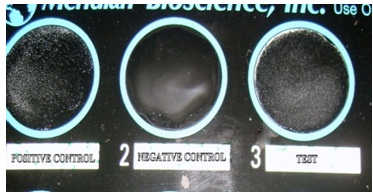
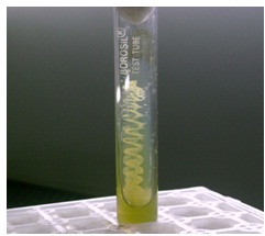
In this prospective study, 66 HIV sero-reactive patients who were admitted with the clinical suspicion of meningitis in the Medicine Ward and at the ART center of KIMS, Bangalore, India, between May 2010 to April 2011, were studied. Separate informed consents were obtained from them. The demographic factors such as age, gender, the date of admission; the info on whether on HAART (Highly Active Antiretroviral Therapy) or not; the risk factors which were involved; the signs and symptoms; a past history of Cryptococcal meningitis and the info on whether on prophylaxis or not; any underlying disease; and the CD4 counts were also noted. CSF and serum were collected and these were taken immediately for processing to the microbiology laboratory. The supernatant of the centrifuged CSF sample was subjected to capsular Ag detection by the Latex agglutination test (CALAS Bioscience, Inc.) [Table/Fig-1]; it was also deposited for direct microscopy (which comprised Gram staining and negative staining with 10% Nigrosin) and Fungal culture. Two sets of SDA (Sabouraud’s Dextrose Agar without Actidione) were inoculated and they were incubated at 25°C and 37°C, separately, over a period of 4 weeks. All the cultures were examined daily during the first week and twice a week during the next 3 weeks. C.neoformans was identified, based on their mucoid colonial appearance on SDA, the presence of spherical budding yeast cells on microscopy, Urease production, Nitrate assimilation and the production of brownish colonies on Niger Seed agar. The serology for the Cryptococcal antigen detection was also employed by using the Latex agglutination test (CALAS Bioscience, Inc.) by pretreating the serum with pronase.
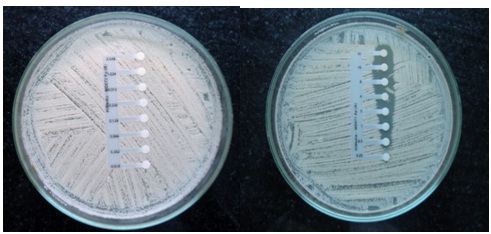
The Cryptococcal isolates were further biotyped by using Canavanine–Glycine–Bromothymol blue agar, [Table/Fig-2] which differentiate Cryptococcus neoformans var. gattii (Serotype B and C), which turn the medium blue, from other Cryptococcusneoformans var., which remain as such i.e., yellow to light green. The minimum inhibitory concentrations of Amphotericin B (AmB) and Fluconazole for the isolates were detected by using E-strips [Table/Fig-3] and they were interpreted as per the CLSI guidelines [7].
CGB medium (Media remained greenish yellow)
E Test- Fluconazole (A) Amphotericin (B)
Statistical Methods
A descriptive statistical analysis was carried out in the present study. The results of the categorical measurements have been presented in Number (%). The Chi-square/ Fisher Exact test was used to find the significance of the study parameters on a categorical scale between two or more groups. The diagnostic statistics viz. the Sensitivity, Specificity, PPV, NPV and the Accuracy were computed to find the correlation of various tests with the incidence of cryptococcal meningitis.
Results
A clinical microbiological study which was done on 66 HIV seropositive patients with the signs and symptoms of meningitis, was undertaken to study the incidence of Cryptococcal meningitis. The incidence of Cryptococcal meningitis was found to be 18.2% [Table/Fig-4]. A majority of the Cryptococcal meningitis cases which were studied, belonged to the age group of 31-40 years (41.7%). Males were predominant (83.3%) [Table/Fig-5]. The common clinical manifestations included headache, fever, vomiting and altered sensorium. 25% of the patients were on HAART. Cryptococcal meningitis were diagnosed, based on the positivity of either–Negative staining/Fungal culture/CSF Latex agglutination test. Gram positive budding yeast cells were seen in 7/12 cases. The negative staining was found to be positive in 9/12 cases. The fungal cultures which were set up at 37oC yielded growth in 10/12 cases. The Latex agglutination Test for the Cryptococcal capsular antigen detection was positive in all the 12 CSF samples and in 11/12 serum samples [Table/Fig-6]. All the isolates belonged to the Cryptococcusneoformans variety other than Gattii. All the isolates were susceptible to AmB (Amphotericin B) and one isolate was found to be resistant to Fluconazole. All the Cryptococcal meningitis patients had CD4 counts of < 200 cells /μl. A majority had CD4 counts of <100 cells /μl (83.3%) [Table/Fig-7]. There was a relapse of the infection in one case and mortality was seen in 3 cases (25%) during the follow up.
Incidence of Cryptococcal meningitis
| Incidence of cryptococcal meningitis | Number of patients (n=66) | % |
|---|
| Positive | 12 | 18.2 |
| Negative | 54 | 81.8 |
| Age in years | Male | Female | Total(%) |
|---|
| 21-30 | 02 | 00 | 02(16.67%) |
| 31-40 | 04 | 01 | 05(41.67%) |
| 41-50 | 03 | 01 | 04(33.33%) |
| 51-60 | 01 | 00 | 01(8.33%) |
| Total | 10(83.30%) | 02(16.70%) | 12(100%) |
Statistical parameters of various Diagnostic tests
| Sensitivity | Specificity | PPV | NPV | Accuracy |
|---|
| Grams Stain | 58.33 | 100.00 | 100.00 | 91.53 | 92.42 |
| Negative Stain | 75.00 | 100.00 | 100.00 | 94.74 | 95.45 |
| Fungal Culture On Sda At 37oc | 83.33 | 100.00 | 100.00 | 96.43 | 96.97 |
| Latex Agglutination |
| Csf | 100.00 | 100.00 | 100.00 | 100.00 | 100.00 |
| Serum | 91.67 | 100.00 | 100.00 | 98.18 | 98.48 |
Correlation of CD4 count with incidence of Cryptococcal meningitis
| CD4 count (cells/micro lt) | Total number of patients(n=66) | Cryptococcal meningitis |
|---|
| Negative (n=54) | Positive (n=12) |
|---|
| 1-50 | 12(18.2%) | 8(66.7%) | 4(33.3%) |
| 51-100 | 11(16.7%) | 5(45.5%) | 6(54.5%) |
| 101-200 | 19(28.8%) | 17(89.5%) | 2(10.5%) |
| 201-300 | 14(21.2%) | 14(100.0%) | 0 |
| 301-500 | 10(15.2%) | 10(100.0%) | 0 |
| Inference | Incidence of cyrptococcal meningitis is significantly associated with lower CD4 count with p<0.001** |
Discussion
The infection with HIV continues to be the risk factor for the development of CNS Cryptococcosis. The present study also shows a higher incidence of C. meningitis among HIV seropositive patients. As the clinical picture may be confusing with viral or tubercular meningitis, a high index of suspicion and a routine mycological surveillance is required to aid in an early diagnosis and for selecting the appropriate therapy.
The incidence of C.meningitis, as was demonstrated in our study-12/66 (18.2%), was well comparable with in other studies- Meena et al., [4] and Rakhmanova AG et al., [7] individually showed an incidence of 17%, whereas Boegerts et al., [8] reported a 19% incidence. A higher incidence which was found in the age group of 20-50 years (91.7%), might reflect a difference of exposure. A similar incidence was shown by Lakshmi et al., [3]. The higher male preponderance which was found in our study (83.33%) was comparable with those in other studies, which was exemplified by Lakshmi et al., [3]. In the present study, 3(25%) patients had concurrent pulmonary tuberculosis. Vasanth et al., [9] also noted concurrent tuberculosis in 28% cases. All these diagnostic tests were found to be 100% specific. In our current study, the overall positivity of microscopy, culture and LAT in CSF, were comparable to the reports which had been found in the literature [11–13] [Table/Fig-8]. In our study, the LAT in CSF was 100% sensitive and specific. The Cryptococcal Ag detection, being rapid and sensitive, became suitable choice for screening for the isolates. All the 10 isolates belonged to the Cryptococcusneoformans species other than variety gattii (Serotype B and C) i.e. either var grubii (Serotype A) or var neoformans (Serotype D). Most of the isolates from the HIV positive patients from India were Serotype A, var. grubii [17]. The further differentiation between var. grubii and var. neoformans was not carried out because of the nonavailability of resources (CDBT Agar). All the isolates were found to be sensitive to Amphotericin B. 10% isolates were resistant to Fluconazole. Similar findings were observed by MR Capoor et al., [12] and Borannsar et al., [18]. All the cases had CD4 counts of ≤200 cells/μl. 10 cases had counts of <100 cells/μl. The incidence of Cryptococcal meningitis was significantly associated with lower CD4 counts in many studies. One such example was the study of S Majumder et al., [19], which showed CD4 counts of <100 cells /μl in 85% of the patients. Low CD4 counts are significantly associated with the development of C.meningitis. The WHO guidelines advocate a Fluconazole prophylaxis for all the patients with CD4 cell counts of <100 cells /μl.
Comparison of sensitivities of different tests with other studies
| Country | No. of patients | No. positive by India ink (%) | No.by CSF culture(%) | No. positive by CSF Ag(%) | No. positive by serum Ag(%) |
|---|
| Australia [14] | 128 | 72.6 | 86.7 | 87.5 | Not reported |
| Brazil [15] | 65 | 93.8 | 100 | 100 | Not reported |
| India [10] | 104 | 69.23 | 76.92 | 100 | 88.4 |
| US [16] | 89 | 73.5 | 100 | 90.9 | 98.6 |
| Present study | 66 | 75 | 83.33 | 100 | 90 |
Conclusion
The high incidence of Cryptococcal meningitis in HIV sero-positive subjects and also the high rate of mortality necessitates the importance of a precise and an early microbiological diagnosis for the better management of such individuals. The detection of the capsular polysaccharide antigen by the Latex agglutination test is of immense value in an early diagnosis and hence, in an early management, as it is a more rapid and sensitive method. In our study, the Antigen detection in Serum by the Latex agglutination test was comparable to that in CSF. Therefore, this test can be used as an screening test for the diagnosis of C. meningitis, where lumbar puncture is not advocated. The Conventional Antifungal susceptibility testing has proven to be difficult to be implemented in the routine clinical laboratory. Although the broth dilution method remains the gold standard for detecting the MIC, it is not convenient to carry it out on a routine basis, as it is time consuming and cumbersome. The E-test is a useful alternative, as the strips are commercially available and as it can be carried out easily in a routine microbiology laboratory. It is as reliable as the microdilution method. Another matter of concern is the increasing resistance to Flucanozole, which also has been documented in our study. Needless to say, the antifungal susceptibility of the isolates has to be carried out wherever a resistance is suspected clinically or epidemiologically.
[1]. Topley and Wilson. Text book of Microbiology, 9th edition; pg no. 461-73 [Google Scholar]
[2]. Chander Jagadish, Text book of Medical Mycology3rd edition:291-313. [Google Scholar]
[3]. Lakshmi V, Sudha T, Prevalence of CNS Cryptococcosis in HIV reactive hospitalized patientsIJMM 2007 25(2):46-49. [Google Scholar]
[4]. Satpute Meena G, Telang Nilima V, Litake Geetanjali M, Prevalence of Cryptococcal Meningitis at tertiary care Centre in Western India (1996-2005)IJMM 2006 55(9):1301-02. [Google Scholar]
[5]. Sivasangutha K, Harissh BN, Sujatha S, Cryptococcal Meningoencephalitis Diagnosed by Blood CultureIJMM 2007 25:282-84. [Google Scholar]
[6]. C. neoformans Cryptococcosis Statistics[Internet],[updated 2012 Jan 5; cited 2013 Feb 22]. Available from: http://WWW.cdc.gov/fungal/Cryptococcosis-neoformans/Statistics [Google Scholar]
[7]. Pfaller Michael A, Chaturvedi Vishnu, Espinel Ana, Ghannoum Linda Frank Reference Method for Broth Dilution Antifungal Susceptibility Testing of Yeasts; Approved Standard-CLSI document M27-A 2002 22nd editionPennsylvania, USA:4-20. [Google Scholar]
[8]. Rakhmanova AG, Giaurgeiva OKH, Clinical course of Cryptococcosis in HIV infectionsKLIN Med(MOSK) 1999 77:39-42. [Google Scholar]
[9]. Bogaerts J, Rouvroy D, Taelman H, Kagame A, Aziz MA, Aids associated Cryptococcal meningitis in Rwanda. Epidemiological and diagnostic featuresJ infect 1999 39(3):329-31. [Google Scholar]
[10]. Baradkar Vasant, Mathur M, De A, Kumar S, Rathi M, Prevalence and clinical presentation of Cryptococcal Meningitis among HIV seropositive patientsIndian Journal of STD and AIDS 2009 30:19-22. [Google Scholar]
[11]. Khanna N, Chandramuki A, Desai A, Ravi V, Cryptococcal infections of the central nervous system: An analysis of predisposing factors, laboratory findings and outcome in patients from South India with special reference to HIV infectionJ MedMicrobiology 1996 45:376-79. [Google Scholar]
[12]. Capoor MR, Nair D, Clinical and Mycoloogical profile of Cryptococcosis in a tertiary care hospitalIndian J Med Microbiol 2007, Oct 25(4):401-04. [Google Scholar]
[13]. Jain N, Wickes BL, Keller SM, Fu J, Casadevall A, Molecular Epidemiology of Clinical Cryptococcus neoformans Strains from IndiaJournal of Clinical Microbiology 2005, Nov 43(11):5733-42. [Google Scholar]
[14]. Chen S, Sorrell T, Nimmo G, Speed B, Currie B, Ellis D, Parr Epidemiology and host- and variety-dependent characteristics of infectiondue to Cryptococcusneoformans in Australia and New ZealandClin.Infect. Dis 2000 31:499-508. [Google Scholar]
[15]. Rozenbaum R, Goncalves AJR, Clinical epidemiological study of 171 cases of cryptococcosisClin. Infect. Dis 1994 18:369-80. [Google Scholar]
[16]. Chuck SL, Sande MA, Infections with Cryptococcusneoformans in the acquired immunodeficiency syndromeN. Engl. J. Med 1989 321:794-99. [Google Scholar]
[17]. Shankar SK, Mahadevan A, Sundaram C, Sarkar C, Chacko G, Pathobiology of fungal infections of the central nervous system with special reference to the Indian scenarioNeurology India 2007, July-September 55:198-212. [Google Scholar]
[18]. Borann Sar, Didier Monchy, Mich Vann, Chantary Keo, Jean Louis Sarthou, Increasing invitro resistance to fluconazole in Cryptococcusneoformans Cambodian isolates: April 2000 – march 2002J Antimicrobial chemotherapy 2004 54:563-65. [Google Scholar]
[19]. Majumder S, Mandal SK, Bandyopadhyay D, Prognostic markers in AIDS related Cryptococcal meningitisJAPI 2011 59:152-54. [Google Scholar]