Giant Inflammatory Variant of Well Differentiated Liposarcoma: A Case Report of a Rare Entity
Manisha Sharma1, Rahul Mannan2, Tejinder Singh Bhasin3, Mridu Manjari4, Rajan Punj5
1 Associate Professor, Department of Pathology, Sri Guru Ram Das Institute of Medical Sciences Research, Amritsar, Punjab India.
2 Associate Professor, Department of Pathology, Sri Guru Ram Das Institute of Medical Sciences Research, Amritsar, Punjab India.
3 Associate Professor, Department of Pathology, Sri Guru Ram Das Institute of Medical Sciences Research, Amritsar, Punjab India.
4 Professor, Department of Pathology, Sri Guru Ram Das Institute of Medical Sciences Research, Amritsar, Punjab India.
5 Consultant Surgeon, New Hope Hospital, Amritsar, Punjab, India.
NAME, ADRESS, E-MAIL ID OF THE CORRESPONDING AUTHOR: Dr. Manisha Sharma, B-241, Ranjit Avenue, Amritsar-143001, Punjab India.
Phone: 9876842942,
E-mail: manisha_salwan@yahoo.com
An inflammatory liposarcoma is a rare variant of a well-differentiated liposarcoma. A case of a giant variety of an inflammatory well–differentiated liposarcoma is being reported. CT scan revealed a large abdomino–pelvic mass which had displaced the gut loops to left and posteriorly and urinary bladder and uterus to right. FNAC yielded mature adipocytes with no evidence of atypical cells. The large bossilated and irregular fibro–fatty mass which weighed 23 kg was excised. Microscopy revealed mature adipocytes with foci of fibrosis and abundant inflammatory cell infiltrates of eosinophils, lymphocytes, plasma cells and few neutrophils, along with few atypical cells. Only a few cases of a giant inflammatory variant of a well–differentiated liposarcoma have been reported in the world literature and this is first of its kind from the Indian sub–continent. A rare giant variant of a inflammatory well–differentiated liposarcoma with abundant eosinophils is being reported here, along with review of literature.
Giant liposarcoma, Retroperitoneal liposarcoma, Inflammatory well differentiated liposarcoma, Eosinophils
Introduction
Liposarcomas are malignant tumours of mesenchymal origin (adipose tissue). The major sites of liposarcomas are the extremities, retroperitoneum and inguinal region. Liposarcomas are remarkable because of their frequently large sizes [1].
Well-differentiated liposarcomas are low grade mesenchymal tumours, which include the sclerosing, spindle cell, inflammatory and adipocytic subtypes [2]. An inflammatory liposarcoma is a rare variant of a well differentiated liposarcoma [3]. A giant variant of this liposarcoma is even rarer. Till date, only few cases (two) of the giant variety of retroperitoneal inflammatory well differentiated liposarcomas have been reported in the Indian subcontinent [4].
Case Report
A 60-years female presented with a gradual increase in weight and abdominal girth, which was there since three years. On examination, a diffuse non–tender, non–mobile mass with ill defined margins, which occupied the whole abdomen was found, which was palpable.
Routine investigations which included haemograms and renal and liver functions test were within normal limits. A CT scan revealed a large fatty retroperitoneal mass which had displaced the gut loops to left and posteriorly, ascending colon medially and urinary bladder and uterus to right. A CT guided FNAC of the mass revealed mature adipocytes, with no evidence of atypical cells. Excision of the retroperitoneal tumour was done through a transperitoneal approach en block and the tumour was sent for a histopathological examination. Post operative follow up was uneventful.
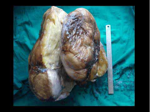
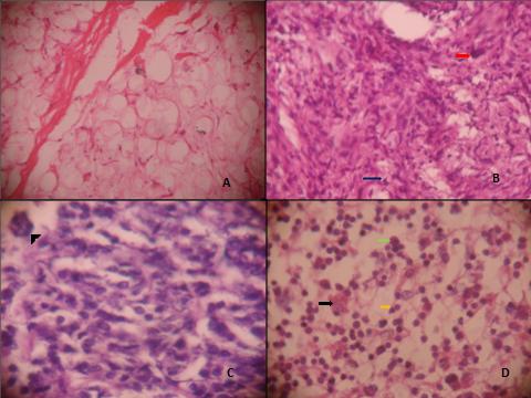
Gross appearance revealed a 47 × 40 × 25 cm, [Table/Fig-1] large, irregular, bossilated fibrofatty mass which weighed 23 kilograms. Cut section showed a homogenous, yellow appearance. Microscopy showed mature adipocytes with foci of necrosis [Table/Fig-2A]. A few atypical tumour cells were present and they had large hyperchromatic pleomorphic nuclei with pale, granular to vacuolated cytoplasm [Table/Fig-2B and C]. Abundant inflammatory cell infiltrates which contained many eosinophils, lymphocytes, plasma cells and few neutrophils were also seen [Table/Fig-2D].
Microscopic findings (a) Sections showing lipomatous area (H& E 400 X); (b)Sections with cellular areas with giant cells and a few atypical tumour cells with large hyperchromatic pleomorphic nuclei with pale, granular to vacoulated cytoplasm (H & E 200 X); (c) Same as 2B with higher magnification (H & E 400 X); (d)Inflammatory cell infiltrate of many eosinophils, lymhocytes, plasma cells and few neutrophils (H & E 400 X)
Resected margins were free of the tumour. The patient has been on regular follow up for the past six months.
Discussion
Liposarcomas are commonly found in lower extremities and the retroperitoneum. The clinical presentation of a inflammatory well differentiated liposarcoma is not significantly different from those of the commoner variants of well differentiated liposarcomas (sclerosing, spindle cell, adipocytic and mixed types), where no inflammatory infiltrate is seen [5]. At times, the extensive lympho-plasmocytic infiltrate mimics an inflammatory pseudotumour [3]. Such tumours have been associated with pyrexia of unknown origin and if they present in unusual locations; they can cause diagnostic dilemmas [4].
Well–differentiated liposarcomas are characterised by repetitive local recurrences, without a metastatic potential. A retroperitoneal localisation is a significant negative prognostic factor for recurrence, along with tumour size, depth and invasion of surrounding structures. Studies have reported a recurrence rate of 27% [6, 7]. Radical surgery is the treatment of choice [1].
Giant retroperitoneal liposarcomas of mixed and de-differentiated types have been reported, but there are only very few reports on giant, inflammatory, well differentiated liposarcomas [6]. The largest, giant, inflammatory, well differentiated liposarcoma which was reported from Indian subcontinent weighed 9 kg [4]. The tumour in the present case weighed 23 kg and it measured 47 × 40 × 25 cms, which is one of the largest reported tumours among inflammatory well differentiated liposarcomas.
Besides inflammatory cells like lymphocytes, plasma cells and neutrophils, the presence of a fair number of eosinophils was an additional finding which was recorded in the present case study, which has not been reported so far in literature. Various case reports have stressed upon the sizes and weights of the tumours, with one of the studies reporting the tumour size to be 32 kgs. In all such case reports, the pathological descriptions were limited to those which were found in well differentiated liposarcomas or emphasis was laid on lympho–plasmacytic infiltrates. Presence of an increased number of eosinophils and the large tumour size made this case unique.
In conclusion, this is one of the largest reported retroperitoneal inflammatory well differentiated liposarcomas especially with abundance of eosinophils.
[1]. Akhoondinasaab MR, Omranifard M, Huge retroperitoneal liposarcomaJ Res Med Sci 2011 April 16(4):565-67. [Google Scholar]
[2]. Dei Tos AP, Liposarcoma: new entities and evolving conceptsAnn Diagn Pathol 2000 4:252-66. [Google Scholar]
[3]. Kraus MD, Fletcher CD, Well-differentiated inflammatory liposarcoma: an uncommon and easily overlooked variant of a common sarcomaAm J Surg Pathol 1997 21:518-27. [Google Scholar]
[4]. Mehrotra PK, Ramachandran CS, Goel D, Vijay A, Inflammatory variant of a well-differentiated retroperitoneal liposarcoma: Case report of a giant varietyIndian Journal of Cancer 2006 43(1):735-40. [Google Scholar]
[5]. Rani U, Sharma MC, Pande GK, Kapoor A, Dey AB, Well differentiated liposarcoma presenting as pyrexia of unknown originAssoc Physic India 2001 49:761-63. [Google Scholar]
[6]. Nijhuis PH, Sars PR, Plaat BE, Molenaar WM, Sluiter WJ, Clinico-pathological data and prognostic factors in completely resected AJCC stage I-III liposarcomasAnn Surg Oncol 2000 7:535-43. [Google Scholar]
[7]. Inoue K, Higaki Y, Yoshida H, Giant retroperitoneal liposarcomaInt J Urol 2005 12:220-22. [Google Scholar]