“Cannon Balls or Pus Balls” in Pap Smears: A Case Report
Vijay Kumar Bodal1, Sarbhjit Kaur2, Ranjiv Bhagat3, Rupinder Kaur4, Manjit Singh Bal5
1 Associate Professor, Department of Pathology, Government Medical college, Patiala, India.
2 Senior Resident, Department of Gynaecology & Obstetrics, Government Medical college, Patiala, India.
3 Lecturer, Department of Pathology, Government Medical College, Patiala, India.
4 Junior Resident, Department of Pathology, Government Medical college, Patiala, India.
5 Professor & Head, Department Pathology, Government Medical college, PatialaIndia, India.
NAME, ADRESS, E-MAIL ID OF THE CORESPONDING AUTHOR: Dr. Vijay Kumar Bodal, Associate Professor, Department of Pathology, Government Medical College, Patiala, India.
Phone: 09814714946,
E-mail: Vijay_bodal@yahoo.com
A 50–year old female presented with the chief complaint of a discharge per vaginum, which was there for the past 15 days. A routine PAP smear was received in the Department of Pathology, Government Medical College, Patiala, India. After its fixation and staining, it was examined under the microscope. It showed the Trichomonas vaginalis infection, with the neutrophils forming cannon balls at places. Neutrophils in the PAP smear are a nonspecific finding, particularly if they are low in numbers or if they are seen in the premenstrual and the menstrual phases. The neutrophils which are adherent to the squamous cells are called “cannon balls” or “pus balls”, which are common in the Chlamydia infection. This case is being presented because of the presence of these rare morphological structures i.e. “cannon balls” or “pus balls”.
PAP, Cannon ball, Trichomonas vaginalis
Introduction
Trichomonas vaginalis is an anaerobic parasitic protozoa. It utilizes glycogen as an energy source. In PAP stained preparations, the protozoa is often found to be attached to the superficial type squamous cells which are rich in glycogen content. The number of pathogens is high in the proliferative phase, while it becomes less prominent during the secretory phase or in pregnancy [1].
Neutrophils in the PAP smear, which are found in infections (bacterial, fungal or viral) – are associated with and are phagocytosed by Trichomonas [2] and Chlamydia [3].
Case History
A 50–years old female presented with the chief complaint of a discharge per vaginum which was there since the past 15 days. She had attained menopause 6 years ago. The patient had one episode of bleeding per vaginum 15 days back (which was in the form of spotting only, which had lasted for one day). After that, she had a discharge per vaginum, for which she came to the Gynaecology Out Patients Department, Rajindra hospital, Patiala, India. The obstetrical history of the patient was G3L1A2. A routine PAP smear was taken and it was sent to the Department of Pathology, Government Medical College, Patiala, India.
Microscopic Examination
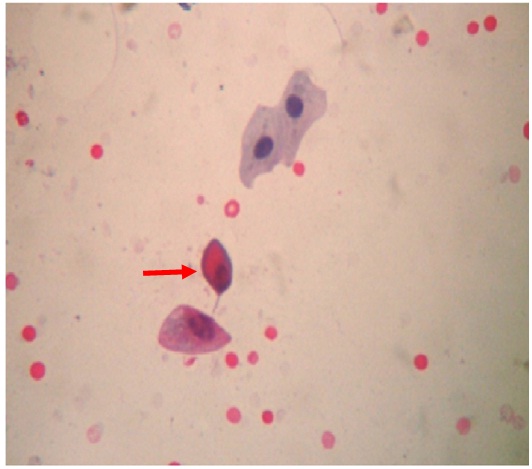
The smear showed grey/blue pear shaped structures (Trichomonas vaginalis) in the background of an inflammatory infiltrate which comprised predominantly of neutrophils, which had formed cannon balls at places [Table/Fig-1].
Red Arrow showing Trichomonas vaginalis (Pap 1800 X 1600)

Discussion and Review of the Literature
Neutrophils in PAP smears is a common finding, which has been reported in 5–25% of the cases [4].
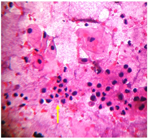
Neutrophils in PAP smears are a non–specific finding, particularly if they are in low numbers or if they are present during the premenstrual and the menstrual phases. The neutrophils which are adherent to the squamous cells are called “cannon balls” or “pus balls”, which are common in the Chlamydia infection [5] [Table/Fig-2].
Yellow arrow showing “cannon ball” appearance- neutrophils adherent to squamous cells (Pap 1800 x 1600)

Typically in the Trichomonal infection, the squamous epithelial cells demonstrate reactive changes with mild nuclear enlargement and small perinuclear halos. There is often a dense neutrophilic infiltrate, and the neutrophils form aggregates which are sometimes referred to as “cannon balls” or “pus balls”; these represent the squamous cells which are covered by the trichomonal organisms that have been phagocytosed by the macrophages and the neutrophils [6].
[1]. Vanacova S, Liston DR, Trachezy J, Johnson PJ, Molecular biology of the amitochondriate Parasites Giardia intestinalis, Entamoeba histilytica and Trichomonas vaginalisInt J Parasitol 2003 33:235-55. [Google Scholar]
[2]. Demirezen S, Safi Z, Beksac S, The interaction of trichomonas vaginalis with epithelial cells, polymorphonuclear leucocytes and erythrocytes on vaginal smears: light microscopic observationCytopathology 2000 11(5):326 [Google Scholar]
[3]. Sellors J, Howard M, Pickard L, Jang D, Mahony J, Chernesky M, Chlamydial cervicitis: testing the practice guidelines for presumptive diagnosisCMAJ 1998 158(1):41 [Google Scholar]
[4]. Bertolino JG, Rangel JE, Blake RL Jr, Silverman D, Ingram E, Inflammation on the cervical Papanicolaou smear: the predictive value for infection in asymptomatic womenFamily Medicine 1992 24(6):447 [Google Scholar]
[5]. Malik SN, Wilkinson EJ, Drew PA, Hardt NS, Benign cellular changes in Pap smears, Causes and significanceActa Cytol 2001 45(1):5 [Google Scholar]
[6]. DeMay Richard Mac, The Pap Test: Exfoliative Gynecologic Cytology(1st Ed)ASCP 2005 :106 [Google Scholar]