The hand is prehensile organ. It is endowed with grasping and precision movements for skilled work and it act as a chief tactile apparatus. Philosophically it may be said that the actions of lumbricals of hand are indices of civilization of a race [1].
The lumbrical muscles are the important part of intrinsic musculature of hand. Although several deviations from normal or variations of lumbricals have been described in the standard textbooks of anatomy and other surgical or anatomical publications, a review of the basic anatomy of lumbricals will be helpful [2].
The lumbrical muscle is unique among the muscles of human body. Its almost parallel fibers arise from tendon of flexor muscle for finger and insert into the tendon of an extensor muscle. This slender muscle has fascinated the anatomists and has been subject of controversy since the days of Galen [3].
The lumbricals arise from tendons of flexor digitorum profundus.
Each tendon passes to radial side of corresponding finger and is attached to the lateral margin of dorsal digital expansion of extensor digitorum that covers the dorsal surfaces of fingers. 1st and 2nd lumbricals are supplied by median nerve (C8 T1). 3rd and 4th are supplied by deep branch of ulnar nerve (C8 T1).
Material and Methods
A total of 50 hands of 25 (formalin embalmed) cadavers of department of Anatomy, Government Medical College, Amritsar were used for this study. Hands were labelled from 1-25 with suffix R (right); L (left); M (male) and F (female) for the sex. For the dissection Cunningham’s Manual of Practical Anatomy was followed [4].
A vertical midline incision was given on palm extending on to the middle finger up to distal phalanx.
Two transverse incision were given:-
On the proximal end of previous incision
On the distal end of palm
Superficial fascia was removed. Palmar aponeurosis was cleared. The apex of Palmar aponeurosis was divided and reflected distally after separating it from flexor retinaculum.
Common tendon of flexor digitorum superficialis was reflected distally after separating the median nerve from its deep surface.
Each lumbrical was observed for its origin, insertion and nerve supply.
Results
First lumbrical: Followed the standard text book pattern in its origin, insertion and nerve supply in all hands.
Second lumbrical: Also was found normal in their origin, insertion and nerve supply in all the 50 hands.
In the present study, third lumbrical was observed normal in 34(68%) out of 50 hands but 16(32%) hands showed variations of third lumbrical. Frequent variations were in the insertions, rather than in origin. In origin and nerve supply third lumbrical was normal in all the 50 hands.
Third lumbrical: Showed variations in insertion in 32 % hands. Rest of the hands were normal.
Commonly seen variations in their insertion were as [Table/Fig-1, 2 and 3]:
Variations seen in 3rd lumbrical
| Lumbrical | 3rd lumbrical |
|---|
| Right | Left | Total |
| Total lumbrical examined | 25 | 25 | 50 |
| Split insertion | 8 (32%) | 6 (24%) | 14 (28%) |
| Misplaced insertion | 1 (4%) | 1 (4%) | 2 (4%) |
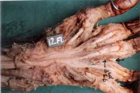
Split insertion of third lumbrical (Lb)
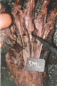
Misplaced insertion of third lumbrical (Lb)
a) Split insertion or bifid insertion in which the tendon divides to insert into extensor expansion of ulnar side of middle finger in addition to the radial side of ring finger and was seen in 14 out of 50 hand, 8(16%) right hands and 6(12%) in left hands i.e split insertion of 3rd lumbrical is common on right side.
b) Misplaced insertion in which the tendon was seen inserted on ulnar side of extensor expansion rather than on its radial side of the corresponding finger. This type of variation was present in 2(4%) hands (1 right and 1 left hand).
Fourth lumbrical: The fourth lumbrical was arising from contiguous sides of tendons for middle and little fingers, that was in consonance with normal text book pattern in 100% cases.
4th lumbrical was normal in origin and nerve supply in all the cases.
4th lumbrical, had its normal insertion (into the radial side of little finger) in 38 (76%) out of 50 hands and rest of 12 (24%) cases showed following variations [Table/Fig-4].
Variations seen in 3rd lumbrical
| Lumbrical | 4th lumbrical |
|---|
| Right | Left | Total |
| Total hands examined | 25 | 25 | 50 |
| Split insertion | 5 (20%) | 3 (12%) | 8 (16%) |
| Misplaced insertion | 3 (12%) | 1 (4%) | 4 (8%) |
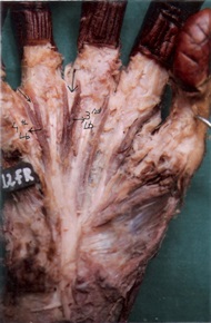
A) Split insertion was found in 8 (16%) out of 50 hands. In this type of variation tendon of the 4th lumbrical split to insert into radial side of expansion of little finger and ulnar side of ring finger [Table/Fig-5].
Split insertion of third and fourth lumbricals (Lb)
B) Misplaced insertion in which fourth lumbrical is inserted into ulnar side of dorsal digital expansion of ring finger, rather than on radial side of little finger [Table/Fig-6].
Misplaced insertion of fourth lumbrical (Lb)
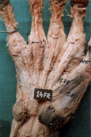
In the present study, misplaced insertion of fourth lumbrical was seen in 4(8%) out of 50 hands in which 3 were right hands and 1 was the left hand [Table/Fig-7].
Comparison of occurrence of split insertion of second lumbrical with previous worker
| Workers | Total no. of hands | Split insertion | %age |
|---|
| Wood (1966) | 72 | 1 | 1.4 |
| Eyler & Markee (1954) | 33 | 0 | 0 |
| Mehta and Gardner (1961) | 75 | 1 | 1.3 |
| Present study | 50 | 0 | 0 |
Discussion
First lumbrical and second lumbrical were seen less variable as compared to third and fourth lumbricals in present study.
Mehta and Gardner [5] have reported their additional origin from flexor digitorum superficialis, the first metacarpal, third metacarpal and also in forearm from flexor pollicis longus [6].
The present study is comparable with the studies of Mehta and Gardner [5] and Eyler & Markee [7] who have reported 100% normal insertion of first lumbrical.
However bipennate origin of second lumbrical was seen by Wood [8] in 6 out of 102 hands, and Mehta and Gardner [5] 6 out of 75 hands.
Split insertion of second lumbrical though rare has been reported by some workers. Wood [9] reported only 1 split insertion, out of 72 hands. Similarly Mehta and Gardner [5] have also reported split insertion of second lumbrical in 1 out of 75 hands. But, Eyler and Markee [7] have reported 100% normal insertion of second lumbrical [Table/Fig-7]. So the present study is comparable with the study of previous workers. A small disparity may be because of difference in the number of hands dissected.
Third lumbrical was seen normal in origin and nerve supply in all cases in present study.
Unipennate origin though rare has been reported by some workers e.g. Wood [8] has reported unipennate origin in 4 out 102 hands. But, Basu and Hazary [2] reported no variation in origin of third lumbrical. So present study is comparable to their study.
In detailed study of human hand lumbricals Mehta and Gardner [5] pointed that insertions of lumbricals were more variable than their origin. In the present study also, lumbricals were seen more variable at their insertion rather than in their origins.
Singh et al., [10] have stated that 3rd and 4th lumbricals are more variable than the first and second lumbricals. First and second lumbricals are known to have a very little variation if any. They have also advocated that amongst the various types of anomalous insertions reported by previous authors, split insertion has been described to be commonest and has been seen most frequently in third lumbrical [Table/Fig-7].
The high incidence of split insertion in case of third and fourth lumbricals may be attributed to their bipennate origin.
In the present study, 3rd lumbrical was abnormal in 16(32%) out of 50 cases with slightly more frequency in right hand. Similarly 4th lumbrical was found abnormal in 12(24%) out of 50 hands with more frequency in the right hand as compared to the left hand. Among the variations of lumbricals ‘split insertion’ was more common type of variation and was seen commonly in the third lumbrical as compared to the fourth lumbrical. In present study, ‘split insertion’ in third lumbrical was seen in 28% and in 16% of cases of fourth lumbrical [Table/Fig-8]. 2nd type of variation in the insertion of lumbricals was ‘misplaced insertion’ which was more common in 4th lumbricals 4/50 (8%) than third lumbricals in 2/50 (4%) hands.
Incidence of split insertion in lumbricals
| Workers | Total no. of hands | 2nd lumbrical | 3rd lumbrical | 4th lumbrical |
|---|
| Eyler & Markee (1954) | 33 | – | 5 (15.6%) | 2 (6.06%) |
| Mehta and Gardner (1961) | 75 | 1 (1.3%) | 29 (38.7%) | 6 (8%) |
| Singh el al., (1975) | 107 | - | 29 (27.1%) | 27 (25.2%) |
| Present study | 50 | - | 14 (28%) | 8 (16%) |
Split insertion may be morphologically comparable to doubling of muscles in some lower mammals such a doubling occurs in virginian oppsum 4th lumbrical is also doubled in some species. This condition may be the atavistic condition [11].
Present study is in line with those of Eyler and Markee [7] and Singh et al., [10] who have reported the misplaced insertion is more common in 4th lumbrical as compared to 3rd lumbrical [Table/Fig-9]. The cause and significance of misplaced insertion is not well understood. Cruveilhier [12] considered the misplaced insertion in EE2 (extensor expansion of middle finger) is normal mode of insertion of third lumbrical.
Comparison of frequency in ‘misplaced insertion’ of 3rd
and 4th lumbricals as reported by various workers
| Workers | Total no. of hands | 3rd lumbrical | 4th Lumbrical |
|---|
| Basu & Hazary (1960) | 72 | 9 (12.5%) | 4 (5.6%) |
| Mehta and Gardner (1961) | 75 | 7 (9.3%) | 6 (8%) |
| Singh et al., (1975) | 107 | 2 (1.86%) | 10 (9.34%) |
| Present study | 50 | 2 (4%) | 4 (8%) |
These variations might have functional implications. In the hand lacking lumbrical to either the ring or the little finger normal extension of interphalangeal joint might not be possible [5].
Conclusion
In the present study lumbricals were found variable in their insertion rather than their origin and nerve supply. The variation was seen in insertion of 3rd (32 %) and 4th lumbrical (24%) and more confined to the right hand.
Variations commonly encountered in insertion were
Split insertion
Misplaced insertion
In split insertion, the tendon of variant lumbrical splits to insert on extensor expansion of the adjacent fingers in addition to its normal insertion. This type of insertion was common than misplaced insertion and more frequent in 3rd lumbrical (28%).
Second type of variation encountered was misplaced insertion, in which the variant lumbrical is inserted on the ulnar side of adjacent finger rather than on the radial side of the corresponding finger. It was more common in 4th (8%) than the 3rd (4%) lumbrical. Both of these variations were seen more common on the right side as compared to the left hand. Awareness of these variations is necessary to avoid complications during radiodiagnostic procedures or hand surgeries [13].