The Mental Foramen (MF) is located in the body of the mandible, midway between the inferior and the alveolar margins of the body [1,2]. It is present between the premolars, in a vertical line with the supraorbital notch. It provides a passage for the exit of the mental nerve and the vessels [3]. Most of the mental foramina are oriented postero-superiorly.
Variations in the position of the MF have been reported by many authors in different ethnic groups [4,5] and various shapes have also been noticed [6].
Any foramen which is in addition to MF is considered as an Accessory Mental Foramen [7] (AMF) and it is usually located below the 1st molar teeth [8]. This accessory mental foramen may transmit the branches of the mental nerve.
The precise knowledge on the variations in the position, shape, and the size of the mental foramen and the presence of the accessory mental foramen would be of much use for dental surgeons while they do surgical procedures on the mandible, such as the curettage of the premolars, filling procedures, dental implants, Root Canal Treatments (RCT), orthognatic surgeries, etc. It is also essential to have an effective and a successful anaesthesia during nerve blocks, prior to the surgical procedure. Many studies have been reported by various authors, which were done in different ethnic groups and on populations of different races, but such studies which have been in the south Indian population are sparse. Hence, an attempt was made in our present study, to determine the most common position and size of the mental foramen in adult south Indian mandibles, which may be useful for the future implications in our south Indian population.
Material and Methods
The mandibles which were used for our study were procured from the Department of Anatomy, Vinayaka Mission Kirupananda Variyar Medical College and from Annapoorna Medical College, Salem, Tamil Nadu, India. About 90 adult, dry South Indian mandibles, irrespective of age and sex, with either all the teeth intact or with preserved alveolar margins, were used for our study. The bones with gross pathological deformities were excluded from our study.
The number, shape and the orientation of the MF were determined by a visual examination.
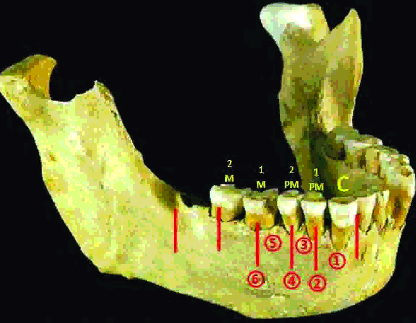
The positions of the mental foramens were measured with respect to the teeth, for which we followed the Tebo and Telford [9] classification [Table/Fig-1]. The positions of the mental foramens with respect to the borders were also measured [Table/Fig-2] with the help of a digital vernier caliper, at a measuring accuracy of 0.01mm. From the transverse and the vertical diameters which were obtained, the size of the MF was calculated.
Position of Mental Foramen in relation to teeth (Tebo & Telford Classification)
1-Foramen lying on a longitudinal axis of passing between the canine and first premolar.
2-Foramen lying on the longitudinal axis of the first premolar.
3-Foramen lying on the longitudinal axis passing between first and second premolar
4-Foramen lying on the longitudinal axis of the second premolar
5-Foramen lying on the longitudinal axis passing between the second premolar and first molar.
6-Foramen lying on the longitudinal axis of first molar.
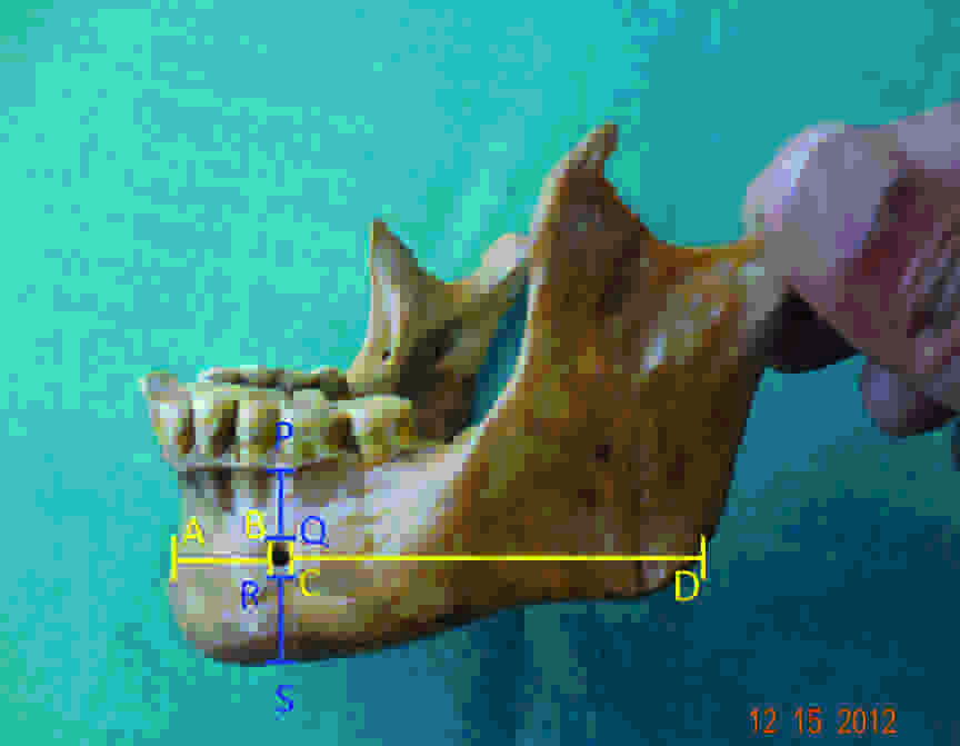
Position of Mental Foramen & its size calculated by transverse & vertical measurements of mandible in relation to borders
AD-Distance from symphysis menti to posterior border of mandible.
AB- Distance from symphysis menti to anterior margin of MF.
CD- Distance from posterior margin of MF to posterior border of mandible.
BC-Transverse Diameter (TD) of MF.
PS- Distance from alveolar margin to base of mandible.
PQ- Distance from alveolar margin to superior margin of MF
RS- Distance from inferior margin of MF to base of mandible.
QR-Vertical Diameter (VD) of MF
The transverse diameters were measured by using various parameters viz., the distance from the symphysis menti, the posterior border of the mandible, the base of the mandible and from the alveolar margins.
AD – The distance from the symphysis menti to the posterior border of the mandible.
AB – The distance from the symphysis menti to the anterior margin of the MF
CD – The distance from the posterior margin of the MF to the posterior border of the mandible.
BC – The transverse diameter (TD) of the MF
The vertical diameters were measured by using the following parameters viz.,
PS – The distance from the alveolar margin to the base of the mandible
PQ – The distance from the alveolar margin to the superior margin of the MF
RS – The distance from the inferior margin of the MF to the base of the mandible.
QR – The vertical diameter (VD) of the MF
All the measurements were recorded by one of the authors to reduce bias. The SPSS, version 15 version software were used for the statistical analysis, to find out the minimum and the maximum incidences, the mean and the standard deviation.
For the accessory mental foramen, the number, location, shape, size and the orientation were recorded [Tables/Fig-3 and 4].
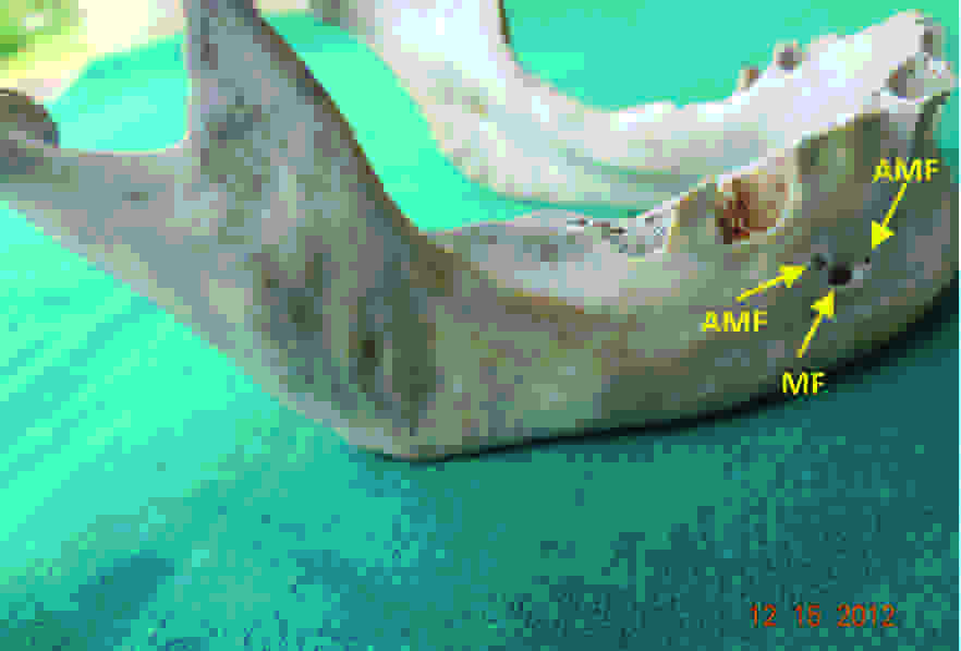
Right side of a mandible showing three mental foramen.
MF-Mental Foramen, AMF-Accessory Mental Foramen
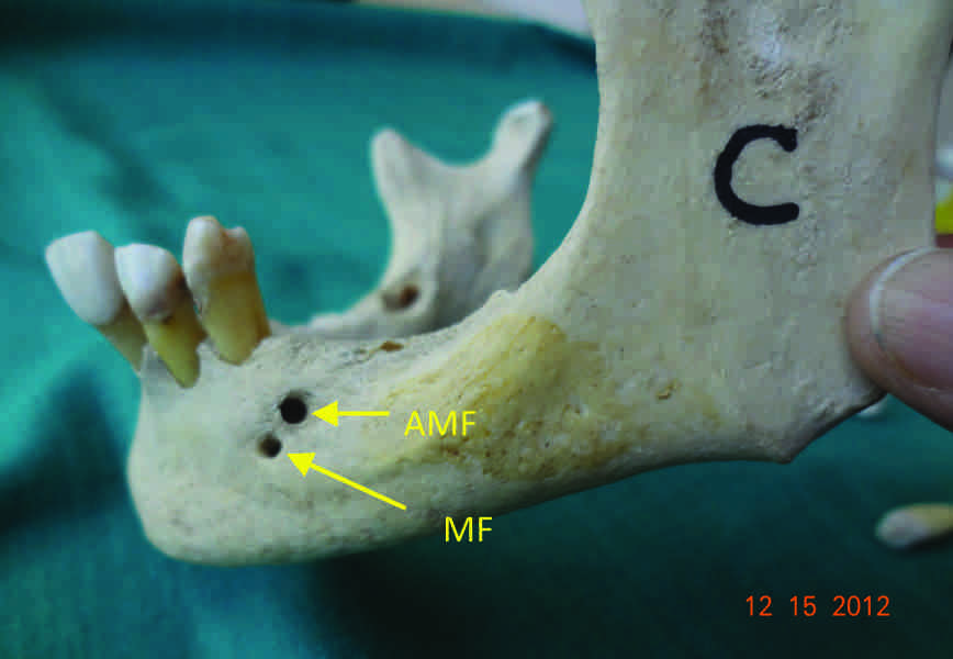
Left side of a mandible showing two mental foramen.
MF-Mental Foramen, AMF-Accessory Mental Foramen
Results
The Mental Foramen
Number
In our present study, 87 bones (96.67%) showed a single mental foramen on the left side and 88 (97.78%) showed a single foramen on the right side.
Position
The position of the MF with respect to the teeth of the lower jaw were classified according to the method of Tebo and Telford, 1950.
The foramen which lay on a longitudinal axis which passed between the canine and the first premolar.
The foramen which lay on the longitudinal axis of the first premolar.
The foramen which lay on the longitudinal axis which passed between the first and second premolars.
The foramen which lay on the longitudinal axis of the second premolar.
The foramen which lay on the longitudinal axis which passed between the second premolar and the first molar.
The foramen which lay on the longitudinal axis of the first molar.
As per this classification, the analysis showed that the most common position in our study was the longitudinal axis of the 2nd premolar (IV), followed by (V) i.e between the 2nd premolar and the first molar, between the 1st and 2nd premolars (III) and the axis of the premolar (II) [Table/Fig-5] respectively. Position I and VI were not observed in any of the mandibles. With respect to the superior and the inferior borders of the mandible, most of the MF (85.56% on the left side and 86.67% on the right side) were found to occupy the mid position [Table/Fig-6].
Position of Mental foramen in relation to teeth
| Valid | Frequency | Percent | Valid Percent | Cumulative Percent |
|---|
| Left Side |
| 1PM | 4 | 4.44 | 4.44 | 4.44 |
| 1PM/2PM | 14 | 15.56 | 15.1 | 19.54 |
| 2PM | 47 | 52.22 | 50.5 | 70.04 |
| 2PM/1M | 25 | 27.78 | 26.9 | 100.0 |
| Total | | 100.0 | 100.0 | |
| Right Side |
| 1PM | 4 | 4.44 | 4.44 | 4.44 |
| 1PM/2PM | 15 | 16.67 | 16.67 | 21.11 |
| 2PM | 46 | 51.11 | 51.11 | 72.22 |
| 2PM/1M | 25 | 27.78 | 27.78 | 100 |
| Total | 90 | 100 | 100 | |
1 PM – 1st Premolar (Position II)
1 PM/2PM–Between 1st Premolar and 2nd Premolar teeth (Position III)
2 PM – 2nd Premolar (Position IV)
2 PM/1M – Between 2nd Premolar and 1st Premolar (Position V)
Position of Mental foramen in relation to borders
| Valid | Frequency | Percent | Valid Percent | Cumulative Percent |
|---|
| Left Side |
| LB | 6 | 6.67 | 6.67 | 6.67 |
| MID | 77 | 85.56 | 85.56 | 92.23 |
| UB | 7 | 7.77 | 7.77 | 100.0 |
| Total | 90 | 100.0 | 100.0 | |
| Right Side |
| LB | 4 | 4.44 | 4.44 | 4.44 |
| MID | 78 | 86.67 | 86.67 | 91.11 |
| UB | 8 | 8.89 | 8.89 | 100 |
| Total | 90 | 100 | 100 | |
LB: Lower border
MID: Midway between upper and lower border
UB: Upper border
The MF was positioned at an average distance of 12.21 ± 2.61 mm from the alveolar margin on the left side, whereas it was positioned at a distance of 12.02 ± 2.48 mm on the right side. From the symphysis menti, the MF was located at a distance of about 25.29 ± 2.29 mm on the left side and at a distance of about 25.79 ± 1.78mm on the right side. The average distance of the MF from the posterior border of the mandible was 63.92 ± 4.26 mm on the left side and it was 64.51 ± 4.06 mm on the right side. From the base of the mandible, it was located at a distance of 12.77 ± 1.73 mm on the left side and at a distance of 12.65 ± 1.59 mm on the right side [Table/Fig-7].
Statistical analysis of position of MF and its size in relation with borders
| Parameters | N | Min | Max | Mean | SD |
|---|
| Descriptive Statistics – LEFT SIDE (mm) |
| (PS) | 90 | 17.97 | 34.9 | 27.51 | 3.32 |
| (PQ) | 90 | 3.87 | 19.11 | 12.21 | 2.61 |
| (RS) | 90 | 7.33 | 17.24 | 12.77 | 1.73 |
| (QR) | 90 | 1.06 | 4.91 | 2.52 | 0.87 |
| (AD) | 90 | 78.24 | 102.61 | 92.15 | 5.28 |
| (AB) | 90 | 20.53 | 31.02 | 25.29 | 2.29 |
| (CD) | 90 | 54.54 | 73.96 | 63.92 | 4.26 |
| (BC) | 90 | 1.77 | 4.77 | 2.95 | 0.68 |
| Descriptive Statistics – RIGHT SIDE (mm) |
| (PS) | 90 | 19.07 | 33.22 | 27.53 | 3.26 |
| (PQ) | 90 | 5.24 | 17.55 | 12.02 | 2.48 |
| (RS) | 90 | 17.97 | 15.28 | 12.65 | 1.59 |
| (QR) | 90 | 1.12 | 4.71 | 2.86 | 0.83 |
| (AD) | 90 | 78.9 | 99.86 | 92.59 | 4.95 |
| (AB) | 90 | 20.63 | 29.64 | 25.79 | 1.78 |
| (CD) | 90 | 54.53 | 71.02 | 64.51 | 4.06 |
| (BC) | 90 | 1.02 | 3.86 | 2.28 | 0.71 |
Min- Minimum, Max-Maximum, SD- Standard Deviation.
In 75 bones, (83.33%) the MF was bilaterally showing an oval shape and in the remaining 15 mandibles (16.67%) the MF was bilaterally showing a round shape [Table/Fig-8].
| Frequency | Percent | Valid Percent | Cumulative Percent |
|---|
| Left Side |
| O | 75 | 83.33 | 83.33 | 83.33 |
| R | 15 | 16.67 | 16.67 | 100.0 |
| Total | 90 | 100.0 | 100.0 | |
| Right Side |
| Valid O | 75 | 83.33 | 83.33 | 83.33 |
| R | 15 | 16.67 | 16.67 | 100 |
| Total | 90 | 100 | 100 | |
O: Oval; R: Round
Most of the MFs (83%) on the left side and 80% on the right side were oriented postero-superiorly and this was followed by a superior and then a horizontal orientation [Table/Fig-9].
Orientation of Mental foramen
| Valid | Frequency | Percent | Valid Percent | Cumulative Percent |
|---|
| Left Side |
| H | 7 | 7.78 | 7.78 | 7.78 |
| PS | 75 | 83.33 | 83.33 | 91.11 |
| S | 8 | 8.89 | 8.89 | 100.0 |
| Total | 90 | 100.0 | 100.0 | |
| Right Side |
| H | 4 | 4.44 | 4.44 | 4.44 |
| PS | 73 | 81.11 | 81.11 | 85.55 |
| S | 13 | 14.45 | 14.45 | 100 |
| Total | 90 | 100 | 100 | |
H-Horizotal; PS- Postero-superior; S – Superior
The Accessory Mental Foramen (AMF)
The incidence of the AMF was more (3.33%) on the left side as compared to that on the right side (2.22%). One mandible out of 90 showed AMF bilaterally. The total incidence of AMF was 5.55% [Table/Fig-10].
| No. of Mental foramen | Frequency | Percent | Valid percent | Cumulative percent |
|---|
| Left Side |
| Valid 1 | 87 | 96.67 | 96.67 | 96.67 |
| 2 | 3 | 3.33 | 3.33 | 100 |
| Total | 90 | 100 | 100 | |
| Right Side |
| Valid 1 | 88 | 97.78 | 97.78 | 97.78 |
| 2 | 1 | 1.11 | 1.11 | 98.89 |
| 3 | 1 | 1.11 | 1.11 | 100 |
| Total | 90 | 100 | 100 | |
1-Single foramen, 2-Double accessory foramen, 3- Triple accessory foramen
Discussion
Our study demonstrated that the most common position of the MF, was position IV, followed by position V, which was similar to that of north Americans [9], north Indians [10], Turks [11], Malawians [12] and Zimbabweans [13] [Table/Fig-11]. Our study findings coincided with those of Agarwal and Gupta [6] and Yesilyurt et al., [14] in different populations. This finding differed significantly from the finding of Gershenson et al., [15] who reported a higher prevalence of the positions I, II and VI between the Indians and the Sinai population. Also, the studies which were done on Nigerians [16] and Kenyans [17] showed the most common position to be III, followed by II. In Malays and in the Srilankan population, it was noticed that position IV was followed by position III [18,19]. Hence, significant differences have been reported in the positions of the MF among different ethnic groups [Table/Fig-11]. Sometimes, an anterior loop of the mental nerve may be present medial to the MF and it may cause a mental nerve injury during a dental implantation [20].
Comparison of position of MF in different ethnic groups (in %)
| S.No | Population | Position (%) |
|---|
| I | II | III | IV | V |
|---|
| 1 | North American [9] | 0 | 3.5 | 23 | 49.4 | 24.1 |
| 2 | Hong kong chinese [4] | 0 | 21 | 51 | 24 | 4 |
| 3 | Chinese* [16] | 0 | 21 | 59 | 19 | 1 |
| 4 | British [5] | 0 | 9.1 | 59.1 | 31..8 | 0 |
| 5 | North Indians [10] | 0 | 2.08 | 17.71 | 68.75 | 11.46 |
| 6 | Asian Indians [16] | 0 | 5.80 | 75.36 | 18.84 | 0 |
| 7 | Turks [11] | 0 | 0 | 44.1 | 55.9 | 0 |
| 8 | East Africans* | 0.30 | 7.57 | 57.88 | 31.52 | 0 |
| 9 | Kenyans [17] | 0 | 31.90 | 56.1 | 12.1 | 0 |
| 10 | Nigerians* | 1.82 | 26.99 | 55.63 | 12..25 | 3.31 |
| 11 | Zimbabweans [13] | 0 | 0 | 12.4 | 56.3 | 31.3 |
| 12 | Malawians [12] | 0 | 2.8 | 10 | 62.9 | 24.3 |
| 13. | Present study | 0 | 4.4 | 16.67 | 52.22 | 27.78 |
%- Percentage, *cited from Shankland [16] (See Reference), [4,5,9,10, 11,12,13,16,17] (See Reference).
I. Foramen lying on a longitudinal axis of passing between the canine and first premolar.
II. Foramen lying on the longitudinal axis of the first premolar.
III. Foramen lying on the longitudinal axis passing between first and second premolar.
IV. Foramen lying on the longitudinal axis of the second premolar.
V. Foramen lying on the longitudinal axis passing between the second premolar and first molar.
VI. Foramen lying on the longitudinal axis of first molar.
Chung et al., [21] reported that the Horizontal Diameter (HD) of the MF was 2.4 mm, whereas Apinhasmit et al., reported a diameter [22] of 2.8 mm. A study which was done on a Turkey population [11] reported the HD to be 2.93 mm on the right side and to be 3.14 mm on the left side; the Vertical Diameter (VD) was 2.38 mm on the right side and it was 2.64 mm on the left side. Rakhi Rastogi et al., [23] described the VD to be 3.58 mm ± 0.17 mm on the right side and to be 3.55 mm ± 0.18 mm on the left side; the horizontal diameter as 4.57 ± 0.19 mm on the right side and it was 4.61 ± 0.17 mm on the left side. Our present study demonstrated the average HD of the MF to be 2.28 ± 0.71 mm on the right side and to be 2.95 ± 0.68 mm on the left side; the vertical diameter was 2.86 ±0.83 mm on the right side and it was 2.52 ± 0.87 mm on the left side [Table/Fig-7].
Our study documented the oval shape as the most common shape of the MF than the round shape. Our study results differed from those of the studies from central India [22] and east India [10], as they noticed more percentages in favour of the round shape than the oval sape. But our results were in close association with those of Gershenson [15], Mbajioru et al., [13] Prabodra and Nanayakkara [24] and Agarwal and Gupta [6] [Table/Fig-12].
Comparison of % of shape of MF by different authors
| S.No | Authors | Shape of MF (%) |
|---|
| Oval | Round |
|---|
| 1 | Gerhenson et al., (1986) | 65.5 | 34.5 |
| 2 | Mbajiorgu et al., (1998) | 56.3 | 43.8 |
| 3 | Prabodha and Nanayakkara (2006) | 66.7 | 33.3 |
| 4 | Singh and Srivatsava | 6.0 | 94.0 |
| 5 | Agarwal and Gupta (2011) | 92.0 | 8.0 |
| 6 | Rakhi Rastogi et al., (2012) | 48.3 | 51.7 |
| 7 | Present study (2013) | 83.33 | 16.67 |
Hauser and De Stefano [25] stated that the different variants may have occurred due to the epigenetic traits, as they could be the products of the genetically determined growth processes of other tissues, which had affected the bone formation. Subsequently, they undergo modifications during ontogeny and variable degrees of expression. Thus, the variations in the position, shape, number and size of the MF depends on the gene modification.
The Accessory Mental Foramen
In our study, 3.33% of the mandible showed AMFs on the left side and 2.22% showed AMFs on the right side. Our results were in contrast with those of Singh and Srivastava [10], where they found 8% AMFs on the left side and 5% on the right side. The incidence of the AMF in the Israeli population [15] was 2.8%, it was 1.8% among the American whites, it was 12.5% among Polynesians.
Cag Irankaya and Kansu [8], Singh et al., [10] reported AMFs below the 1st molar. But in our study, each AMF showed a variable position viz., between the 2nd premolar and the 1st molar, followed by between the 1st molar and the 2nd premolar (left side); between the 2nd premolar and the 1st molar and then between the 1st premolar and the 2nd premolar (right side). The literature on this are very sparse in Indian studies.
The literature on the size of the AMF is hardly available, to compare with ours. Singh et al., [10] recorded an average diameter of 1mm, but our study recorded a vertical diameter of 2.22 mm with an HD of 1.58 mm on the left side. Similarly, a VD of 1.76 mm and an HD of 3.9 mm on the right side were recorded.
Conclusion
The knowledge on the variations of the mental foramen is important for dental surgeons while they perform endodontic and periodontal surgeries, dental implantations, orthognatic surgeries, etc. Also, the verification on the presence of the AMF would prevent an accessory mental nerve injury during surgery and inadequate paraesthaesia. Our present study on the variations in the position, size, shape and the existence of the AMF would of use, for preventing complications and for better outcomes of the surgical procedures which are related to the mental foramen and the mental nerve.
%- Percentage, *cited from Shankland [16] (See Reference), [4,5,9,10, 11,12,13,16,17] (See Reference).I. Foramen lying on a longitudinal axis of passing between the canine and first premolar.II. Foramen lying on the longitudinal axis of the first premolar.III. Foramen lying on the longitudinal axis passing between first and second premolar.IV. Foramen lying on the longitudinal axis of the second premolar.V. Foramen lying on the longitudinal axis passing between the second premolar and first molar.VI. Foramen lying on the longitudinal axis of first molar.