Evaluation of the Variant Anatomical Disposition of the Renal Hilar Structures in South Indian Adult Human Cadavers and Its Cinical Implications
Naveen Kumar1, Anitha Guru2, Ashwini Aithal P.3, Surekha D. Shetty4, Satheesha Nayak B.5, Narendra Pamidi6
1Lecture,Department of Anatomy, Melaka Manipal Medical College (Manipal Campus), Manipal University, Madhav Nagar, Manipal, Karnataka–576104, India.
2Lecture,Department of Anatomy, Melaka Manipal Medical College (Manipal Campus), Manipal University, Madhav Nagar, Manipal, Karnataka–576104, India.
3Lecture,Department of Anatomy, Melaka Manipal Medical College (Manipal Campus), Manipal University, Madhav Nagar, Manipal, Karnataka–576104, India.
4Lecture,Department of Anatomy, Melaka Manipal Medical College (Manipal Campus), Manipal University, Madhav Nagar, Manipal, Karnataka–576104, India.
5Professor and Head, Department of Anatomy, Melaka Manipal Medical College (Manipal Campus), Manipal University, Madhav Nagar, Manipal, Karnataka–576104, India.
6Assistant Professor,Faculty in Anatomy, School of Medicine and Health sciences, Monash University, Sunway Campus, Kuala Lumpur-46150, Malaysia.
NAME, ADDRESS, E-MAIL ID OF THE CORRESPONDING AUTHOR: Mrs. Ashwini Aithal P., Department of Anatomy, Melaka Manipal Medical College (Manipal Campus), Manipal University, Manipal, Karnataka–576104, India.
Phone: +91 9738928345, Email: ashwini.anat@gmail.com
Aim: To evaluate the anatomical disposition of the renal hilar structures in human cadavers of south Indian origin, considering their antero–posterior distribution.
Material and Methods: Ninty–six renal hila of the isolated kidneys from adult south Indian cadavers were observed for the branching patterns and the distributions of the renal hilar structures. The number of branches of the renal artery and the divisions of the renal vein in the pre hilar region were noted, along with their pattern of arrangement with respect to the renal pelvis.
Results: In the present study on the pre hilar region, we observed that the highest division of the renal artery was 8 and that the highest incidence was of 4 divisions of the renal artery in 30.2% cases. The highest number of venous divisions which was observed was 7. The highest incidence of 40.6 % cases showed 2 divisions of the veins. Regarding the patterns of arrangement of these structures, we observed 12 patterns of arrangement, with a higher incidence (45.8%) of the classical arrangement (V-A-P), as has been described in the standard text books of anatomy, which was followed by the A-V-P pattern (28.1%).
Conclusion: An anatomical knowledge on the possible variant topography of the renal hilar structures is of great importance when urological surgical procedures are performed.
Renal anatomy, Renal hilum, Renal artery, Renal vein, Renal pelvis
Introduction
The renal hilum is a vertical slit on the medial border of the kidney, which is bound by the thick lips of the renal substance [1]. Classically, the topographic arrangement of the hilar structures is referred to in the antero-posterior sense, as its vein-artery-pelvis [2].
Various kidney disorders pose fatal complications such as a risk of cardio morbidity, hospitalisation or even death [3]. Nephrectomy is being used as a choice of a therapeutic procedure towards certain kidney disorders in which the functional units of the nephrons are spared [4]. A Laparoscopic Partial Nephrectomy (LPN) minimises the risk of a radical nephrectomy. However, the LPN procedure is a very complicated and a technically challenging task for the urologists, as it requires the skill of ligation or clamping of the vessels which are present in the narrow spaced hilum [5].
However, clamping of the individual structures is beneficial than the en-bloc clamping procedures [6]. Hence, it is necessary to have an ample knowledge on the arrangements of the renal hilar structures before making a surgical approach, as these arrangements and the number of structures in the hilum are highly variable than the actual patterns which are given in the standard text books. On reaching the hilum, the renal arteries usually divide into the anterior and the posterior divisions. The posterior division of the renal artery and the posterior tributary of the renal vein may generally enter the kidney tissue, posterior to the pelvis, in some cases [2, 7].
The surgeons who perform endopyelotomies should be aware of the arrangements of the structures at the hilum of the kidney. Many of the investigative imaging and angiographic procedures have described the abnormal anatomies of the hilar structures, which were detected mainly as incidental findings. Many studies have been done on the anatomical analysis of the arrangements of the structures in the renal hilum. However, studies which have been done on the evaluation of the topographic disposition of the renal hilar structures are scanty. So, the present study was undertaken to observe the various branching patterns as well as the arrangements of the structures in the prehilar region of the kidneys in the south Indian population.
Material and Methods
The hila of a total number of 96 (42 right and 54 left side) isolated cadaveric kidneys were examined. The hila and the adjacent pre-hilar area of each kidney were dissected carefully, to visualize the arrangement and the division pattern of the renal vessels.
The topographic arrangements of the structures in the renal hilum were analysed at approximately 0.5 cm (0.2 inches) from the anterior lip of the renal hilum. They have been documented anteroposteriorly, considering only the renal artery, the renal vein and the renal pelvis. The division patterns of the renal vessels, just before they entered the corresponding hilum, were examined carefully.
This was then followed by the observation of their arrangements in the hilum and their correlation with the renal pelvis. The total incidences of the presence of aberrant arteries were also noted. Their positions and formations were examined.
Any incidence where it was formed outside the hilum, was documented. However, the distribution and the disposition of these structures inside the organ were not studied. The aberrant arteries were not included in the arterial segmentation pattern analysis.
Results
The number of arterial sub divisions and their percentage incidences have been given in [Table/Fig-1]. The higher incidence of the presence of 4 branches of the renal artery before they entered the hilum was noted in 30.2% cases (29 kidneys, 13 right, 16 left) [Table/Fig-2]. However, the maximum of 8 branches of the renal artery were observed in 2 kidneys (2.1%). The hila of those kidneys were congested with the renal structures [Table/Fig-3].
Arterial division pattern in the hilum and their percentage of incidence
| No. arterial branches | Number of kidneys (n=96) | Incidence | Side |
|---|
| Right (n=42) | Left (n=54) |
|---|
| One | 3 | 3.1% | 1 | 2 |
| Two | 8 | 8.3% | 5 | 3 |
| Three | 16 | 16.7% | 10 | 6 |
| Four | 29 | 30.2% | 13 | 16 |
| Five | 26 | 27.1% | 9 | 17 |
| Six | 8 | 8.3% | 3 | 5 |
| Seven | 4 | 4.2% | 1 | 3 |
| Eight | 2 | 2.1% | 0 | 2 |
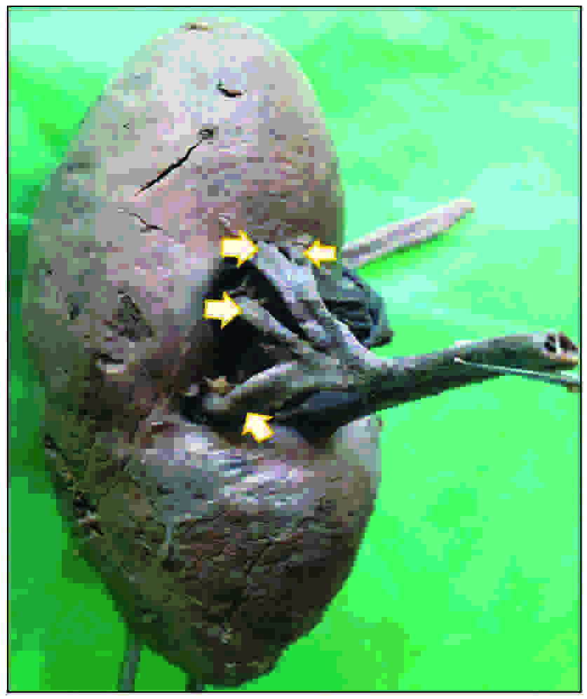
Hilum of kidney showing 4 branches of renal artery
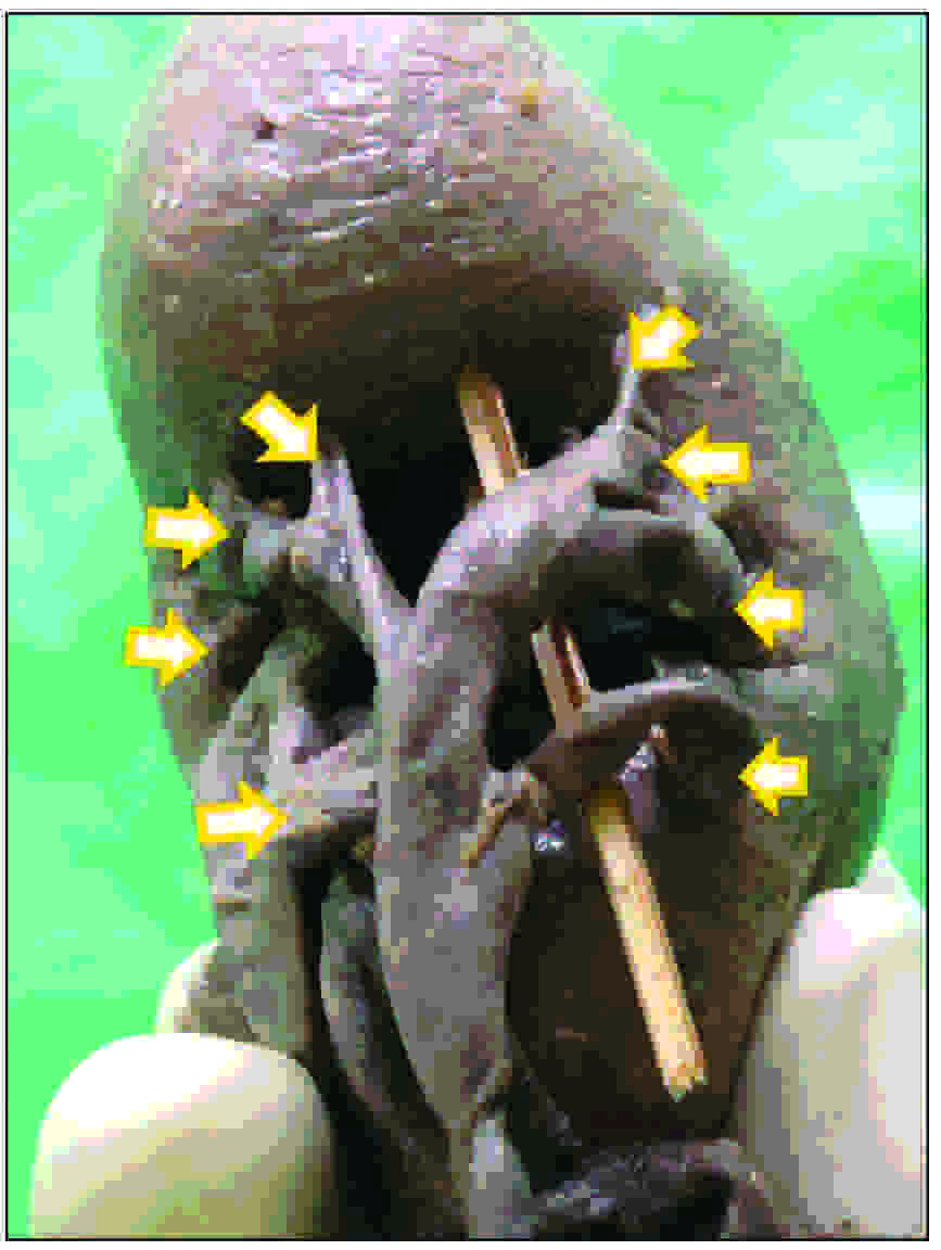
Hilum of kidney showing 8 branches of renal artery
The number of venous tributaries and their percentage incidences have been given in [Table/Fig-4]. The highest incidence of the presence of 2 divisions of the renal vein which emerged from the hilum separately and then united to form a single renal vein outside the hilum, was noted in 40.6% cases (39 kidneys, 14 right, 25 left) [Table/Fig-2]. However, the maximum of 7 divisions of a renal vein were also observed [Table/Fig-5].
Venous division pattern in the hilum and their percentage of incidence
| No. of venous divisions | Number of kidneys (n=96) | Incidence | Side |
|---|
| Right (n=42) | Left (n=54) |
|---|
| One | 28 | 29.2% | 19 | 9 |
| Two | 39 | 40.6% | 14 | 25 |
| Three | 19 | 19.8% | 6 | 13 |
| Four | 6 | 6.3% | 2 | 4 |
| Five | 2 | 2.1% | 1 | 1 |
| Six | 1 | 1% | 0 | 1 |
| Seven | 1 | 1% | 0 | 1 |
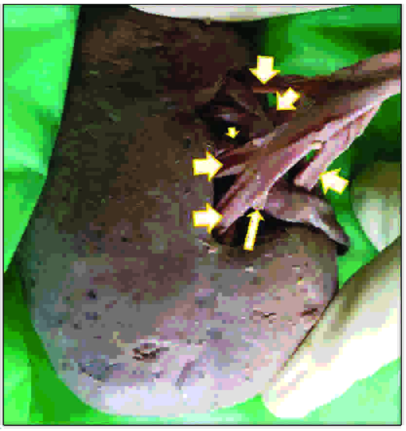
Hilum of kidney showing multiple divisions of renal vein
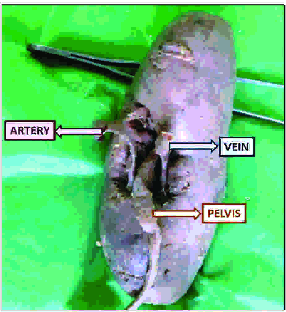
The patterns of arrangement of the hilar structures were carefully noted and about 12 major patterns and their percentages of occurrence have been shown in [Table/Fig-6]. The classical arrangement (V-A-P), as is given in the standard text books, was observed in 44 out of 96 cases, which accounted for the highest (45.8%) incidence in our study. It was followed by the next highest incidence (28.1%) which was seen as the A-V-P type of arrangement. We also noted a total of 3.1% cases wherein the renal pelvis was entrapped between the renal vessels, either in the pattern of the V-P-A (2.1%) or the A-P-V (1%) arrangement. [Table/Fig-7] However, the renal pelvis in between the divisions of the renal vessels was also observed and its detailed pattern has been shown in [Table/Fig-8].
Anatomical arrangement of hilar structures (antero-postero sense)
| Structural arrangement in the hilum | Number of kidneys (n=96) | Incidence | Side |
|---|
| Right (n=42) | Left (n=54) |
|---|
| V-A-P | 44 | 45.8% | 20 | 24 |
| A-V-P | 27 | 28.1% | 14 | 13 |
| V-A1-P-A2 | 8 | 8.3% | 3 | 5 |
| A1-V-P-A2 | 4 | 4.2% | 1 | 3 |
| A-V1-P-V2 | 3 | 3.1% | 0 | 3 |
| A1-V-A2-P | 2 | 2.1% | 1 | 1 |
| V1-A1-P-A2-V2 | 2 | 2.1% | 1 | 1 |
| V-P-A | 2 | 2.1% | 1 | 1 |
| A-P-V | 1 | 1% | 1 | 0 |
| A1-P-V-A1 | 1 | 1% | 0 | 1 |
| A1-V1-P-A1-V2 | 1 | 1% | 0 | 1 |
| V1-A-P-V2 | 1 | 1% | 0 | 1 |
V: renal vein, A: Renal artery, P: Renal pelvis, A1: anterior division of renal artery, A2: Posterior division of renal artery, V1: anterior division of renal vein, V2: Posterior division of renal vein
Hilum of kidney showing renal pelvis entrapped between renal vessels
Comparison of pattern of arrangement of renal structures in the hilum with other previous studies
| Hilar structure arrangement pattern (antero-posterior) | Present study | Study by Trivedi S et al., (MP, India) | Study by Joao A et al., (Brazil) |
|---|
| V-A-P | 45.8% | 19% | 83% |
| V-A-A-P | NA | 8% | NA |
| A-V-A-P | 2.1% | 23% | NA |
| V-A-P-V-A | 2.1% | 22% | NA |
| V-A-P-A | 8 .3% | 20% | NA |
| A-V-P-A | 4.2% | 8% | NA |
| V-P-A | 2.1% | NA | 3% |
| A-V-P | 28.1% | NA | 3% |
| A-P-V | 1% | NA | 1% |
In addition to its vascular disposition, we also noticed the formation of the renal pelvis outside the hilum in 8 (8.5%) kidneys out of the 96 kidneys which were studied [Table/Fig-9].
Formation of renal pelvis outside the renal hilum

Discussion
Possessing the knowledge on the distribution of the renal hilar structures is of crucial importance for urological surgical procedures which involve the hilar vessel clamping. Therefore, a prior knowledge on various possibilities of the branching patterns of the renal vessels in the hilar region, as well as their topographic arrangements, come in handy for the urologists before they perform various surgical procedures in the hilar region.
The results of the previous studies which were done on the patterns of the hilar structures were compared with those of the present study, as shown in [Table/Fig-8]. In the study which was conducted by Joao et al., [8] they observed that the main trunk of the renal artery had divided into 3 segmental branches before it entered the renal tissue. Similarly, Arora et al., observed 2 segmental arteries on the right side [9]. However, in our study, we observed as many as 8 divisions emerging from the main arterial trunk. Such variations in the varied pattern of the divisions of the renal artery in the hilar region are generally associated with renal malformations in the embryo [10].
A study which was conducted in 2008 by Kaneko et al., presented 25% multiple renal arteries which included the polar or accessory renal arteries. However, some authors [11, 12] believe that this aspect may be a misinterpreting factor for the true number of renal arterial divisions. In our study, we observed these aberrant arteries in 6 (6.3%) kidneys, which we did not include in the arterial segmentation pattern analysis.
Considering the distribution of the extra–parenchymal renal vein, Joao A et al., observed that 2.6% of the kidneys had more than one renal vein and that 7% had bifurcated renal veins. In our study, we observed a maximum of 7 divisions of the veins which had emerged from the hilum of the kidney. The highest incidence of 40.6% cases which we documented here, with a minimum of 2 divisions of the renal veins, coincided with the findings of the study which was conducted by Satyapal [13] in south Africa. In our study and in the study which was conducted by Trivedi S et al., [14] there was a common observation that; the incidence of the variant patterns of the renal veins was more common on the left kidney. This might be because of the embryological basis which differs on both sides. The deviant development of the venous channels, which is attributed to the formation of the left renal vein, may change the interrelationship between the renal hilar structures [14].
Understanding the uretero–pelvic anatomy is very important, as any obstruction at its junction is considered to be the commonest form of an upper urinary tract obstruction [15,16]. Rouviere et al., [17] reported that, the 29-65% incidence of the cases which had presented with anomalous courses of the renal vessels which had crossed the renal pelvis, was caused by ureteropelvic obstructions. Obstructions, strictures and stenosis may be caused by any external compression. The most reliable reason for the extrinsic obstruction which is caused by a renal vessel could be an incomplete rotation of the kidney [18]. Hence, the rotational defects of the kidney may result in the anomalous placements of the structures in the hilum [19]. The surgical interventions which require hilar dissections, need a separate clamping of the vessels and the renal pelvis, which is preferred over an en–bloc mass stapling of the renal hilum. This is because the en-block clamping may result in an arterio–venous fistula as a late complication of a nephrectomy. A difficult hilar dissection results in the conversion of a laparoscopic operation to an open procedure [6]. With the presence of anomalous hilar structural arrangements, it is advised to make a lateral deep incision alongside the ureteropelvic junction, rather than approaching its anterior or posterior aspects during endopyelotomies [16]. The variations in the branching patterns of the renal vessels are critical issues and a challenging task for a radiologists who interpret renal angiograms and for the urologists who perform laparoscopies [20]. With the increase in the number of cases of kidney transplantations, living donor grafts have become the major source for maintaining the donor pool, and successful allografts with multiple arteries have become a necessity. The variations in the origin and the course of the renal arteries occur frequently and they are of special interest to the urologists with respect to the diseases which are associated with them [21].
Conclusion
An anatomical knowledge on the patterns of the structures in the renal hilum is of paramount importance for various urological surgical procedures, such as in laparoscopic nephrectomies, anatrophic nephrolithotomies and renal transplantations, in which clamping of the hilar vessels is the prerequisite. Hence, a prior knowledge on the various possibilities of the branching patterns of the renal vessels in and adjacent to the hilar region, as well as their unusual patterns of arrangement are of utmost importance for the urologists before they perform any kind of surgical procedures which pertain to the kidney.
V: renal vein, A: Renal artery, P: Renal pelvis, A1: anterior division of renal artery, A2: Posterior division of renal artery, V1: anterior division of renal vein, V2: Posterior division of renal vein
[1]. Snell Richard S, Clinical anatomy by regions8th editionWalters KluwerLippincot Williams and Wilkins:260-64. [Google Scholar]
[2]. Standrings Gray’s AnatomyThe Anatomical Basis of Clinical Practice38th editionNew YorkElsevier Churchill Livingstone:1271-74. [Google Scholar]
[3]. Huang W. C, Levey A. S, Serio A. M, Chronic kidney disease after nephrectomy in patients with renal cortical tumours: a retrospective cohort studyLancet Oncol 2006 7(9):735-40. [Google Scholar]
[4]. Nuygen M. M, Gill I. S, Ellison L. M, The evolving presentation of renal carcinoma in the United States: trends from the Surveillance, Epidemiology, and End Results programJ. Urol. 2006 176(6 Pt1):2397-400. [Google Scholar]
[5]. Desai M. M, Gill I. S, Laparoscopic partial nephrectomy for tumour: current status at the Cleveland ClinicBJU Int. 2005 95(2):41-45. [Google Scholar]
[6]. Rapp D. E, Orvieto M. A, Gerber G. S, En bloc stapling of renal hilum during laparoscopic nephrectomy and nephroureterectomyUrology 2004 64(4):655-59. [Google Scholar]
[7]. Sinnatamby C. S., Last’s anatomy: regional and applied in abdomen11th editionChurchill LivingstoneElsevier:293-96. [Google Scholar]
[8]. Pereira-Correia Joao A, Lucas S, Analysis of renal hilum extraparenchymal structures in Brazilian adult human cadaversEuro J. Anat 2009 13(3):145-53. [Google Scholar]
[9]. Arora Anterpreet K, Verma Poonam, Lalit Monika, Variant segmental renal arteries in the right kidney-Clinical correlationsAnat Physiol 2012 2:3 [Google Scholar]
[10]. Bayramoglu A, Demiryurek D, Erbil KM, Bilateral additional renal arteries and an additional right renal vein associated with unrotated kidneysSaudi Med Journal 2003 24:535-37. [Google Scholar]
[11]. Ross JA, Samuel E, Miller DR, Variations in the renal vascular pedicle (an anatomical and radiological study with particular reference to renal transplantation)Br J Urol 1961 33:478-85. [Google Scholar]
[12]. Harrison LH JR, Flye MW, Seigler HF, Incidence of anatomical variants in renal vasculature in the presence of normal renal functionAnn Surg. 1978 188:83-89. [Google Scholar]
[13]. Satyapal KS, Classification of the drainage patterns of the renal veinsJ Anat 1995 186:329-33. [Google Scholar]
[14]. Trivedi S, Athavale S, Kotgiriwar S, Normal and variant anatomy of renal hilar structures and its clinical significanceInternational journal of Morphology 2011 29(4):1379-83. [Google Scholar]
[15]. Snyder HM, Lebowitz RL, Colodny AH, Ureteropelvic junction obstruction in childrenUrol Clin North Am 1980 7:273-290.PMID: 7404869 [Google Scholar]
[16]. Sampaio FJ, Favorito LA, Ureteropelvic junction stenosis: vascular anatomical background for endopyelotomyJ Urol 1993 150:1787-91. [Google Scholar]
[17]. Rouviere O, Lyonnet D, Berger P, Ureteropelvic junction obstruction: Use of helical CT for preoperative assessment: comparison with intra-arterial angiographyRadiology 1999 213:668-73. [Google Scholar]
[18]. Barnett JS, Stephens FD, The role of the lower segmental vessel in the aetiology of hydronephrosisAus NZ J Surg 1962 31:201-13. [Google Scholar]
[19]. Das S, Paul S, Variation of renal hilar structures: A cadaveric caseEuropean journal of Anatomy 2006 10(1):41-4. [Google Scholar]
[20]. Loh Hitendra Kumar, Gupta Vanitha, Arora Jyoti, Atypical twin renal arteries with altered hilar anatomyInternational Journal of Anatomical Variations 2009 2:124-26. [Google Scholar]
[21]. Kommuru Hemanth, Lekha Sree D, Jothi S.S, Presence of renal artery variations and its surgical correlationInternational Journal of Medical and Clinical Research 2012 3:176-79. [Google Scholar]