Study of Mastoid Canals and Grooves in North Karnataka Human Skulls
Gavishiddappa Andanappa Hadimani1, Ishwar Basavantappa Bagoji2
1 Lecturer, Department of Anatomy, Shri BM Patil Medical College Hospital and Research Centre,BLDE University Bijapur, Karnataka, India.
2 Lecturer Department of Anatomy, Shri BM Patil Medical College Hospital and Research Centre, BLDE University Bijapur, Karnataka, India.
NAME, ADDRESS, E-MAIL ID OF THE CORESPONDING AUTHOR: Dr. Gavishiddappa A. Hadimani, Lecturer, Department of Anatomy, Shri BM Patil Medical College, BLDE University Bijapur, Karnataka, India.
Phone: +919901352604,
E-mail: gavish.hadimani@yahoo.com
Introduction: This study was undertaken to observe the frequency of mastoid canals and grooves in north Karnataka dry human skulls. 100 dry human skulls of unknown age and sex from the department of Anatomy were selected and observed for the present study.
Material and Methods: The mastoid regions of dry skulls were observed for the presence of mastoid canals and grooves, if any. A metallic wire was passed through the canal for its confirmation and then the length was measured.
Results: The Mastoid canals were present in 53% of the total 100 skulls observed either bilaterally or unilaterally. Mastoid grooves were present in 18% of the total skulls (100) observed. Double mastoid canal was found in 01% of total skull studied and both Mastoid canals & Mastoid grooves together were present in 02% of the total skulls (100) observed.
Conclusion: The knowledge of mastoid canals and grooves is very important for otolaryngologists and neurosurgeons. Because they contain an arterial branch of occipital artery with its accompanying vein which is liable to injury resulting into severe bleeding.
Mastoid canal, Mastoid groove, Occipital artery
Introduction
Mastoid canals are the canals which are formed in the mastoid region of temporal bone of skull, which are located on the outer surface of mastoid processes, posterior and parallel to the petrosquamous suture, anterior to the occipitomastoid suture and anteroinferior to the asterion. Perforated lateral walls of mastoid canals are called as mastoid grooves. Some authors have mentioned that blood vessels lie in this area [1, 2]. Hollinshead WH described an ascending or auricular branch of the occipital artery at this site [2]. These vascular canals and grooves are of varying calibres and lengths, which have not been described in any of the textbooks of Anatomy, though these are of significant importance for neurosurgeons and otolaryngologists.
Material and Methods
This study was undertaken to observe the frequency of mastoid canals and grooves in dry human skulls in north Karnataka. 100 dry human skulls from the Department of Anatomy of Shri B M Patil Medical College, BLDE University, Bijapur, India which were of unknown age and sex, were selected and observed in the present study. The mastoid regions of dry skulls were observed for the presence of mastoid canals and grooves, if any. A metallic wire was passed through the canal for its confirmation and then the length of mastoid canal and groove was measured. In some of the skulls, grooves led to the openings of the canals.
Only those skulls which had canals patent to metal wire were considered as possessing mastoid canals. The length of the canal was measured with the help of a thread by passing a metal wire. The diameter of the mastoid canal could be measured in only few skulls where mastoid canal was larger, but in most of the skulls, it was not possible to measure the diameter. Length of the mastoid grooves was measured with the help of a thread.
Results
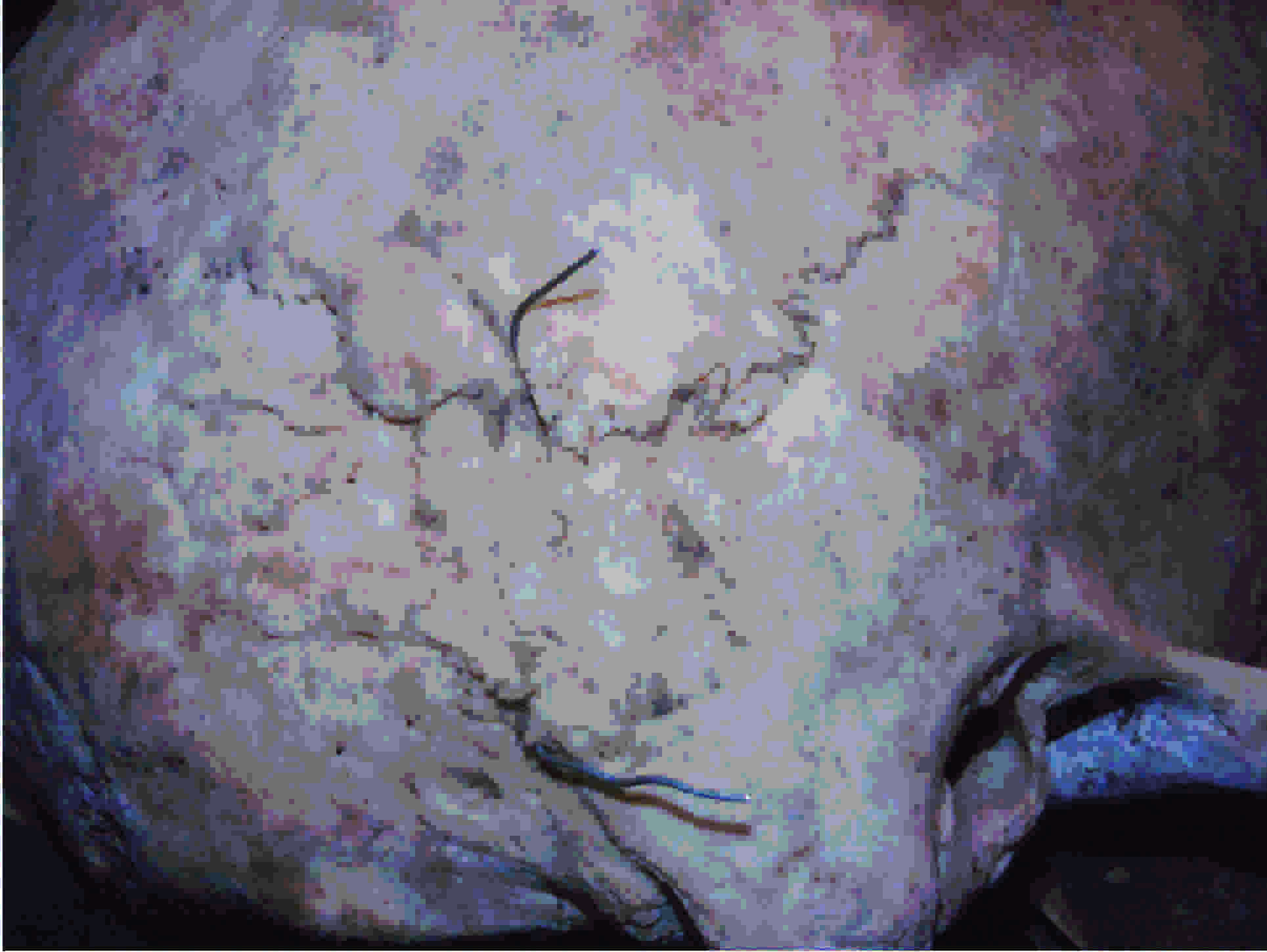
Mastoid canals of varying lengths were present in 53(53%) of the total 100 skulls which were observed, either bilaterally or unilaterally [Table/Fig-1 and 2]. Among the mastoid canals which were found in 53 skulls, 14(14%) were bilateral, 36(36%) were unilateral, 02(2%) were both mastoid canals and grooves together [Table/Fig-3] and 01(01%) was a double mastoid canal [Table/Fig-4]. Among the 36(36%) unilateral canals, 20(20%) were on the left side and 16(16%) on the right side. Mastoid grooves were present in 18(18%) of the total skulls (100) which were observed. Among the mastoid grooves which were found in 18 skulls, 03(3%) were bilateral 15(15%) were unilateral. Among the 15 unilateral grooves, 06(6%) were on the right side and 09(9%) were on left side [Table/Fig-5].The distances between the 2 openings of mastoid canal ranged from 1 to 31 mm. The lengths of mastoid grooves ranged from 9 to 21 mm. The diameter of the mastoid canal was measured; diameter was less than 5 mm in most of the skulls.
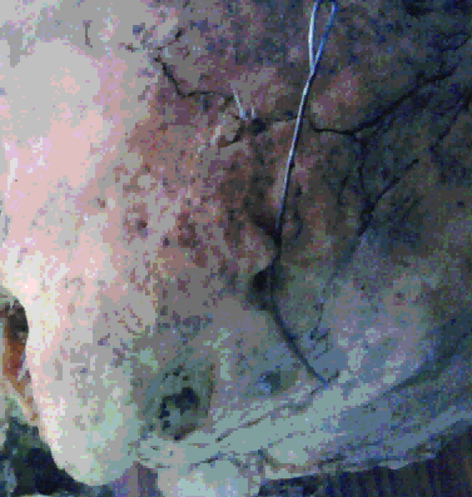
Longest Mastoid Canal, Metal wire in the canal
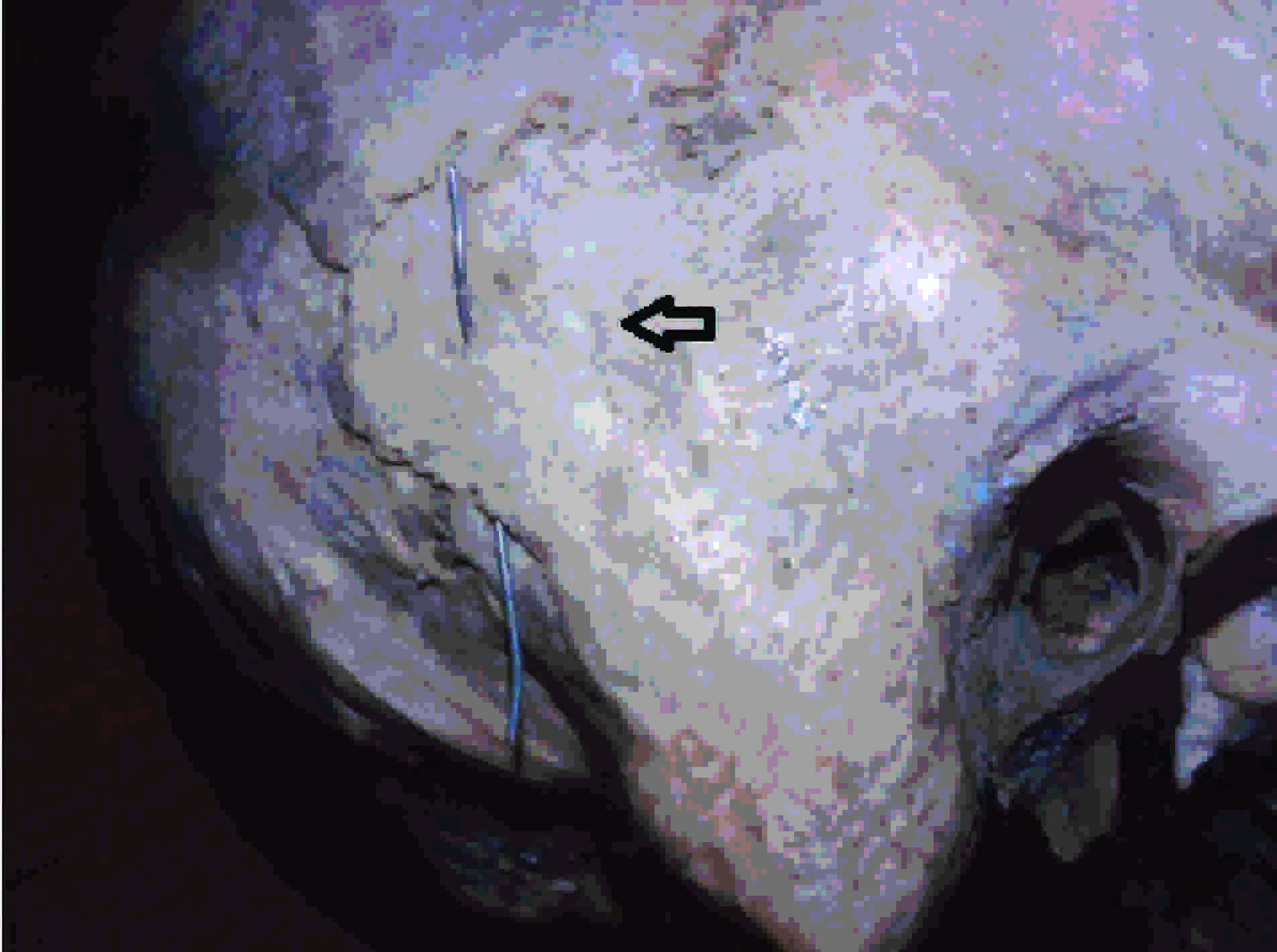
Both Mastoid Canal & Groove, Aero shows mastoid groove, metal wire shows mastoid canal
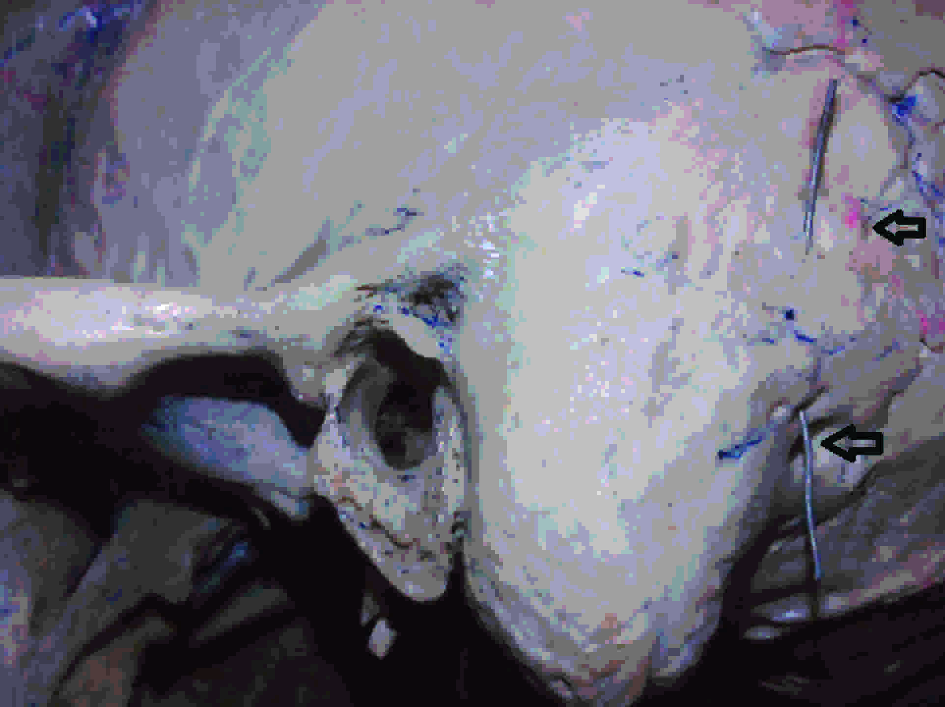
Double Mastoid Canal, metal wire shows 1st mastoid canal, Aero’s showing both the ends of second mastoid canal
Distribution of Mastoid Canals & Grooves
| Total skulls studied | Bilateral | Unilateral Right side | Unilateral Left side | Double Mastoid canal | Mastoid Canal & groove both | Total |
|---|
| Mastoid Canals | 100 | 14 (14%) | 16 (16%) | 20 (20%) | 01 (1%) Left Unilateral | 02 (02%) | 53 (53%) |
| Mastoid Grooves | 03(3%) | 06(6%) | 09(9%) | - | - | 18(18%) |
Knowledge on mastoid canals and grooves is very important for otolaryngologists and neurosurgeons, because they contain an arterial branch of occipital artery with its accompanying vein, which is liable to injuries which can result in severe bleeding.
Discussion
In an earlier study which was done by Choudary et al., it has already been proved that mastoid canals were present in Indian human skulls [3]. Singh M proved the presence of mastoid canals and mastoid grooves in Japanese skulls. The percentage frequency of mastoid canals in Japanese population was observed to be higher than that in Indian population [4]. Another study which was done by Hussain et al., showed the presence of mastoid canals and mastoid grooves in south Indian skulls [5]. Present study revealed that percentage frequency of mastoid canals in north Karnataka population was observed to be higher than that in Japanese population.
Warwick et al., reported two occipital branches of the occipital artery at this site [6]. In the present study, the presence of double mastoid canals in one region and presence of a mastoid canal and a groove together on one side were also reported; this indicated presence of two branches of occipital artery at this site, this supported the findings of Warwick et al.
Percentage frequency of mastoid grooves in north Karnataka population was observed to be higher than that in Japanese population [Table/Fig-6].
Comparison of result of present study with other studies
| No of Skulls | | Bilateral | Unilateral Right side | Unilateral Left side | Double Mastoid canal | Mastoid Canal & groove both | Total |
|---|
| Choudary | 265 | Canals | 8 | 12 | 21 | - | - | 41 |
| Grooves | - | - | - | - | - | |
| Singh M | 435 | Canals | 109 (25.05%) | 49 (11.26%) | 70 (16.09%) | - | - | 228 (52.40%) |
| Grooves | 21.1 (4.8%) | 20 (4.5%) | 18 (4.13%) | - | - | 59 (13.56%) |
| Hussian S | 125 | Canals | 35 (28%) | 20 (16%) | 19 (15.2%) | - | - | 74 (59.2%) |
| Grooves | 10 (8%) | 8 (6.4%) | 7 (5.6%) | - | - | 25 (20%) |
| Present study | 100 | Canals | 14 (14%) | 16 (16%) | 20 (20%) | 01 (1%) Unilateral Lt | 02 (02%) | 53 (53%) |
| Grooves | 03 (3%) | 06 (6%) | 09 (9%) | - | - | 18 (18%) |
Mastoid canals which contain vessels may be attributable to the mode of development of this part of the temporal bone. In the embryo, the bone develops from two components. The squamous part arises in mesenchyme in the 8th week of foetal life and it forms the anterosuperior part. The petro mastoid part develops from the cartilaginous epiotic centre at 5-6th months of foetal life and it forms the posteroinferior part by 1 year of age [1]. These are demarcated on the external surface as the petrosquamous suture and are directed downwards and forwards into the mastoid process. In the adult skull, these may barely be distinguishable or are seen as series of irregular depressions or well marked fissures. The squamous plate grows posterior and it covers a large area of the lateral surface of the petro mastoid bone. The ‘junction’ between these two components of the temporal bone is often separated by a heavy plate of bone in many adults, which is referred to as Korner’s septum or a ‘false bottom’ and is a remnant of the suture [7]. The ascending branch of the occipital artery, which lies on the developing petro mastoid in foetal life, is likely to be buried by the ossifying squamotemporal bone. In other words, this ascending branch of the artery in some skulls is ‘trapped’ between two growing bones.
Since the presence of this artery and its accompanying vein in this region is liable to injury, as it may be undetected, it is necessary for surgeons who operate this area, to be aware of this vascular arrangement, to avoid troublesome bleeding. The importance of structures in the mastoid area has increased due to the increasing use of the transtemporal route for surgical procedures which are performed by neurosurgeons and otolaryngologists, which involve access to structures in the posterior fossa and the mastoid air system.
[1]. Schaeefer JP, Morris’ Human Anatomy 1953 11th edNew YorkBlakiston:166:650 [Google Scholar]
[2]. Hollinshead WH, Anatomy for Surgeons the Head and Neck 1982 Vol. 13rd edNew YorkHarper and Row:183 [Google Scholar]
[3]. Choudhry R, Raheja S, Gaur U, Anand C, Mastoid canals in adult human skullsJournal of Anatomy 1996 188:217-19. [Google Scholar]
[4]. Singh M, Mishra A, Nagashima M, Mastoid Canals and Grooves in adult Japanese human skullsJ. Anat. Soc. India 2004 53(2):40-43. [Google Scholar]
[5]. Hussain SS, Muralidhar PS, Desai SD, Thomas ST, Maavishettar GF, Haseena S, Study of Mastoid canals and grooves in South Indian skullsIJMHC 2012 1(1):32-33. [Google Scholar]
[6]. Warwick PL, Williams R, Dyson M, Bannister LH, Gray’s Anatomy38th EdEdinburghChurchill Livingstone:591 [Google Scholar]
[7]. Wright D, Scott-Brown’s Otolaryngology 1978 5th ednLondonButterworths:10 [Google Scholar]