Frequency of Foetal Anomalies in a Tertiary Care Centre
Rameswarapu Suman Babu1, Sujatha Pasula2
1 Associate Professor, Department of Anatomy, Mediciti Institute of Medical Sciences, Ghanpur, Ranga Reddy District-501401, Andhra Pradesh, India.
2 Assistant Professor, Department of Biochemistry, D.D. Institute of Medical Science and Research, Thiruvalluri, Tamilnadu, India.
Name, Address, E-Mail Id of The Corresponding Author: Dr Sujatha Pasula, H. No:16-2-700/D/1, Palton, Malakpet, Hyderabad – 500036, Andhra Pradesh, India.
Phone: 9966355528
E-mail: drsujathapasula@gmail.com
Objective: The present study was undertaken to explore the incidence of congenital foetal anomalies and the advantages of ultrasonography in detecting the foetal anomalies during the antenatal period.
Method: We focused our study on 1000 consecutive pregnancies that came for check up in the second and third trimesters, with major or minor clinically relevant malformations which were detectable by ultrasonography.
Results: The analysis revealed that they were 38 foetal anomalies in 37 foetuses. One had multiple anomalies, with the highest incidence of neural tube defects. There was also significant correlation with consanguinity.
Conclusion: The overall incidence of congenital foetal anomalies in the present study was 3.8%.This might be probably due to environmental pollution, radiation, exposure to different chemicals and teratogenic drugs.
Foetal anomalies, Ultrasonography, Premarital counseling
Introduction
Human evolution from a single cell, ‘zygote’ to a multi cellular organism, is an intricate and a complex process. Lucky are those foeti which travel through this wonderful journey without encountering any hindrance. The birth of a malformed baby is an unfortunate event for any family and equally for the society too! Influence of teratogens in the form of pathogens, extensive use of chemicals, causes of environmental pollution and use of drugs by the mothers indiscriminately in their day to day life, have resulted in an increased incidence of congenital abnormalities in the newly born children, [1]. Congenital anomalies are the vital causes for prenatal mortality and morbidity. Therefore, an antenatal diagnosis and foetal therapy have attained importance in the field of human embryology. According to Dolk, [2] “Environmental factors include any non – genetic factor that increases the risk of a birth defect for the exposed individual. Such factors are nutritional excesses or deficiencies (e.g. folic acid), maternal illnesses or infections (e.g. diabetes, rubella), drugs which are taken during pregnancy (e.g. thalidomide), chemical exposure in the workplace or home (e.g., to solvents or pesticides) and radiation (e.g., medical X–rays).” Scientific literature is interested in the association between congenital anomalies and the possible role of chemical contaminants [1], and foetuses are thought to be a further subgroup of the population who could be vulnerable to the effects of air pollutants [1,3].
Official records and studies on congenital malformations have confirmed the fact that, most of the common anomalies or birth defects occur in 2.5% of live births. Gaining hands on experiences show that birth defects pose multifarious social, economic and cultural problems, as well as mental trauma to the whole humanity. It has been widely noticed that many mothers are not aware of the impact of factors in causing congenital defects in their foetuses. For a better understanding of the aetiological factors of congenital anomalies, a knowledge on embryology, teratology, clinical genetics and diagnostic ultrasonography is very important [4,5]. Highly advanced imaging techniques such as 3-D and 4-D ultrasound have been helping largely in the diagnosis and treatment of birth defects in foetuses during the antenatal period. It has been strongly advised that antenatal ultrasonography has to be conducted compulsorily for a minimum of two times in all antenatal mothers. The first antenatal ultrasonography, preferably a transvaginal one, could be done between 14 and 16 weeks of gestation period. The second one, preferably a transabdominal one, could be done after the 26th week of pregnancy. This is because ultrasound imaging during antenatal period produces an anatomical record of embryological development of the human embryo. Further, an early detection of neural tube defects and an excellent medical management, which include termination of pregnancy and counseling the eligible couple, will result in betterment of the society by attaining eugenics.
An ultrasound examination will not, of course detect chromosomal anomalies, inherited disorders of metabolism or gene defects such as sickle cell disease. Detection of anomalies by ultrasound is based upon the direct visualisation of a structural defect (e.g. anencephaly), the demonstration of abnormal growth rates (e.g. short limbs in dwarfism), or the demonstration of a pathological process which is caused by the defect (e.g. a dilated stomach and duodenum in cases of duodenal atresia).
Some congenital abnormalities can be curable if they are detected early in the antenatal period (e.g. Cardiac anomalies). In – utero surgical interventions have been made possible by the advancement in the field of medicine. Pregnant women who carry anomalous foetuses can be counseled regarding the foetal anomalies and they can be sent to neonatal paediatricians for an early management or they can be advised to go for termination of pregnancy if the anomalies are of a incurable variety (e.g. anencephaly).
In view of the above, we were prompted to take up the present study “Frequency of foetal anomalies in a tertiary care centre”. Apart from determining the incidence of congenital foetal abnormalities, such a study might bring to light certain factors which could possibly play an aetiological role in the production of foetal anomalies.
Material and Methods
The present study was based on the ultrasonographic diagnoses of 1000 consecutive pregnancies which were studied in the Department of Radiology, Government Maternity Hospital, which was attached to Osmania Medical College, Hyderabad, India, which is a tertiary care centre, in the years 2012-13 . The cases were selected from among women who were in the second and third trimesters, who were of the age group of 20-35 years, who had come for obstetric services.
The inclusion criteria were a history of suspected anomalies on clinical examination and women who were in their 2nd and 3rd trimesters, who have came for routine check ups. The women were selected, based on their ages as per criteria and parity and from all socio-economic classes. A detailed history of anomalies in other siblings, a history of consanguinity and age of the mother were also noted. A majority of the cases which were referred for scanning were referred for confirmation of the gestational ages and for exclusion of associated pathologies and anomalies. A more specific indication was the disproportionate uterine size as compared to the period of amenorrhoea.
Results
The data was analysed by using SPSS, version 15.0 and the Microsoft Excel software. A p value of <0.05 was considered as significant. The results have been represented in the form of tables. Figures of cases which were followed up with terminations and autopsies which were done in Osmania Medical College were included.
Foetal anomalies are significantly associated with consanguinity.
Overall, incidence of congenital anomalies was 3.8%. highest incidence of CNS anomalies observed.
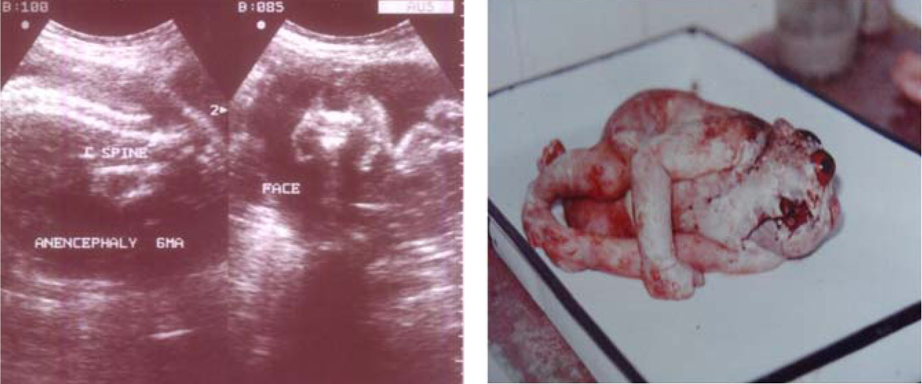
Total 5 anencephaly were noted in the observation.
These are the overall findings which were observed in the present study.
Discussion
[Table/Fig-1,2,3,4,5,6,7,8,9,10,11]
Showing the systemic distribution of anomalies
| Systems involved | No. of fetuses affected | Percentage (%) |
|---|
| CNS | 17 | 45.94 |
| URINARY SYSTEM | 08 | 21.62 |
| SKELETAL SYSTEM | 03 | 8.10 |
| GIT | 06 | 16.22 |
| CVS | 03 | 8.10 |
| TOTAL | 37 | 3.8 % (overall) |
Distribution of fetal anomalies in accordance with consanguinity
| Total No cases | Without fetal anomalies | With fetal anomalies | p-value |
|---|
| Second trimester | 11 | 10 | <0.05 |
| Third trimester | 7 | 9 | <0.05 |
| Total | 18 | 19 | <0.05 |
The distribution of the CNS anomalies
| CNS anomalies | No. of fetuses affected | Percentage (%) |
|---|
| Anencephaly | 05 | 13.51 |
| Meningo\ myelocele | 02 | 5.41 |
| Spina bifida | 01 | 2.70 |
| Encephalocele | 01 | 2.70 |
| Hydrocephalus | 04 | 10.81 |
| Holoprosencephaly | 01 | 2.70 |
| Dilatation of ventricles | 03 | 8.10 |
| Total | 17 | 45.94 % |
Showing anencephaly in USG and autopsy
Distribution of urinary system anomalies
| Urinary system anomalies | No. of fetuses affected | Percentage (%) |
|---|
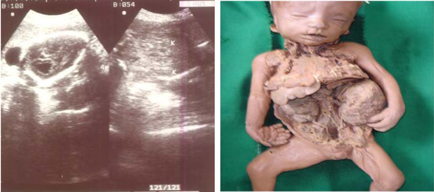
| Hydronephrosis | 05 | 13.51 |
| Cystic kidneys | 02 | 5.41 |
| Renal cyst | 01 | 2.70 |
| TOTAL | 08 | 21.62 % |
Picture showing polycystic kidney with hydronephrosis
The distribution of skeletal system anomalies
| SKELETAL anomalies | No. of fetuses affected | Percentage (%) |
|---|
| Limb bone shortening | 01 | 2.70 |
| Achondroplasia | 01 | 2.70 |
| Club foot | 01 | 2.70 |
| TOTAL | 03 | 8.10 % |
The distribution of GIT anomalies
| GIT anomalies | No. of fetuses affected | Percentage (%) |
|---|
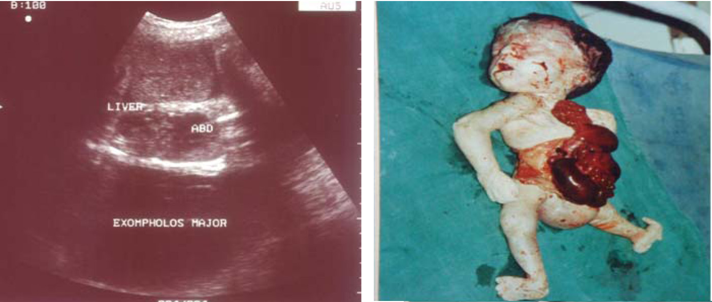
| Duodenal atresia | 01 | 2.70 |
| Diaphragmatic hernia | 02 | 5.41 |
| Cleft lip / palate | 02 | 5.41 |
| Omphalocele/Exomphalus | 01 | 2.70 |
| TOTAL | 06 | 16.22 % |
Showing exompholos major in USG and autopsy
The distribution of CVS anomalies
| CVS anomalies | No. of fetuses affected | Percentage (%) |
|---|
| Echogenic left ventricle | 01 | 2.70 |
| Twin twin transfusion | 01 | 2.70 |
| Single umbilical artery | 01 | 2.70 |
| TOTAL | 03 | 8.10 % |
The overall incidence of the fetal anamolies according
| 1 | Ogunyemi et al., [7] | 2000 | 6877 | 3.1 |
| 2 | Rashid SQ [8] | 2002 | 5841 | 1.7 |
| 3 | Tripale P et al., [9] | 2003 | 20465 | 1.5 |
| 4 | Nakling J et al., [10] | 2005 | 18181 | 1.5 |
| 5 | Salvador J et al., [11] | 2005 | 99753 | 1.9 |
| 6 | Sonka Ap et al., [12] | 2006 | 1148 | 1.2 |
| 7 | Becker R et al., [13] | 2006 | 3094 | 2.8 |
| 8 | Emilio et al., [14] | 2012 | 8503 | 2.3 |
| 9 | Present study | 2013 | 1000 | 3.8 |
During the study period, 38 foetal malformations were identified in ultrasound, in the 1000 pregnancies, which corresponded to a prevalence of 3.8%. Among all anomalies, one foetus had multiple anomalies. The clinically relevant major and minor malformations [Table/Fig-1] were arranged system wise and tabulated. A detailed history, period of gestation and results of the present study were noted in a proforma.
It was seen that there was significant correlation (p<0.05) between the foetal anomalies and consanguinity [Table/Fig-2], which was inconsistent with the findings of Naeimeh Tayebi et al., [6].
The findings of the present study correlated with those of Lee k et al., [15], Pagnotta G et al., [16] and Khronf N et al., [17]. Our values were higher as compared to those of the other studies. One of the possible reasons for this increased rate was that ours being a tertiary care centre, cases were referred here for further follow up and also radiation, environmental pollution and teratogens would have been other causes.
The most frequently observed abnormalities involved those of the central nervous system (17/38; 45%); 5/38 (13%) cases which were associated with anencephaly were detected in the prenatal period. A possible reason could be inheritance of abnormal genes from the parents, as well as new mutations in one of the germ cells that could have given rise to foetuses with such anomalies. This finding was in correlation with those of Vial Y et al., and Stefost et al., [18, 19].
Occurrence of other anomalies like Urinary anomalies [Table/Fig-5 and 6] and also skeletal, [Table/Fig-7] GIT [Table/Fig-8 and 9] and CVS anomalies [Table/Fig-10] have been represented in the descending order of their incidence in present study. Incidence of congenital anomalies in various areas is in consistent with other studies represented [Table/Fig-11].
Congenital foetal anomalies are a major cause of antenatal mortality and morbidity. The diagnosis of an anomalous foetus has been a challenging task over the years. Detection of foetal anomalies in the antenatal period is very important for the prognosis of foetuses, because they result in abortions, stillbirths and other foetal defects [Table/Fig-3 and 4]. An accurate and early diagnosis of congenital foetal anomalies will help in an early intervention of the foetus and this can prevent late irreparable damages. Prenatal ultrasonography is the best means for diagnosing malformed foetuses [20]. However, published results concerning the sensitivity of the screening vary greatly, depending on the population which was studied (high versus low risk), the quality of the equipment and in particular, the ultrasonographers’ experience. Nowadays, high-resolution equipment allows the detection of minor malformations which are considered to be sonographic markers of specific conditions. These were not included in our study, to maintain the homogeneity of the population which was screened. We focused our attention on the severe abnormalities which could be detected by prenatal ultrasonography.
In conclusion, we share the view of Bucher and Schmidt [21], who in their meta – analysis insisted that, “a routine ultrasound screening in pregnancy is indicated only if it is explicitly performed to exclude congenital malformations”. According to the results of the present study, we also recommend that all pregnancies, especially of consanguineous marriages, should be thoroughly examined and investigated for congenital anomalies. Premarital counseling, especially on the subject of parental consanguinity, is advised.
Summary and Conclusion
Congenital anomalies of foetuses are a great concern since time immemorial. Aetiological factors for these anomalies are plenty. There is a strong correlation between the congenital anomalies of the foetuses and chromosomal abnormalities, either structural or numerical. The overall incidence of congenital foetal anomalies in the present study was 3.8%. This correlated with the findings of some of the previous authors. But it was higher as compared to the findings of other authors. This might probably be due to environmental pollution, radiation and exposure to different chemicals and teratogenic drugs
[1]. Dolk H, Epidemiological approaches to identifying environmental causes of birth defectsAm J Med Genet C Semin Med Genet 2004 125C(1):4-11. [Google Scholar]
[2]. Dolk H, Epidemiological evidence regarding environmental causes of congenital anomalies: interpretational issuesIn special report: A Review of Environmental Risk Factors for Congenital Anomalies 2004 Co Antrim Northern IrelandUniversity of Ulster Newtownabbey:30-47. [Google Scholar]
[3]. Pope CA 3rd, Epidemiology of fine particulate air pollution and human health: biologic mechanisms and who’s at risk?Environ Health Perspect 2000 108(Suppl. 4):713-23. [Google Scholar]
[4]. Dolk H, Loane M, Garne E, The prevalence of congenital anomalies in EuropeAdv Exp Med Biol 2010 686:349-64. [Google Scholar]
[5]. Perera FP, Jedrychowski W, Rauh V, Whyatt R, Molecular epidemiologic research on the effects of environmental pollutants on fetusEnviron Health Perspect 1999 107(suppl):451-60. [Google Scholar]
[6]. Tayebi Naeimeh, Yazdani Katayon, Naghshin Nazila, The Prevalence of Congenital Malformations and its Correlation with Consanguineous MarriagesOMJ 2010 25:37-40. [Google Scholar]
[7]. Ogunyemi Prenatal diagnosis of fetal anomalies in a regional tertiary center: the role of a maternal fetal medicine unit a review of 6,877 deliveriesJ Matern Fetal Med 2000 Jul-Aug 9(4):219-23. [Google Scholar]
[8]. Rashid SQ, A study of fetal anomalies detected by ultrasound in Bangladesh and their relative frequenciesJ R Soc Health 2002 Mar 22(1):55-57. [Google Scholar]
[9]. Tripale P, Two-statge ultrasonography in screening for fetal anomalies at 13 14 and 18-22 weeks of gestationActa Obstet Gynecol Scand 2004 Dec 83(12):1141-46. [Google Scholar]
[10]. Nakling J, Routine ultrasound screening and detection of congenital anomalies outside a university settingActa Obstet Gynecol Scand 2005 Nov 84(11):1042-48. [Google Scholar]
[11]. Salvador J, Increasing detection rates of birth defects by prenatal ultrasound leading to apparent increasing prevalence.Lessons learned from the population-based registry of birth defects of BarcelonaPrenatal Diagn 2005 Nov 25(11):991-96. [Google Scholar]
[12]. Sonka Ap, Screening for major structural abnormalities at the 11-to-14-week ultrasound scanAm J Obstet Gynecol 2006 Feb 194(2):393-96. [Google Scholar]
[13]. Becker R, Detailed screening for fetal anomalies and cardiac defects at the 11-13-week scanUltrasound Obset Gynecol 2006 Jun 27(6):613-18. [Google Scholar]
[14]. Emilio Antonio Luca Gianicolo, Congenital anomalies among live births in a polluted area. A ten-year retrospective studyBMC Pregnancy and Childbirth 2012 12:165 [Google Scholar]
[15]. Leek Effectiveness of prenatal ultrasonography in detecting fetal anomalies and perinatal outcome of anomalous fetusesYousej Med J 1998 Aug 39(4):372-82. [Google Scholar]
[16]. Pagnotta G, Antenatal sonographic diagnosis of clubfoot: a six – year experienceJ Foot Surg 1996 Jan-Feb 35(1):67-71. [Google Scholar]
[17]. Khronf N, Malformation in 10,000 consecutive births in TunisActa Pediatr Scand 1986 Jul 75(4):534-39. [Google Scholar]
[18]. Vial y, Screening for foetal malformations: performance of routine ultrasonography in the population of the Swiss Canton of VaudSwiss Med Wkly 2001 Aug 25 131(33-34):490-94. [Google Scholar]
[19]. Stefost Routine obstetrical ultrasound at 18-22 weeks: our experience on 7,236 fetusesJ Maternal Fetal med 1999 Mar-Apr 8(2):64 [Google Scholar]
[20]. Shirley IM, Bottomley F, Robinson VP, Routine radiographer screening for foetal abnormalities by ultrasound in an unselected, low-risk populationBr J Radiol 1992 65:564-69. [Google Scholar]
[21]. Bucher HC, Schmidt JG, Does routine ultrasound scanning improve outcome in pregnancy? Meta-analysis of various outcome measuresBr Med J 1993 307:13-17. [Google Scholar]