An Analysis of the Vertical Bone Loss in Edentulous Mandibles by Using the Mental Foramen as a Reference: A Radiographic Study
Anjali Sofat1, Virat Galhotra2, Ramandeep Singh Gambhir3, Shushant K. Garg4
1 Senior Lecturer, Department of Prosthodontics, Gian Sagar Dental College and Hospital, Rajpura, Punjab, India.
2 Professor, Department of Pedodontics, Gian Sagar Dental College and Hospital, Rajpura, Punjab, India.
3 Sr. Lecturer, Department of Public Health Dentistry, Gian Sagar Dental College and Hospital, Rajpura, Punjab, India.
4 Professor, Department of Prosthodontics, MM University Mullana, Ambala, India.
Name, Address, E-Mail Id of The Corresponding Author: Dr Ramandeep Singh Gambhir, Senior Lecturer, Gian Sagar Dental College, Rajpura, Punjab, India-140601,
Phone: +91-99156-46007. Fax- +91-1762 520011
E-mail: raman1g@yahoo.co.in
Objectives: To analyze the amount of vertical bone loss in edentulous mandibles by using the mental foramen as a reference.
Method: Ninety subjects who consisted of thirty dentulous (males I MD and females II FD) and sixty edentulous (males I ME and females II FE) subjects were selected and they were subjected to digital panoramic radiography. Among the dentulous subjects, in both the groups, the ratio between the height of the mandible and the distance from the mental foramen and the lower border of the mandible (A/B) were calculated by using inbuilt software. This ratio A/B was applied in the edentulous subjects to measure the original height of the mandible before resorption. The amount of vertical bone loss was then calculated.
Results: In the dentulous subjects, the A/B ratio in males was 2.94 and in females, it was 3.09. In the edentulous subjects, the vertical bone loss in males was 3.55mm and in females, it was 6.59mm.
Conclusion: The ratio of the distance from the inferior border of the mandible to the superior border of the mandible(A) and the distance from the inferior border of the mandible to the inferior margin of the mental foramen (B) were less in the dentulous males than in the dentulous females. The comparison of the ratios was nonsignificant. The mean bone loss in edentulous females was found to be greater than the mean bone loss in edentulous males on comparison.
Dentulous, Edentulous, Comparison, Vertical bone loss
Introduction
The preservation of which remains is of utmost importance and not the meticulous replacement of that which has been lost [1]. In the light of the present statement, the mere replacement of the lost teeth in completely edentulous patients, does not amount to the complete prosthetic rehabilitation of the patients, but the steps which have to be taken towards the preservation of the underlying supporting tissues are to be borne in mind. In a prosthetic sense, the bone is considered to be the base which provides support for dentures. After the extraction of teeth, the alveolar portion of the jaw starts to atrophy; this is also referred to as Residual Ridge Resorption (RRR) [2]. According to G.P.T (2005) [3], residual ridge resorption is a term which is used for the diminishing quantity and the quality of the residual ridge after the teeth are removed. Residual ridge resorption is a chronic, progressive and an irreversible process; the rate is fastest in the first 6 months after the extraction [4].
The goal of modern dentistry is to restore the patient’s normal contour, function, comfort, aesthetics, speech, and health, regardless of the atrophy, disease, or injury of the stomatognathic system [5]. Panoramic radiographs were developed as a rapid, effective and a simple method for recording the oral and associated structures for the screening of edentulous patients, especially to evaluate the vertical bone loss [6]. Therefore, the present study was carried out to determine the vertical bone loss in edentulous mandibles by using the mental foramen as a reference.
Material and Methods
This study was carried out on ninety subjects who consisted of thirty dentulous and sixty edentulous subjects who were selected from the Department of Prosthodontics and also from Crown and Bridge, M.M. College of Dental Sciences and Research, Mullana (Ambala, Haryana, India). The thirty dentulous subjects were divided into two groups according to their sexes (male-I MD and female-II FD). Similarly, the sixty edentulous subjects were divided into two groups according to their sexes (male-I ME and female-II FE). All the subjects, both dentulous and edentulous, were then subjected to digital panoramic radiography.
The measurements which were taken on the radiographs
I. For the dentulous subjects [Table/Fig-1]: One dentulous subject was chosen for the measurement from group I MD(male) and various markings were made on the specified anatomical landmarks of the mandible, as were viewed on the panoramic radiographs:-
The superior border: It was marked by drawing a line between the mesial of the 1st premolar and the distal of the 1st molar, at the crestal level, with the help of a program which was inbuilt within the software.
The inferior border: It was marked by drawing a line along the tangent, to the body of the mandible.
A line was drawn perpendicular from the inferior border up to the superior border, to measure the distance (existing height) and this was denoted by - A.
Another measurement was taken from the inferior border of the mandible to the inferior margin of the mental foramen and this was denoted by - B.
Dentulous subject-Measurement taken on Radiograph
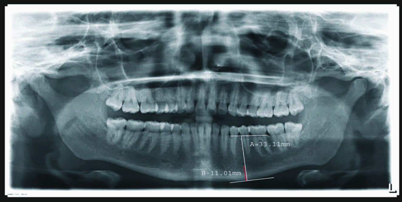
Then, the ratio was calculated as: A/B = X
All the measurements were taken with the help of a scale which was inbuilt in the program module.
The same procedure was repeated for the remaining 14 subjects from group IMD(males) and for 15 subjects of group II FD(females)
II. For the Edentulous subjects [Table/Fig-2]:
Edentulous Subject-Measurement taken on Radiograph
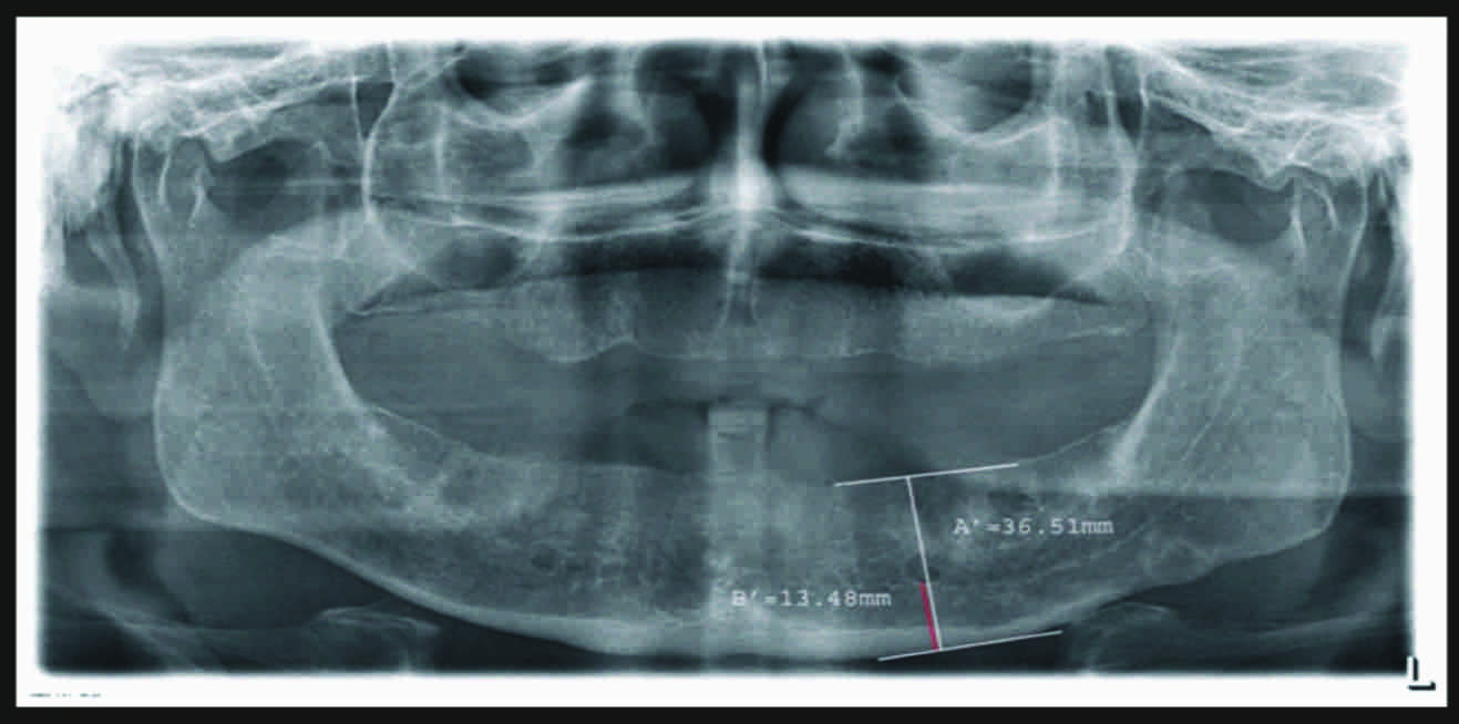
One edentulous subject was chosen for the measurement from group IME (male) and various markings were made on the specified anatomical landmarks of the mandible, as were viewed on the panoramic radiographs:-
The superior border: It was marked by drawing a tangent to the highest point of the alveolar bone in the premolar and the molar regions, with the help of a program which was inbuilt within the software.
The inferior border: It was marked by drawing a line along the tangent, to the body of the mandible.
A line was drawn perpendicular from the inferior border up to the superior border, to measure the distance (existing height) and this was denoted by A’.
Another measurement was taken from the inferior border of the mandible to the inferior margin of the mental foramen and this was denoted by B’ . This measurement was used to determine the original height (before resorption) i.e. Z.
III. Measurement of the vertical bone loss in the edentulous subjects (Mandible).
The above values were used as follows:
In the Dentulous patients, the Ratio was A/B.
The average ratio in the Dentulous patients was X.
In the Edentulous patients,
The original height (before resorption) Z = B’ multiplied by X.
Bone loss = Original height (Z) – Existing height (A’).
The same procedure was repeated for the remaining 29 subjects from group I ME and for 30 subjects from group II FE. The statistical analysis of the differences between men and women was done by using the Student’s t-test. A 5% significance level was used for assessing the statistical significance.
Results
In the dentulous male group (IMD), the mean ratio (A/B) was calculated as 2.94:1 and in the dentulous female group (IIFD), the mean ratio (A/B) was calculated as 3.09:1 [Table/Fig-3]. Comparison of the mean ratios in the dentulous males and females showed that it was non significant [Table/Fig-4]. The mean bone loss in the edentulous male group (IME) was 3.5555mm and the mean bone loss in the edentulous female group (IIFE) was 6.5905mm [Table/Fig-5]. Comparison of the mean bone loss in the edentulous males and females was highly significant [Table/Fig-6].
Mean Ratio (A/B) OF Dentulous Males (MD) and Dentulous Females (FD)
| Group | M | SD | SE | 95%Confidence Interval for Mean | Minimum | Maximum |
|---|
| Lower | Upper |
|---|
| Males (MD) | 2.9400 | 0.27723 | 0.07158 | 2.7865 | 3.0935 | 2.40 | 3.50 |
| Females (FD) | 3.0933 | 0.42505 | .10975 | 2.8579 | 3.3287 | 2.40 | 3.90 |
M: Mean(in mm) ; SD: Standard Deviation; SE : Standard Error
Comparison of mean ratio of Dentulous Male and Female subjects
| GROUP | t | df | Sig. (2tailed) | ‘p’ – value |
|---|
| MD: FD | -1.170 | 28 | 0.252 | > 0.05 NS |
Mean bone loss of edentulous Males (ME) and edentulous Females (FE)
| Group | M | SD | SE | 95%Confidence Interval for Mean | Minimum | Maximum |
|---|
| Lower | Upper |
|---|
| Males (ME) | 3.5555 | 2.29450 | 0.41892 | 2.6987 | 4.4123 | 0.87 | 12.42 |
| Females (FE) | 6.5905 | 2.85696 | 0.52161 | 5.5237 | 7.6573 | 2.56 | 14.85 |
M: Mean(in mm); SD : Standard Deviation; SE : Standard Error
Comparison of Mean bone loss of edentulous Male and Female subjects
| Group | T | Df | Sig. (2-tailed) | ‘p’ – value |
|---|
| ME: FE | -4.537 | 58 | 0.001** | < 0.05** |
Discussion
The panoramic radiographs provided an informative picture of the bone loss. Their importance in evaluating the residual bone resorption had been described by several authors. Axelsson G. [7] described the growing popularity of panoramic radiography because of the following reasons; it provided radiographs of the entire maxilla and the mandible on a single film, it was time saving, the film was not inserted into the mouth and the radiation dose to which the patient was exposed was smaller than that which was involved when periapical films were used.
In this study, digital panoramic radiographs were used. The basic technique of digital panoramic radiography was the same as that which was used in the conventional machines. In conventional panoramic radiography, the true dimensions could be determined easily and precisely with calipers. In contrast, a software measurement algorithm had to be used in digital panoramic radiography. According to some authors [8], the digital measurements which were made with the digital panoramic radiographs had shown adequate reproducibility. The relation of the mental foramen with the inferior border of the mandible appeared to be consistent, which justified the use of the mental foramen as a reliable landmark in evaluating the bone loss. Also, the ratios [2.94 (in males)] and [3.09 (in females)] could be used for determining the original height and the proportion of the bone height which had undergone resorption. According to Von Wowern [9], the incidence of the mandibular residual ridge resorption in edentulous patients who were above 70 years of age varied with the gender and it amounted to 1.5% in females and to 0.9% in males. Such results were also reported by Rusiniak-Kubik et al., [9] which indicated an increase in the mandibular residual ridge resorption in the course of the life of an edentulous patient and double the incidence of severe atrophy in females as compared to that in males. A correlation with sex was also shown by Kordatzis et al., [9] and Solar et al., who revealed that the female gender was an independent risk factor for severe bone resorption.
The significant bone loss in females could be attributed to the following reasons:
Females have shorter faces and smaller skeletons as compared to males [10].
Women tend to loose teeth faster as compared to men because of the aesthetic reasons and periodontal problems which arise in pregnancy [10].
After the age of 40 years, there is a decline in the density of the bone. The decreased physical activity and decreased oestrogen levels may account for this [11].
Conclusion
It was concluded that:
The ratio A/B in dentulous males was 2.94:1 and in dentulous females, it was 3.09:1; the comparison of the ratios was nonsignificant.
The mean bone loss in edentulous males was 3.5555mm and the mean bone loss in edentulous females was 6.5905mm; the comparison of the ratios was highly significant.
M: Mean(in mm) ; SD: Standard Deviation; SE : Standard Error
M: Mean(in mm); SD : Standard Deviation; SE : Standard Error
[1]. Veeraiyan DN, Ramalingam K, Bhatt V, Textbook of Prosthodontics 2003 1st edJaypee Brothers, Medical Publishers(P) Ltd:65 [Google Scholar]
[2]. Klemetti E, A review of residual ridge resorption and bone densityJ Prosthet Dent 1996 75(5):512-14. [Google Scholar]
[3]. Residual Ridge ResorptionGlossary of Prosthodontic termsJ Prosthet Dent 2005 945th ed(1):68 [Google Scholar]
[4]. Atwood DA, Reduction of residual ridges: A major oral disease entityJ Prosthet Dent 1971 26(3):266-79. [Google Scholar]
[5]. Guler AU, Sumer M, Sumer P, Bicer I, The evaluation of vertical heights of maxillary and mandibular bones and the location of anatomic landmarks in panoramic radiographs of edentulous patients for implant dentistryJ Oral Rehab 2005 32:741-46. [Google Scholar]
[6]. Wical KE, Swoope CC, Studies of residual ridge resorption. Part I. Use of panoramic radiographs for evaluation and classification of mandibular resorptionJ Prosthet Dent 1974 32(1):7-12. [Google Scholar]
[7]. Axelsson G, Orthopantomographic examination of edentulous mouthJ Prosthet Dent 1988 59(5):592-97. [Google Scholar]
[8]. Schulze R, Krummenauer F, Schalldach F, Hoedt B, Precision and accuracy of measurements in digital panoramic radiographyDentomaxillofac Radiol 2000 29:52-56. [Google Scholar]
[9]. Vonwowern N, Stoltze K, Sex and age differences in bone morphology of mandiblesScand J Dent Res 1978 86:478-85. [Google Scholar]
[10]. Mercier P, Ridge reconstruction with hydroxyl apatite Part-1 Anatomy of residual ridgeOral Surg Oral Med Oral Pathol 1988 65:505-10. [Google Scholar]
[11]. Baylink DJ, Wergedal JE, Yamamoto K, Manzke E, Systemic factors in alveolar bone lossJ Prosthet Dent 1974 31(5):486-505. [Google Scholar]