A Pure Epidural Spinal Cavernous Hemangioma — With an Innocuous Face But A Perilous Behaviour!!
Hemalatha A L1, Ravikumar T2, Neelima P Chamarthy3, Kunal Puri4
1 Professor & Head, Department of Pathology,
2 PG Student, Department of Pathology,
3 PG Student, Department of Pathology,
4 PG Student, Department of Pathology, Mysore Medical College & Research Institute, Karnataka, India.
Name, Address, E-Mail Id of The Corresponding Author: Dr Hemalatha A L, Professor & Head, Department of Pathology, No. 156, 12th Cross, 2nd Main, Jayanagar, Mysore – 570014, Karnataka, India.
Phone: +91-8453399335
E-mail: halingappa@gmail.com
Cavernous hemangiomas occur frequently in the intracranial structures but they are rare in the spine, with an incidence of 0.22 cases/million/year, which account for 5 – 12% of the spinal vascular lesions, 51% of which are extradural. Most of the epidural hemangiomas are secondary extensions from the vertebral lesions. The spinal cavernous hemangiomas which do not involve the vertebrae are referred to as “pure” types. The pure epidural hemangiomas are rare, which account for only 4% of all the epidural lesions.
A case of a Pure spinal epidural cavernous hemangioma in a 50 year old male, with the clinical picture of a slowly progressive compressive myelopathy, has been presented here.
The imaging studies showed a well-defined, enhancing epidural lesion at the T7 – T8 level, with dorsal cordedema and myelomalacic changes. A radiological diagnosis of a meningioma was considered. Histopathologically, the lesion was diagnosed as a hemangioma. The patient improved dramatically after the excision of the lesion.
Cavernous hemangioma, Spinal, Epidural
Introduction
Cavernous hemangiomas are vascular malformations which appear frequently in the intra-cranial structures. They are rare in the spine and they account for an incidence of 0.22 cases/million/year and 5 – 12% of the spinal vascular lesions, of which 51% are extradural [1]. Most of the epidural hemangiomas are secondary extensions from the vertebral lesions. The pure epidural hemangiomas are rare and they account for 4% of all the epidural lesions [2]. The review of the literature has revealed only 80 reported cases till date [3,4].
Case Report
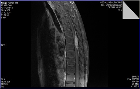
A 50 year old male presented with a history of progressive weakness in both the lower limbs since 1 year. He also complained of inability in walking, lifting or appreciating sensations in the lower limbs, as well as loss of the bowel and bladder control. The general examination revealed no significant abnormalities. A detailed Central Nervous System (CNS) examination showed no neurological deficit in the upper limbs. The lower limbs were flaccid, with a reduced bulk and a power of grade 1/5. A loss of sensation was observed upto the groin crease. An examination of the spine revealed no tenderness or deformity. The systemic examination showed no abnormality. The routine haematological investigations showed mild anaemia. Magnetic Resonance Imaging (MRI) of the dorsal spine showed a well-defined, oval, enhancing lesion which measured 4.4 x 1.3 x 2.0 cm, in the posterior epidural compartment, at the T7 – T8 level, with dorsal cord oedema and early myelomalacia. The lesion was homogenously iso-intense in the T1 images and it was hyperintense in the T2 images, with effacement of the subarachnoid space posteriorly. The spinal cord showed mild extension into the neural foramina. The radiological diagnosis which was offered was meningioma [Table/Fig-1].
MRI (Lateral view): Homogenously isointense lesion at T7-T8 level
The patient underwent laminectomy from the T7-T8 level. Per-operatively, an epidural reddish brown mass which measured 6x2 cm at the T7-T8 level, with no dural attachment, or spinal extension or bony involvement, was observed. The excised mass was subjected to a histopathological examination.
The histopathological examination findings were as follows:
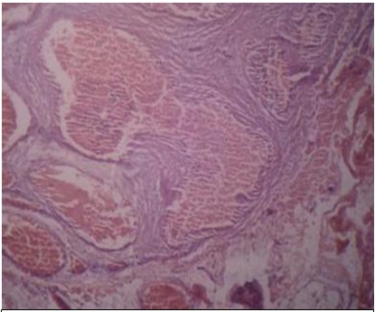
The gross examination of the specimen showed a single, brownish black, soft tissue mass which measured 3.5 x 1.5 x 0.5 cm. The cut section showed a brown black colour with a spongy appearance. The microscopic examination showed a benign tumour which was composed of vascular spaces of variable sizes, with fibrocollagenous walls which were lined by a single layer of flattened endothelial cells. The lumina contained red blood cells. A fibrofatty connective tissue was seen in between the lobules of the vascular spaces [Table/Fig-2]. Considering these features, the histopathological diagnosis of a Cavernous hemangioma was arrived at. The final diagnosis was an epidural spinal cavernous hemangioma. The differential diagnoses which were considered, included meningioma, benign schwannoma and chordoma. Meningioma was ruled out, since vascular spaces were present. The absence of the Antoni A and B areas helped in ruling out a Benign Schwannoma. A chordoma was ruled out due to the absence of the physalliferous cells.
H&E.40x: Showing blood filled interconnecting spaces with intervening fibrous septae
Discussion
A pure epidural cavernous hemangioma was first reported in 1929 [5]. In contrast to the present case, which was a male patient, a majority of these tumours occur in women [2]. The mean age of onset is between 30 to 60 years, as in the present case [6]. The thoracic spine is involved in 54-60% of the cases, as in the present case, while the cervical spine is involved in 30% of the cases and the lumbar spine is involved in 10% of the cases [7]. An epidural mass with a large posterior component in the spinal canal on MRI indicates an epidural hemangioma, as in this case [7]. The clinical manifestations depend on the location, growth rate and the biological behaviour of the tumour [8]. Disturbances of the sphincter functions, as in the present case, have been reported [2]. Though the tumour in the present case was morphologically an innocent and a benign lesion, the behaviour was perilous in that, it caused serious neurological manifestations like paraplegia and loss of the autonomic functions of the bowel and bladder. Besides this, there was a high per-operative risk of an intra-lesional haemorrhage, since it was a tumour which had a vascular origin. On the other hand, the failure of a timely intervention and an excision could have resulted in permanent neurological deficits. Spinal cavernous hemangiomas may be associated with cutaneous vascular lesions and fibromatosis, which were not present in this case [9].
Neuroimaging findings are invaluable in the diagnosis and the evaluation of the relationship between the tumour and the surrounding anatomical structures [10,11]. MRI is the most reliable technique which can be used for its diagnosis, with the key diagnostic features being the lobulated contour of the tumour and a cystic change [2].
The treatment of choice is total excision [2,12]. Most of the patients achieve a good recovery with the improvement of the neurological deficits and a complete resolution of the tumour after a total extirpation, as in this case [7]. Radiotherapy is advocated for the patients in whom a complete resection is not possible. The patients with acute and paraplegic conditions have poor outcomes. Our patient recovered completely from the paraperesis, following a total excision of the tumour. There was an immediate and a tremendous neurological improvement.
Conclusion
The pure epidural spinal cavernous hemangiomas exhibit the characteristic MR imaging and histopathological findings, which help in making an accurate diagnosis. However, caution should be exercised in ruling out the differential diagnoses. Complete excision of the lesion bears a good prognosis with no mortality, if a total extirpation is achieved.
[1]. Cosgrove GR, Bertrand G, Fontaine S, Robitaille Y, Melanson D, Cavernous angiomas of the spinal cordJ Neurosurg 1998 68:31-36. [Google Scholar]
[2]. Hatiboglu MA, Iplikeioglu AC, Ozcan D, Epidural spinal cavernous angiomaNeurol Med Chir (Tokyo) 2006 46:455-58. [Google Scholar]
[3]. Satpathy DK, Das S, Das BS, Spinal epidural cavernous hemangioma with myelopathy: A rare lesionNeurol India 2009 57:88-90. [Google Scholar]
[4]. Aoyagi N, Kojima K, Kasai H, Review of spinal epidural cavernous hemangiomaNeurol Med Chir. (Tokyo) 2003 43:471-75. [Google Scholar]
[5]. Goyal A, Singh AK, Gupta V, Tatke M, Spinal epidural cavernous haemangioma: a case report and review of literatureSpinal Cord 2002 Apr 40(4):200-02. [Google Scholar]
[6]. Pagni CA, Canavero S, Forni M, Report of a cavernoma of the cauda equine and review of the literatureSurg Neurol 1990 33:124-31. [Google Scholar]
[7]. Shin JH, Lee HK, Rhim SC, Park SH, Choi CG, Suh DC, Spinal epidural cavernous hemangioma. MR findingsJ Comput Assist Tomogr 2001 25:257-61. [Google Scholar]
[8]. Hillman J, Bynke O, Solitary extradural cavernous hemangiomas in the spinal canal: Report of five casesSurg Neurol 1991 36:19-34. [Google Scholar]
[9]. Lee J, Wang AD, Wai Y, Ho Y, Spinal extradural cavernous hemangiomaSurg Neurol 1990 34:345-51. [Google Scholar]
[10]. Sanghvi D, Munshi M, Kulkarni B, Kumar A, Dorsal spinal epidural cavernous hemangiomaJ Craniovertebr Junction Spine 2010 Jul-Dec 1(2):122-25. [Google Scholar]
[11]. Zevgaridis D, Büttner A, Weis S, Hamburger C, Reulen HJ, Spinal epidural cavernous hemangiomas. Report of three cases and review of the literatureJ Neurosurg 1998 May 88(5):903-08. [Google Scholar]
[12]. Jo BJ, Lee SH, Chung SE, Paeng SS, Kim HS, Yoon SW, Pure epidural cavernous hemangioma of the cervical spine that presented with an acute sensory deficit caused by hemorrhageYonsei Med J 2006 47:877-80. [Google Scholar]