A Ruputured Left Cornual Pregnancy: A Case Report
Surekha S M1, Chamaraja T2, Nabakishore Singh N3, Bimolchandra Singh N4, Neeraja T S5
1 Junior Resident, Department of Obstetrics and Gynaecology, Regional Institute of Medical Sciences, Imphal, Manipur-795004, India.
2 Junior Resident, Department of Obstetrics and Gynaecology, Regional Institute of Medical Sciences, Imphal, Manipur-795004, India.
3 Professor and Head, Department of Obstetrics and Gynaecology, Regional Institute of Medical Sciences, Imphal, Manipur-795004, India.
4 Assistant Professor, Department of Obstetrics and Gynaecology, Regional Institute of Medical Sciences, Imphal, Manipur-795004, India.
5 Junior Resident, Department of Obstetrics and Gynaecology, Regional Institute of Medical Sciences, Imphal, Manipur-795004, India.
Name, Address, E-Mail Id of The Corresponding Author: Dr Surekha S M, Junior Resident, 3rd year, Department of Obstetrics and Gynaecology, Regional Institute of Medical Sciences, Imphal, Manipur-795004, India.
Phone: 9774867510
E-mail: drsiri.sm@gmail.com
A cornual gestation is one of the most hazardous types of ectopic gestations, which accounts for 2 – 4% of all the ectopic pregnancies and it has a mortality rate which is 6 – 7 times higher than that of the ectopics in general. The diagnosis and the treatment of such a pregnancy is challenging and it constitutes an urgent medical situation. Because of the myometrial stretch ability, they tend to present relatively late, at 7 – 12 weeks of gestation. A significant maternal haemorrhage which can lead to hypovolaemia and shock, can rapidly result from a cornual rupture. We are reporting a case of 28 year old woman who presented to the emergency obstetrical room in a state of hypovolaemic shock. The diagnosis of a ruptured ectopic pregnancy was confirmed in view of the history of 10 weeks of amenorrhoea, with a positive urine pregnancy test. She was shifted for emergency exploratory laparotomy. Intraoperatively, we encountered a left lateral wall ruptured uterus with a 10 week old foetus in the peritoneal cavity, which suggested a left cornual ectopic pregnancy which had ended up as a catastrophic event. A cornual resection and repair was done successfully.
Cornual pregnancy, Ectopic, Resection
Case History
A 28 years old female gravida5, para4 with a history of 10 weeks of amenorrhoea, who was urine pregnancy test positive, was admitted in a state of shock. There was a history of an acute onset of generalized lower abdominal pain of about 12 hours duration. The pain was colicky in nature, which was aggravated on lying down, which radiated to both the shoulders. There was no other significant past, obstetric, or surgical history.
On examination, a moderately built and a moderately nourished middle aged female was seen, with a severe degree of pallor, with the feature of shock, with a pulse of 110 bpm and a blood pressure of 90/60mm of Hg. Her abdominal examination revealed generalised tenderness with distension. Her vaginal examination revealed cervical tenderness, her uterus size could not be assessed because of this tenderness and her fornices were full. Culdocentesis showed a haemoperitoneum and all the laboratory investigations were within normal limits, except haemoglobin, which was 5.8 gm%.
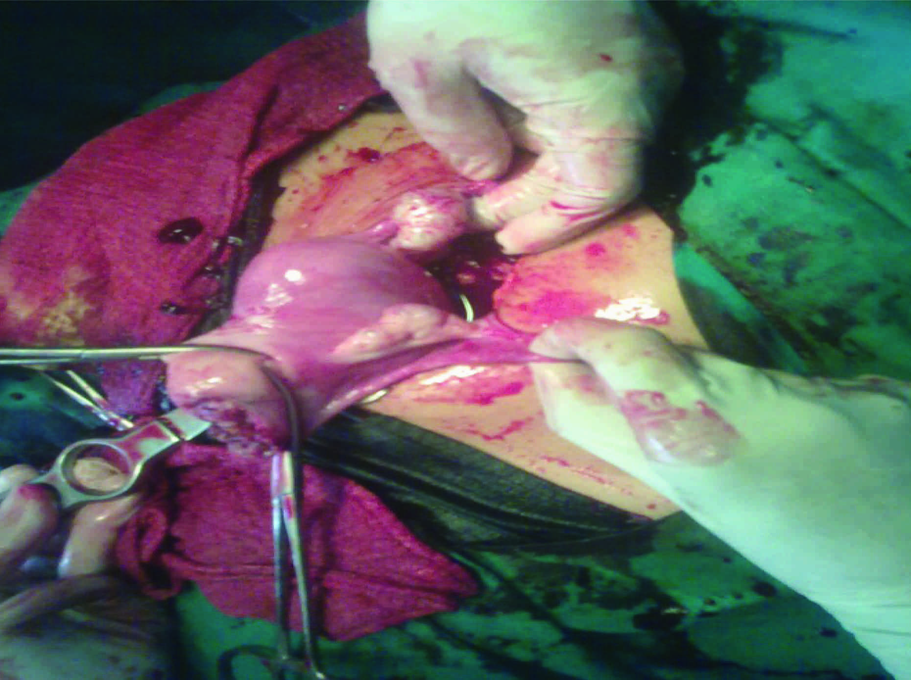
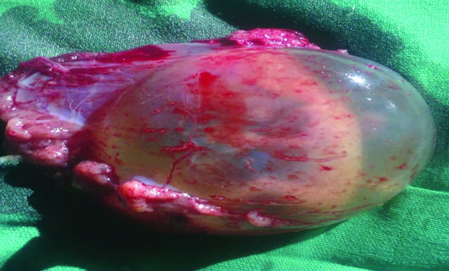
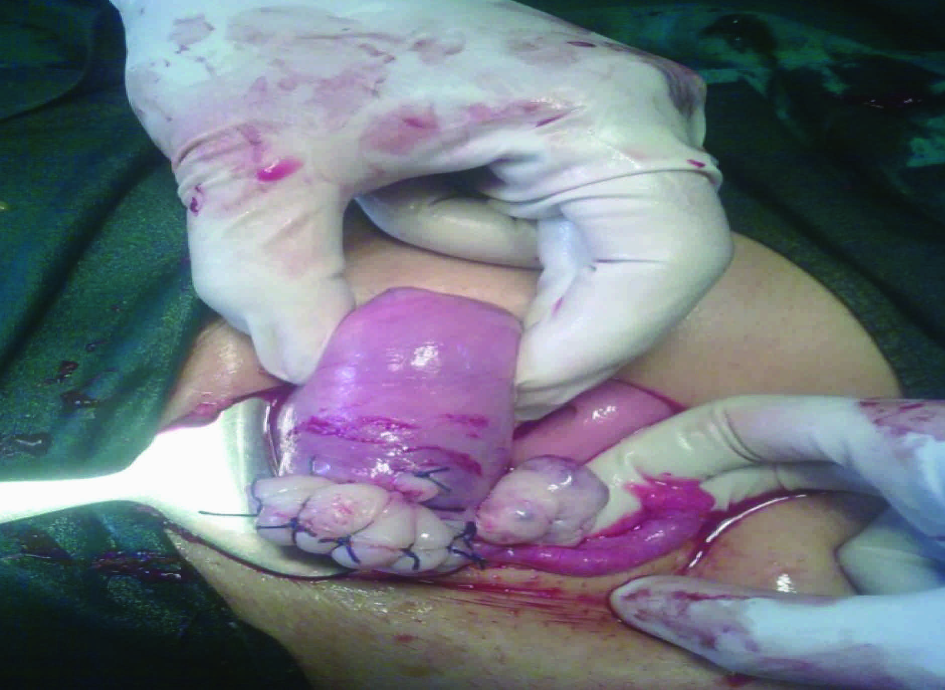
A provisional diagnosis of a ruptured ectopic pregnancy was made, two wide bore intravenous accesses were made and compatible whole blood and fluid resuscitations were started. The patient’s consent taken for an emergency exploratory laparotomy and it revealed a massive haemoperitoneum with a 10 week old foetus with an intact gestational sac in the peritoneal cavity and with a ruptured left cornual pregnancy [Table/Fig-1 and 2]. The site of the bleeding was first clamped and a cornual resection and repair was done [Table/Fig-3]. The postoperative period was uneventful. She was followed up after 1 month and she did not have any complaints. The patient’s consent and the institutional ethical board’s permission taken for the publication of this case report.
Showing the site of rupture

Showing gestational sac found in the peritoneal cavity

After repair of the left cornual ruptured site

Discussion
A cornual pregnancy is one of the most hazardous types of ectopic gestations. Cornual pregnancies account for 2 – 4% of the ectopic pregnancies or for 1 in 2500 – 5000 live births and they have a mortality rate of 2.0 – 2.5%. The mortality rate is 6 – 7 times higher than that in ectopic pregnancies [1].
A cornual pregnancy is a uterine but an ectopic pregnancy, as it is located outside the uterine cavity in that part of the fallopian tube that penetrates the muscular layer of the uterus. An interstitial pregnancy is sometimes used as a synonymous name. This part of the fallopian tube is 1 – 2 cm in length and 0.7 cm in width, which is supplied by Sampson’s artery, which is connected to both the ovarian and the uterine arteries. Cornual (interstitial) pregnancies can be confused with angular pregnancies; the latter, however, are located within the endometrial cavity, in the corner where the tube connects; typically those pregnancies are viable, although a high miscarriage rate has been reported [2].
A cornual pregnancy is a rare type of pregnancy and it is more dangerous than other types of ectopic pregnancies, as they tend to rupture later because of the myometrial stretch ability and with potentially devastating haemorrhages. 4 out of 11 deaths from ectopic pregnancies result from cornual ruptures. The risk factors include assisted reproductive techniques, previous tubal pregnancies, tubal surgeries, a history of pelvic inflammatory disease and sexually transmitted diseases [1].
The typical symptoms include abdominal pain and vaginal bleeding. Haemorrhagic shock is found in almost a quarter of the patients. This explains the relatively high mortality rate of cornual pregnancies [3]. An early diagnosis is important and today, it is facilitated by the use of ultrasonography and the quantitative HCG assay. The USG criteria for making a diagnosis includes:
An empty uterine cavity
A gestational sac which is separate from the uterine cavity
A myometrial thinning of less than 5mm around the gestational sac, typically the interstitial line sign-an echogenic line from the endometrial cavity to the corner which is next to the gestational mass is seen [4].
The paucity of the myometrium around the gestational sac is diagnostic, while, in contrast, an angular pregnancy has at least 5 mm of myometrium on all its sides [3]. The traditional treatment includes a surgical cornual resection. Sometimes, a hysterectomy is done due to the haemorrhage. Currently, a more conservative laparoscopic treatment and even a medical treatment can be accomplished with great success and with less unfavourable effects on the future pregnancies [4]. In the patients with asymptomatic interstitial (cornual) pregnancies, methotrexate has been successfully used. However, this approach may fail and it may result in a cornual rupture of the pregnancy [5]. A selective uterine artery embolisation has been successfully performed to treat these pregnancies [6].
Conclusion
A cornual pregnancy poses a significant diagnostic and a therapeutic challenge. It still remains the significant cause of the maternal death in women of the childbearing ages, despite the advances in both the diagnosis and the treatment. An appropriate individual counselling is needed regarding the risk of future pregnancies and the mode of delivery.
[1]. Tulandi T, AI-Jaroudi D, Interstitial pregnancy: results generated from the society of reproductive surgeons registryObstet Gynecol 2004 103:47-50. [Google Scholar]
[2]. Maowad NS, Mahajan ST, Moinz MD, Tayler SE, Hurd WW, Current diagnosis and treatment of interstitial pregnancyAmerican Journal Obstetrics and Gynaecology 2010 202:15-29. [Google Scholar]
[3]. Soriano D, Viscus D, Mashiach R, Schiff E, Seidman D, Goldenberg M, Laparoscopic treatment of corneal pregnancy: a series of 20 consecutive casesJournal of Reproductive Immunology 2008 90:839-43. [Google Scholar]
[4]. Radwan Faraj, Martin Steel, Management of cornual (interstitial) pregnancyRoyal College of Obstetricians and Gynaecologist 2007 9:249-55. [Google Scholar]
[5]. Jerny K, Thomas J, Doo A, Bourne T, The conservative treatment of interstitial pregnancyBJOG 2004 111:1283-88. [Google Scholar]
[6]. Dervelle P, Closset E, Management of interstitial pregnancy using selective uterine artery embolizationObstet and Gynecol 2006 107:427-28. [Google Scholar]