The infratemporal fossa is a surgically significant region, in which osseous part of pterygoid process extends vertically downwards from the junction of the root of greater wing and body of sphenoid. Each pterygoid process comprises of a lateral and a medial pterygoid plate. The lateral pterygoid plate consists of anterior and posterior margins. The free anterior margin forms the posterior boundary of pterygomaxillary fissure. The posterior free margin presents a small spur at the root or in the centre, which is called spine of civinini. Thickening of fascia between the lateral and medial pterygoid muscles leads to the formation of Pterygospinous ligament, which extends from the bony spur to spine of sphenoid. The ligament may ossify or calcify to form pterygospinous bar. The ossification may be complete or incomplete. The complete ossification of the bar results in an individual foramen which is called Foramen of civinini. This foramen is completed by an upward continuation of the interpterygoid fascia or aponeurosis, which is called cribiform fascia. This fascia is pierced by accessory meningeal artery, nerve to medial pterygoid and tensor velli palatine muscle. Sometimes, pterygoid venous plexus may pass through the pterygospinous ligament [1]. So, ossification of ligament may entrap the neurovascular structures and this may be an obstacle for the mandibular nerve block. An incomplete foramen exists because of partial ossification of ligaments.
Pterygospinous bar completely differs from the pterygoalar bar because of its attachments. Pterygoalar ligament extends from the root of lateral pterygoid plate to the infratemporal surface of greater wing of sphenoid. A complete foramen which is called porus crotaphitico-buccinatorius is formed from the complete ossification of pterygoalar ligament [2,3]. On the basis of attachments and passage of ligaments, either medial or lateral to foramen spinousm or ovale [4,5], serves to distinguish pterygospinous bar.
Material and Methods
A total number 160 cases of unknown age and sex which consisted of 50 dried skulls and 30 dried sphenoid bones of both the sides, were studied for the presence of Pterygospinous bar. Measurements were taken by using a sliding digital vernier caliper. X-rays were used as a tool in order to determine the view of foramen.
Results
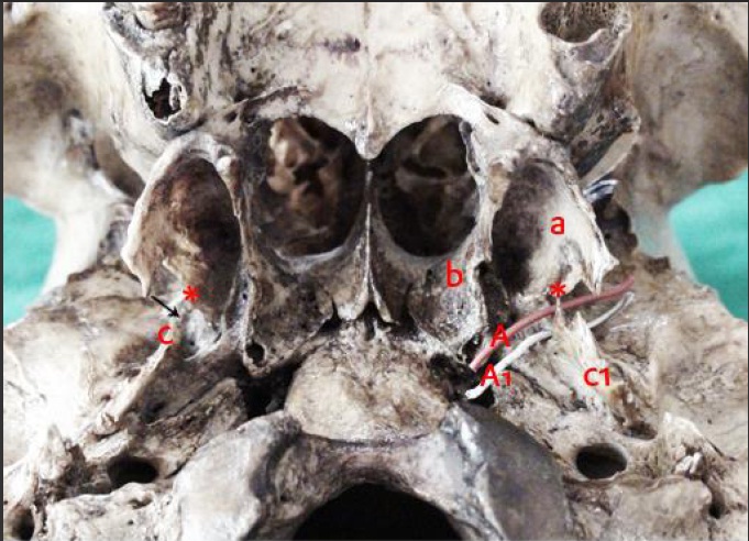
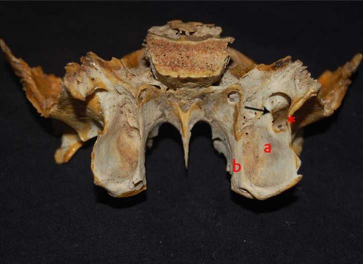
A bilateral incomplete foramen of civinini was noticed in three (6 cases) bones and a unilateral incomplete foramen of civinini was observed in four cases of bones, of which three were left sided and one was a right sided bone. Particularly, one of the skulls showed a right sided incomplete formen, a complete left sided one and an accessory foramen of civinini [Table/Fig-1]. An individual sphenoid bone showed a unilateral complete foramen of civinini on the right side and an incomplete foramen on the left side and it also represented the absence of foramen spinosum on right side [Table/Fig-2]. As a result, the complete ossified bar that formed a complete foramen of civinini was noticed in two (1.25%) cases, one on left side of the skull and other on right of the sphenoid bone. In both the cases, pterygospinous bar was placed across or inferiorly to the foramen ovale [Table/Fig-1 and 3] [6–9]. Incomplete foramen of civinini were noticed in 12 (7.5%) cases. The peculiar sphenoid and a skull bone which showed a larger area and accessory foramen of civinini were considered, to be discussed. In a sphenoid bone, the maximum width of lateral pterygoid plate which was measured at the level of spine of civinini and root was approximately 19.6mm and 17mm and the length was 12mm and area of foramen of civinini in a sphenoid bone was 94.2mm2. The transverse and vertical diameters were 10mm and 12mm [Table/Fig-4]. The length and width of the pterygospinous bar were 12mm and 3.5mm respectively. The same sphenoid bone also showed complete ossification of carotico-clinoid and interclinoid ligament, which was significant. On left sided skull bone, it was difficult to measure the length and width of pterygospinous bar and area of foramen, because the pterygospinous bar was not present in a single stretch, from one point to other. Rather, pterygospinous ligament may spread as a thin sheet from spine of sphenoid to spine of civinini. The sheet may be ossified around the perforated branches of mandibular nerve, resulting in one very small complete foramen and one accessory foramen of civinini in 0.625% of the cases.
Basal new - Left side complete & accessory foramen. Right side3 foramen a - Lateral pterygoid plate; b - Medial pterygoid plate; c - Spine of sphenoid (right side) cl - Spine of sphenoid (illdefined left side); A - Foramen of civiaini Al - Accessory foramen of civinini, * - Spine of civivnini Arrow shows incomplete Foramen of civinini
Poserior view - sphenoid bone exhibits incomplete foramen of leftside and complete foramen on right side a - Lateral pterygoid plate, b - Medial pterygoid plate, * - Pterygospinous bar Arrow shows foramen of civinini
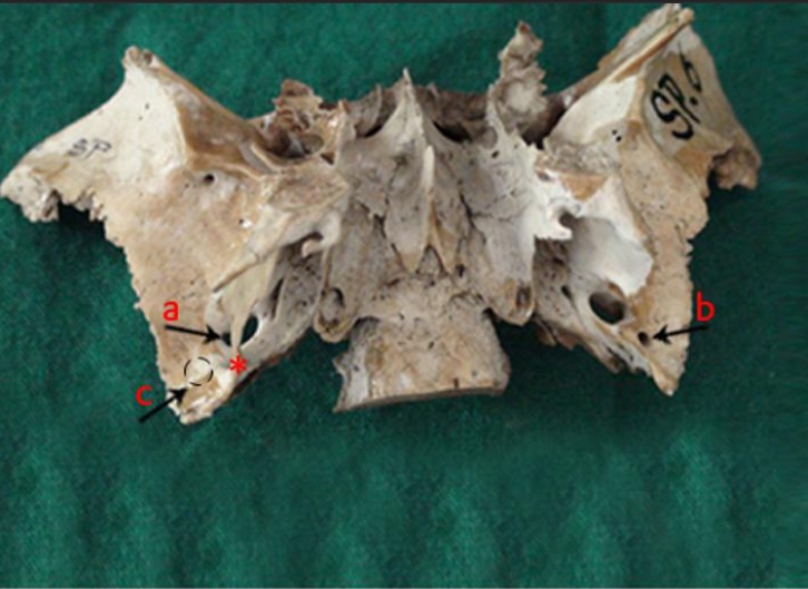
Basal view PSB inferiority across the foramen ovale a - Foramen ovale; b - Foramen spinosum; c- Absence of foramen spinosum; * - Pterygospinous bar
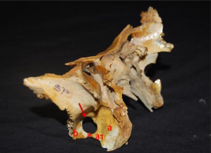
Lateral view, sphenoid bone with large area of foramen of civinini (right side) a - Lateral pterygoid; al - Spine of civinini; b - Spine of sphenoid. * - Pterygospinous bar; Arrow shows large area of foramen of civinini
Discussion
The thickened cranial part of fascia, between the lateral and medial pterygoid muscles, results in fibrous bands which are named as pterygospinous ligaments. Italian anatomist F Civinini (1805-1844), first described and coined the term “pterygospinous ligament” or “ligament of civinini, but earlier, Pterygospinous bar used to be named as ala Ingrassiae, after its invertor GF Ingrassia (1510-1580), who is also known as “Hippocrates of Sicily”. Ossified ligament projects as a bar, medial or lateral or sometimes across the foramen ovale in the submento vertical projection and it may interfere with percutaneous injection of the mandibular nerve. Pterygospinous bar passed medial to foramen spinosum and crossed the foramen ovale at an angle of 20° – 40° degree to the sagittal plane [9]. In the present report, the complete pterygospinous bar was parallel across the foramen ovale, with the absence of foramen spinosum in an individual sphenoid bone. A foramen which is created between the base of skull and the complete ossified bar transmits neurovascular structures of the medial pterygoid muscles [6,9]. The size of the foramen varies from 2 to 12 mm [9]. In this study, the maximum transverse and vertical diameters of the complete foramen on right side of the individual sphenoid bone were approximately 10mm and 12mm.
Von Ludinghansen et al., studied the pterygospinous bar in 100 human dried skulls from Japan and in 54 cadaveric cases from German and they noticed complete osseous bar in 6 of the human dry skulls, Pterygospinous ligament in 11 (20.4%) cases, pterygospinous muscle in 5 (9.2%) cases, which gets inserted into medial wall capsule and the articular disc of temporo – mandibular joint and is considered to be the third head of lateral pterygoid muscle. In a cadaveric study, coexistence of pterygospinous bar with pterygospinous muscle and pterygospinous ligament with pterygospinous muscle was observed. The existence of such muscle or fascia which accompanied the pterygospinous ligament has been described earlier by Testut and Latarjet [10]. Nathan et al., specified that pterygospinous muscle, as an atavistic remnant of one of the many pterygoid muscles, was present in reptiles. Among 50 Greek dry skulls, complete ossifications were found in only one skull bilaterally and incomplete ossifications were found in 25 cases [11]. Among 154 adult human dry skulls, only one foramen of civinini with complete ossification of pterygospinous bar was reported [12]. In a series of 1000 skulls, the incidence of complete ossification was noted to be 4.3% [9]. The percentage of complete ossification of pterygospinous bar was less as compared to that in this study. A higher percentage of ossification of pterygospinous bar was noticed in the following reports. A radiographic study which was done on 93 dry skulls from Brazil, detected the presence of pterygospinous ligament in 27.97% skulls, of which 19.36% were partially ossified and 8.61% were completely ossified [13]. Among the 452 dried skulls of the Anatolians population, complete pterygospinous osseous bridges were found in 8.8% [14]. A total of 416 skulls of Indian origin showed that pterygospinous bar was found in 9.61%, of which 5.76% was complete and 3.84% was incomplete. The pterygospinous ligament was completely ossified in 3% and there was a partial ossification in 8% of the dry skulls [9]. A wide survey which was done on 454 skulls from different racial groups showed the presence of pterygospinous foramen in 10% of the individuals [15]. When age factor was considered, complete or incomplete ossification of pterygospinous bar was noticed in the age group of more than 40-50 years, in 1.31% and 3.93% from 305 skulls from Croats respectively [16]. In 1999, Krmpotic stated that in specimens from younger individuals, the pterygospinous foramen couldn’t be found [17], but in contrary, pterygospinous bar was observed in the skulls of younger individuals [10,11]. Ps foramen was found in 5.46% of 2,745 skulls of American and Negroes and it was more frequent in whites [7]. Among 312 dry human skulls from major Brazilians, Ivan et al., classified the complete and incomplete ossification of pterygospinous ligament as types 1 (1.6%) and 2 and notified that the maximum diameter of the foramen of civinini was 10.626mm [18]. Group of basal cranial studies for the presence of pterygospinous bar from various regions like Europe, North pacific coast and Japan projected 4.5%, 5.9% and 4% [19, 20]. Complete ossification of pterygospinous bar was found to be 3-4% in the skulls of sociopaths and criminals [21], to be 2-7% in Caucasians and to be 12-13% in African skulls [22]. On considering the number of bones or cadavers, prevalence and incidence outcomes from the study were found to vary with respect to the races. There were reports that Ossification of the pterygospinous ligament was genetically controlled and that it showed a racial variation in frequency [23].
Tubbs et al., stated that the area of complete foramen of civinini was 16.72mm [21]. In this present study, maximum area of foramen civinini was found to be 94.2mm2 respectively. The width of pterygospinous bar was 4.5mm and its length (AP) was 11mm [24, 25]. In our study, the maximum width of the pterygospinous bar was 3.5mm and its length was 12mm respectively. The maximum width of lateral pterygoid plate from the spine of civinini was 19.6mm, which was 0.6 mm higher than that which was reported from studies which were done on 50 bones [26]. 21% skulls from various regions showed the width of lateral pterygoid plate to be higher than 20mm [4]. In this present case, the gap between the projections of spine of civinini and spine of sphenoid from the right sided skull was 0.3mm, whereas in the previous reports, it was noted as 1mm [26] and 3mm [27].
There are wide variations in the mammals with respect to the development and disposition of the lateral and medial pterygoid process [23]. The wider lateral pterygoid plates should be considered for the possible embracement of neurovascular structures and they may difficult for the administration of mandibular anaesthesia. A variable ossification at the posterior border of lateral pterygoid plate may be an obstacle for conductive anaesthesia on mandibular nerve via sub zygomatic route [16]. The lingual nerve and the inferior alveolar nerve are forced to take a long curved course in presence of a large pterygoid plate and the mandibular nerve gets fixed between the foramen ovale and mandibular foramen. So, during contraction of pterygoid muscles, there may be pain, that may lead to trigeminal neuralgia. Similar symptoms could be provoked by the foramen of civinini, since it lies across the foramen ovale. A bilateral foramen was noticed in 2 skulls and three were unilateral [17].
The area and the vertical and transverse diameters of the foramen civinini and the length and width of the pterygospinous bar in this present study were clinically much significant . Because among the 160 cases, a sphenoid bone showed one complete foramen with a large area and one accessory foramen civinini was noted in skull unilaterally.
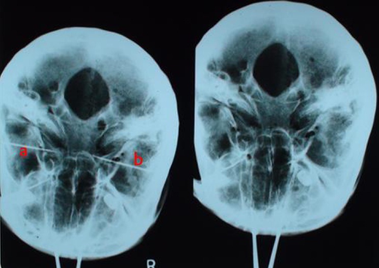
Pterygospinous bar was clearly notable in normal basal view (mentocoronal) and described its applied aspects and observed unilateral PSB in 7.05% and bilateral in 0.89% of cases [28]. Pterygospinous bar is visible in the basal view as a 1 to 2mm [9]. In a study of 50 sphenoid bones, unilateral incomplete foramen of pterygospinous bar was seen in oblique view of skiagram [25]. Hirtz axial or submentovertex technique is an excellent tool for the observation of a complete or an incomplete ossification of pterygospinous ligaments [13]. Pterygospinous bar also is visible on Panorax and Trans maxillary views [9]. Similarly, in this study, it was noted that normal basal view [Table/Fig- 5] and lateral radiological view demonstrated the pterygospinous bar and foramen clearly. In radiographic anatomic picture, the existence of foramen of civinini or a complete pterygospinous bar may superimpose the Para and retro maxillary space. Pterygospinous ligaments can be an obstacle in a radiographically guided trigeminal ganglion blockage [9–11, 15].
Skiagrtam - Basal view a - complete foramen of civini; b - Incomplete foramen of civinini Basal view clearly shows pterygospinous bar on left side
As the pterygospinous bar is more medially oriented, it doesn’t compromise the foramen ovale and therefore, it has no clinical significance [29]. Only pterygoalar bar interfere with injection of mandibular nerve. Contrary to this, the literature has proved that Pterygospinous bar is clinically significant. Developmental basis for the formation of the ligaments was described by James [2,30]. Possibilities of entrapment or compression of lingual nerve from the ossified pterygospinous ligament or between the ossified ligament and medial pterygoid muscle have been reported [31,32]. Chorda tympani branch of facial nerve may also get compressed by an ossified ligament [26] and it may result in impairment of taste sensation to the anterior two thirds of tongue. The course of the branches of mandibular nerve and maxillary artery may change during development, because of the hindrance in development of ligament or bar and even the branches of mandibular nerve, that innervate the muscles of mastication, also get compressed or a difficulty in approaching these structures is highly accidental. The presence of ossified pterygospinous ligament may cause trouble in performing thermo coagulation and anaesthesia for trigeminal neuralgia and it may cause a surgical difficulty in a lateral transzygomatic infratemporal fossa approach to the para and retro pharyngeal space [10,15].
Entrapment and compression of lingual nerve may occur due to one of the following causes: partial or complete ossification of pterygospinous ligaments. Pterygospinous bar ridge separates the trunk of lingual into anterior and posterior branches. Anterior fibres may get compressed because of their course between the tensor veli palatini muscle and the bony ridge [31] or because of a large lateral pterygoid plate [32–34].
Existence of osseous bar between the lateral pterygoid plate and
the spine of sphenoid has been considered as a phylogenetic remnant in human beings. A wide pterygospinous bar was noted in all the skulls of herbivores, carnivores and old world monkeys and a small pterygospinous bar was noted in rodents but never in new world monkeys. So, in humans, this pterygospinous bar represents a phylogenetic remnant [10]. In lemurs, the bar passes medial to foramen ovale, but in pithecoid condition, the pterygospinous bar is complete and it passes lateral to foramen ovale. In man and anthropoids, the pterygospinous bar is incomplete. If it is present in varying degrees of completion, it forms porus Crotaphiticobuccinatorius & andforamen of civinini [35]. In 100 Hawaiian skulls, 8% showed the unilateral completion of pterygospinous bar, which passed medial to foramen ovale and in no case was it bilateral [23].
In platyrrhines, a small spine, the spina civinini, projects from the middle portion of the posterior border of the short lateral pterygoid plate. A wide semilunar space, the insicura civinini, is present between the plate and anterior border of auditory bulla. In Aotus, an incomplete or a complete ossification of the ligamentum pterygospinous bridges the gap between the spina civinini and the posteroventral process of the sphenoid bone. In tarsius, lemurids and galagids, the plate is long and widely expanded, and the posterior border has intimate contact with the outer wall of the auditory bulla. In cercopithecoid condition, the ossification of the ligament may be complete and also in colobines, the foramen of civinini is usually established and it serves as a passage for the vessels, which include the internal pterygoid nerve. but In pongids, spine of civinini is well defined, insicura civinini is well opened and the sphenoidal spine for attachment of ligamentum pterygospinosum is little or only moderately developed. In humans, lateral pterygoid plate resembles pongids, but spine of sphenoid is well developed [36].
Conclusion
Various studies have stressed about the significance of this pterygospinous bar. The present study report on occurrence of larger area and accessory foramen of civinini may be the additional variations which have to be noted. So, the presence of an anatomical rare variant of pterygospinous ligament or bar or muscle, inadvertently affects the neurovascular structures of the foramen ovale and causes a problem during anaesthesia or surgeries. Pterygoalar or pterygospinous bar is never a hindrance for injecting semilunar ganglion via inframandibular approach, but an ossified pterygoalar bar is truly significant in a supramandibular approach. Importance of the foramen of civinini and structures which pass through it, may guide the dental and maxillofacial surgeons in doing safe and effective procedures. From the present study, it is concluded that when the area is more, the risk of the entrapment is less, as compared to the smaller foramen, where the risk is higher. But in both the circumstances, a hindrance against mandibular anaesthesia is common.