A Study on the Nutrient Foramina of Adult Humerii
Shanta Chandrasekaran1, K.C. Shanthi2
1 Associate Professor, Department of Anatomy, Vinayaka Missions Kirupananda Variyar Medical College, Salem, Tamil Nadu, India.
2 Associate Professor, Department of Anatomy, Vinayaka Missions Kirupananda Variyar Medical College, Salem, Tamil Nadu, India.
NAME, ADDRESS, E-MAIL ID OF THE CORRESPONDING AUTHOR: Dr. Shanta Chandrasekaran, Associate Professor, Department of Anatomy, Vinayaka Missions Kirupananda Variyar Medical College, Salem, Tamil Nadu, India.
Phone: 09585463102
E-mail: drshantasekaran@yahoo.co.in
Background: It is not uncommon to see the non union of the fractures in the long bones. Among the various reasons for the nonunion of the fractured bones, the nutrient artery plays an important role.
Objectives: This study was aimed at analyzing the nutrient foramina in dry adult humerii ,with regards to the number, size and the location of the nutrient foramen with respect to the surfaces and the zones and its distance from the mid point of the humerus.
Materials: This was an analytical study. All the humerii from various medical colleges of the Vinayaka Missions University in Salem were included in the study. Those which were damaged and those which had pathological abnormalities were excluded. The following parameters were noted; namely, the length of the humerus, the number and the size of the nutrient foramen and the location of the nutrient foramen with respect to the surfaces and the zones of the humerus and its distance from the mid-point of the humerus were analyzed. The length of the humerus and the mid point of the humerus were measured by using the osteometric board, whereas the size of the nutrient foramen was measured by using various size of hypodermic needles. When more than one foramen was found, the larger one was taken as the dominant foramen and its size was measured. All the data were noted and the statistical analysis was done by calculating the mean, the range and the standard deviation.
Results: Totally, 258 adult dry humerii were studied. The mean length of the humerii was 27.96 cm, with a SD of 2.18. The mean size of the nutrient foramen was 0.828 mm, with a SD of 0.26. The mean distance of th dominant nutrient foramen from the mid point of the humerus was 2.31 cm, with a SD of 1.25 cm. In majority of the humerii (86.43%), the nutrient foramen was located in the middle 1/3rd of the bone and in 13.57% of the bones, it was located in the lower 1/3rd of the bone. The location of the nutrient foramen in the anteromedial surface was 89.92%, that in the posterior surface was 8.53% and that in the anterolateral surface was 1.55%.
Nutrient foramen, Humerus, Nutrient artery, Humerus fracture
INTRODUCTION
The fractures of the long bones are increasing in numbers, due to an increase in the numbers of industrial and road traffic accidents, sports injuries and pathological fractures in osteoporotic victims. The non union of a fractured bone can be a complication of a closed or an open reduction.
When the blood supply is not established well, it can be complicated by a delayed union or a nonunion of the fracture and this reveals that the medullary arterial system plays an important role in the revascularization of the necrozing cortex and the uniting callus of the fracture site [1].
On this basis, having knowledge on the location of the nutrient foramen and the relevant anatomy, the surgeon can prevent a damage to the nutrient artery and can minimize the formation of a delayed union or a non union of the fracture [2]. Hence, this study was aimed at analyzing the nutrient foramen of the humerus.
OBJECTIVES
This study was aimed at analyzing the nutrient foramen in dry adult humerii, with regards to the number, size and the location of the nutrient foramen with respect to the surfaces and zones and its distance from the mid point of the humerus.
MATERIALS AND METHODS
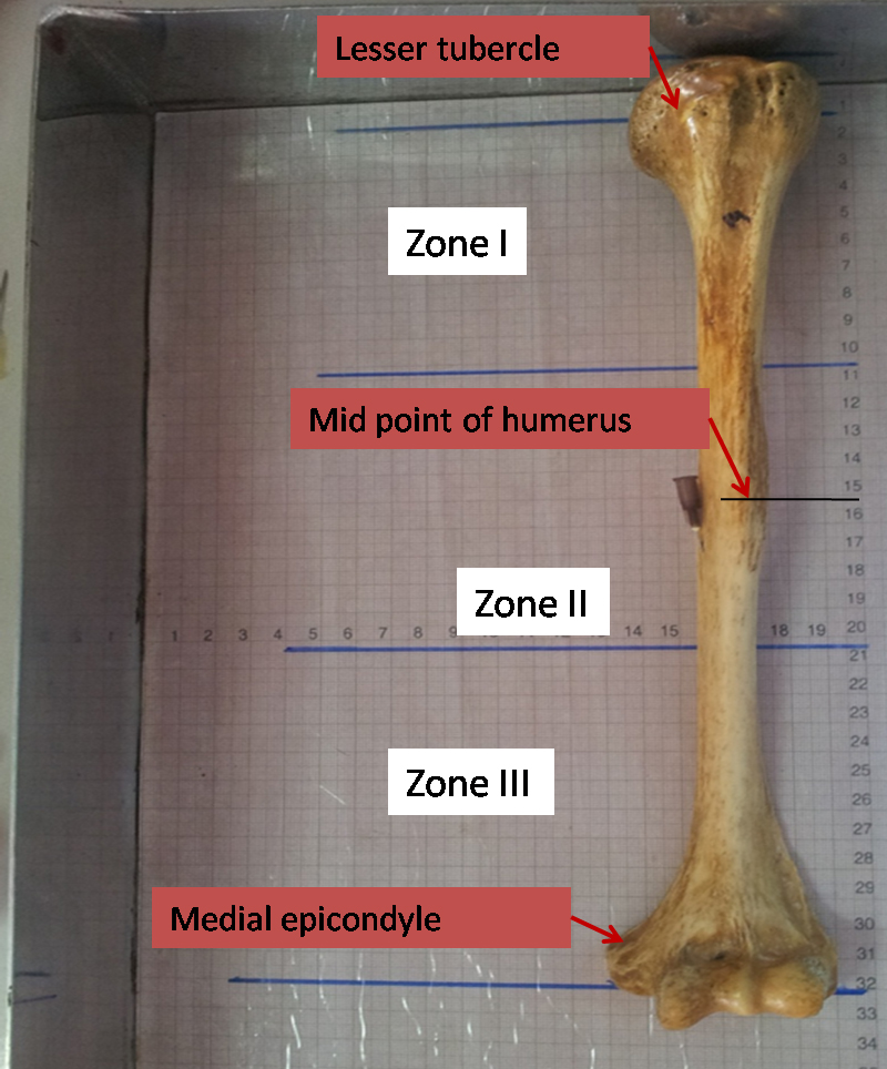
All the humerii from various medical colleges of the Vinayaka Missions University in Salem, namely, Vinayaka Missions Kirupananda Variyar Medical College, Annapoorna Medical College, and Vinayaka Missions Homeopathy Medical College, were included in the study. The bones which were damaged and those which had pathological abnormalities were excluded. All the adult humerii which were not necessarily paired and those of unknown age and sex were examined. The following parameters were noted; namely, the length of the humerus, the number of the nutrient foramen, the size of the nutrient foramen and the location of the nutrient foramen with respect to the surfaces and the zones and the distance of the nutrient foramen from the mid-point of the humerus were analyzed. The length of each humerus bone was measured from the superior aspect of the lesser tubercle to the inferior surface of the medial epicondyle of the humerus by using an osteometric board [2]. Further, the humerus bone was divided into three equal zones, as zone I (upper 1/3rd), zone II (middle 1/3rd) and zone III (lower 1/3rd) [Table/Fig-1]. The midpoint of the humerus was calculated by dividing the length of the humerus by two and the same was identified and marked on the humerus with the help of the osteometric board. The location of the nutrient foramen was noted with respect to the three surfaces, namely, the anteromedial, the anterolateral and the posterior and the three zones, namely, zone I, zone II and zone III. The distance of the nutrient foramen from the midpoint of the humerus was measured by using a vernier caliper. The size of the nutrient foramen was determined by using hypodermic needles of various sizes, which ranged from 18 Gauge to 26 Gauge, of known diameter s (18 Gauge =1.2 mm, 20 Gauge = 0.9mm, 24Gauge = 0.55mm and 26 Gauge = 0.45mm). When more than one foramen was found, the larger nutrient foramen was considered as the dominant foramen and its size was measured. All the data were noted and the statistical analysis was done by calculating the percentage, mean, range and the standard deviation.
Measuring the length of humerus using osteometric board and dividing into zones - (I, II and III)
RESULTS
Totally, 258 adult dry humerii were studied, of which 51.16% were left sided bones and 48.84% were right sided bones. The mean length of the humerii was 27.96 cm, with a SD of 2.18, which ranged from 18 to 33 cm. The median length was 28cm. Though some humerii had two or more nutrient foramina, a majority of the humerii had a single nutrient foramen [Table/Fig-2].
Number of nutrient foramen in humerii (n=258)
| Number of foramen | Number of humerii | Percentage |
|---|
| 1 | 198 | 76.74% |
| 2 | 53 | 20.54% |
| 3 | 07 | 02.71% |
The sizes of the foramina ranged from 0.45 to 1.2 mm, with a mean of 0.828 mm and a SD of 0.26. The distance of the dominant nutrient foramen from the mid point of the humerus ranged from 0.5- 8 cm, with a mean distance of 2.31 cm and a SD of 1.25 cm. 88.76% of the nutrient foramina were located above the mid points of the humerii and 11.24% were located below the mid points of the humerii. A majority of the nutrient foramina were located in the anteromedial surfaces of the humerii. [Table/Fig-3] Similarly, a majority of the humerii had the nutrient foramina in zone II (middle 1/3) of the bone [Table/Fig-4].
Location of number of nutrient foramen with regard to surfaces of humerus ( n-258)
| Location of nutrient foramen | Number of humerii | Percentage |
|---|
| Anteromedial surface of humerus | 232 | 89.92% |
| Anterolateral surface of humerus | 4 | 01.55% |
| Posterior surface of humerus | 22 | 08.53% |
Location of number of nutrient foramen with regard to zones of humerus ( n-258)
| Number of foramen | Number of humerii | Percentage |
|---|
| Zone-I | 0 | 0% |
| Zone-II | 223 | 86.43% |
| Zone-III | 35 | 13.57% |
DISCUSSION
The blood supply is the main factor in the healing of fractures [3–5]. Any damage to the nutrient artery during surgical fixations or subsequent manipulations, is a significant factor which predisposes to delayed unions or nonunions [6–10]. The humerus is supplied by the nutrient artery, the metaphyseal artery and the periosteal vessels from the axillary and the brachial arteries and their branches. The periosteal and the metaphyseal arteries supply the outer cortex and the metaphysis of the bone, but the inner half of the cortex and the medulla of the shaft are predominantly dependent on the nutrient artery. The study on the blood supply of the shaft will help in knowing about the healing of fractures, delayed unions and non unions of the bone following fractures and bone transplants [11]. Laing studied the vascularity of the humerus and he opined that the main nutrient artery of the humerus must be protected from injuries during operations which are done on the humeral shaft [6]. Carroll stated that the nutrient artery enters through the restricted anteromedial surface, in the middle 1/3rd of the humerus and that the surgeries which are done on the middle 1/3rd of the shaft of the humerus should be handled well without causing damage to the nutrient foramen, in order to prevent delayed unions or non unions of the fractures. Our study results correlated well with those of Carroll's study [2]. Many studies which have been done on the humerus bone have revealed that a majority of the humerii had one nutrient artery and that some had additional accessory arteries. Our study results correlated well with Manjunath S Halagatti's and Pramod's study results and they differed from those of others. This could be because of the variation in the population [Table/Fig-5] [2,6,12–13]. There is no correlation between the length of the humerus and the number of the nutrient foramina according to the studies which were done by Manjunath S Halagatti and Chhatrapati DN [13,14]. Hence, we did not attempt to correlate the same. This study adds a message to the existing knowledge, that a variation in the population can result in a variation in the number of nutrient foramina.
Comparison of various studies with regard to number of nutrient foramina in humerus
| S.E. Carroll | Manjunath & Pramod | Hamang Joshi | P.G. Laing | Percent Study |
|---|
| Number of humerii analysed | 71 | 200 | 200 | 30 | 258 |
| Humerus with single nutrient foramen | 48(68%) | 161(80.5%) | 126(63%) | 28(93%) | 198 (76.74%) |
| Humerus with double nutrient foramina | 20(28%) | 35(17.5%) | 66(33%) | 02(07%) | 53 (20.54%) |
| Humerus with three nutrient foramina | 03(04%) | 4(2%) | 08(04%) | ---- | 07 (02.71%) |
CONCLUSIONS
This study concludes that the nutrient foramina of the humerii were not only located on the anteromedial surfaces but also on the anterolateral and the posterior surfaces. Similarly, the nutrient foramen of the humerus was found on both the middle and the lower third of the shaft. A majority of the humerii had a single nutrient foramen, though some humerii had more than one nutrient foramen.
[1]. Rhinelander FW, The normal microcirculation of diaphyseal cortex and its response to fractureJournal of Bone and Joint Surgery 1927 50A:643-62. [Google Scholar]
[2]. Carroll SE, A Study of Nutrient Foramina of the Humeral DiaphysisThe Journal of Bone and Joint Surgery 1963 45B:176-81. [Google Scholar]
[3]. Williams Peter L, Bannister Dyson, Gray's Textbook of Anatomy 1993 37th editionLondonElsevier Churchill Livingstone:299-300.:759 [Google Scholar]
[4]. Johnson RW, A Physiological study of the Blood Supply of the DiaphysisJournal of Bone & Joint Surgery 1927 9:153 [Google Scholar]
[5]. Coolbaugh CC, Effects of Reduced Blood Supply of boneAmerican Journal of Physiology 1952 169:26 [Google Scholar]
[6]. Laing PG, The Arterial supply of Adult HumerusThe Journal of Bone and Joint Surgery 1956 38A:1105-16. [Google Scholar]
[7]. Mercer Sir W, Orthopaedic Surgery 1959 5th editionLondonEdward Arnold Ltd. [Google Scholar]
[8]. Stewart MJ, Hundley J M, Fractures of the Humerus; a Comparative Study in Methods of TreatmentJournal of Bone and Joint Surgery 1955 37-A:681 [Google Scholar]
[9]. Kennedy JC, Wyatt JK, An Evaluation of the Management of Fractures through the Middle Third of the HumerusCanadian Journal of Surgery 1957 1:26 [Google Scholar]
[10]. Watson-Jones Sir R, Fractures and Joint Injuries 1955 4th EditionEdinburgh and LondonE. and S. Livingstone Ltd [Google Scholar]
[11]. Robert WA, A physiological study of the blood supply of the diaphysisJ Bone Joint surg 1927 9:153-54. [Google Scholar]
[12]. Joshi Hemang, Doshi Bhavik, Malukar Ojaswini, A Study of the Nutrient Foramina NJIRM 2011 2(2):14-17. [Google Scholar]
[13]. Halagatti Manjunath S, Rangasubhe Pramod, A study of nutrient foramina in dry adult humerii of south Indian subjectsNational Journal of Clinical Anatomy 2011 1(2):76-80. [Google Scholar]
[14]. Chattrapati DN, Mishra BD, Text book of Anatomy 1984 244th edPhiladelphiaHarper and Row publishers:171-74. [Google Scholar]