Breast cancer is a malignant disease with a heterogenous prognosis. Evaluation of the possible prognostic parameters is a growing interest. Assessment of the biological aggressiveness of the cancer without removing it, would be valuable.
FNAB is not only helpful in the diagnosis and the further planning of the treatment, but it is also helpful in the prognostification of the tumour factors like the nuclear grading, the mitotic index, the hormone receptor status and the DNA contents [4]. Nuclear grading is an important prognostic factor [6].
Here, an attempt was made to study the accuracy of the grading and typing of the fine needle aspiration cytology of malignant breast lesions with histopathological correlations and to study the lymph node statuses in various cytological grades of malignant breast lesions.
MATERIAL AND METHODS
This was a retrospective and a prospective study which was done from January 2004 to December 2007 of 60 cases of cytologically suspicious and malignant breast lesions which had histopathological correlations. These cases were selected from the Department of Pathology, Kempegowda Institute of Medical Science, Bangalore, Karnataka, India. The cases which were diagnosed as benign lesions on cytology were excluded from the study. The cytological and histopathological slides of few retrospective cases were retrieved.
Fine needle aspiration of breast lumps was performed with 22 gauge needles which were attached to 10ml syringes. The samples which were obtained were smeared onto glass slides and fixed in 95% ethyl alcohol. They were stained by the Papanicoloau [7] and the haemotoxylin-eosin (H and E) method.
The cytological grading was done by Robinsons' method, in which the cell dissociation, nuclear size, cell uniformity, nucleoli, nuclear margins and the chromatin patterns were studied [8, 9]. A value which was between 1 and 3 were given to each factor which was analyzed. The scores of each of the 6 cytological features were added together to give a total score for each case. In each case, the final score ranged between 6 and 18. The scores were added and the grading was done. Grade I: 6-11, Grade II: 12-14 and Grade III: 15-18. The cytological typing was also done.
The subsequent mastectomy specimens which were received were adequately fixed in 10% formalin. Grossing was done and the tissues were sampled to include the nipple and areola, the tumour proper, the deep surgical margins and all the lymph nodes that were procured from the specimens. The tissues were processed and the blocked sections were subsequently stained with the H and E stain.
The histopathological typing was done and the grading was done by the modified Scarff Bloom Richardson criteria, taking into consideration, the tubule formation, the nuclear pleomorphism and the mitotic count [10–12].
The statistical analysis was done with the SPSS software in which the Chi-square test was used and a contingency tale analysis (Cross Tabulation Procedure) was done.
RESULTS
The aspirated samples were graded according to Robinson categories and the following results were obtained:
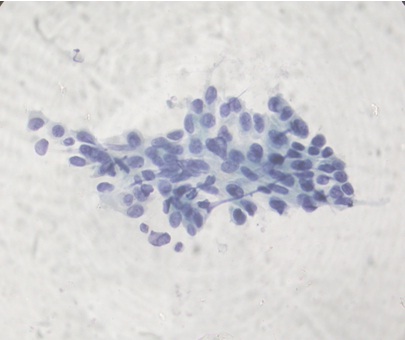
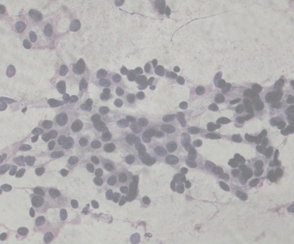
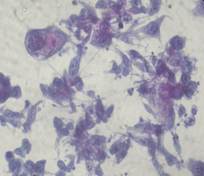
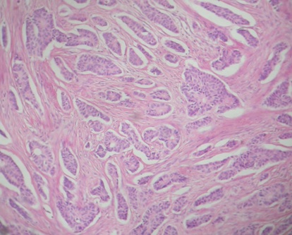
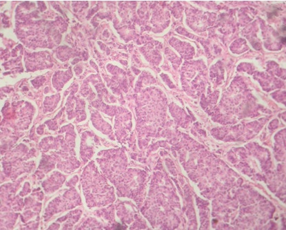
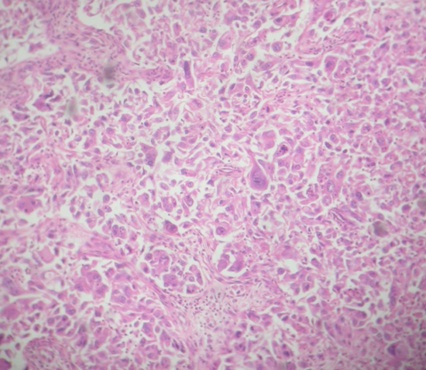
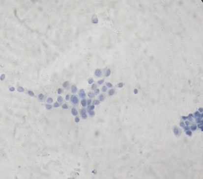
In the present study, 13 cases (22%) were grade 1 [Table/Fig-1], 16 cases (27.1%) were grade 2 [Table/Fig-2] and 30 cases (50.8%) were grade 3 [Table/Fig-3]. The histopathological grading was done by the modified Scarff Bloom Richardson grading method. 7 cases (11.9%) were grade 1 [Table/Fig-4], 21 cases (35.6%) were grade 2 [Table/Fig-5] and 31 cases (52.5%) were grade 3 [Table/Fig-6]. The grading of one case of malignant phyllodes was not done, as the Scarff Bloom Richardson grading was done only for adenocarcinomas. Correlation of both the systems was done and the statistical value was calculated.
Cytological Grade 1 (Papanicolaou Stain x 400)
Cytological Grade 2 (Papanicolaou Stain x 400)
Cytological Grade 3 (Papanicolaou Stain x 400)
Histological Grade 1 (H&E x 100)
Histological Grade 2 (H&E x 100)
Histological grade-3 (H and E x 100)
[Table/Fig-7] demonstrates the relationship between the cytological grade and the histological grade. The cytohistological grading correlation was accurate in 7 cases (100%) of grade 1, in 22 cases (71%) of grade 3 and in 9 cases (42.9%) of grade 2. The histological grade correlated positively with the cytological grade.
Showing Correlation of Cytological and Histopathological Grading in Present Study
| Cytological grading | Histopathological grading | Total |
|---|
| 1 | 2 | 3 | |
| 1 | 7 (100%) | 4 (19%) | 2 (6.5%) | 13 (22%) |
| 2 | | 9 (42.9%) | 7 (22.6%) | 16 (27.1%) |
| 3 | | 8 (38.1%) | 22 (71%) | 30 (50.8%) |
| Total | 7 | 21 | 31 | 59 |
The contingency co-efficient (CC) was 0.602, the x2 test P value was < 0.001 and the sensitivity was 98.3%. The specificity could not be calculated as only positive cases were taken up for study. In the present study, 33 cases (56.7%) has metastatic deposits in the lymph nodes. 14 cases (23.3%) were reactive and 12 cases (20%) had no metastases.
[Table/Fig-8] shows the relationship between the cytological grade and the nodal metastasis. CC = 0.399, P < 0.006. A higher cytological grade was associated with a nodal metastasis (Lumpectomies and quadrantectomy cases were not included).
Showing Cytological Grade and Nodal Metastasis
| Robinson grade | No metastasis | Metastasis | Total cases |
|---|
| 1 | 7 (53.9%) | 6 (46.2%) | 13 |
| 2 | 0 | 14 (100%) | 14 |
| 3 | 12 (42.9%) | 16 (57.1%) | 28 |
| Total | 19 (34.5%) | 36 (65.5%) | 55 |
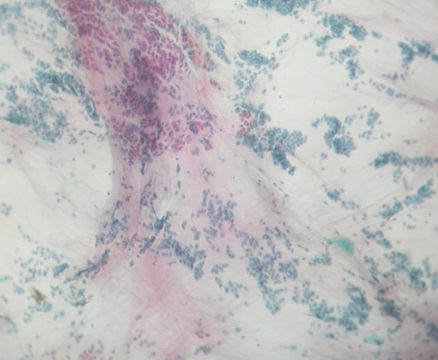
In the present study, the cytological typing was accurate in 44 cases out of the 60 cases [Table/Fig-9]. The cytological typing was accurate in 40 cases (78.4%) of IDC, 2 cases of lobular carcinomas (66.6%) [Table/Fig-10], in 1 case of mucinous carcinoma (100%) [Table/Fig-11] and in 1 case of malignant phyllodes (100%).
Showing Correlation of Cytological Typing and Histological Typing
| Cytological diagnosis | Histopathological diagnosis | Total |
|---|
| IDC | ILC | Medullary carcinoma | Colloid carcino-ma | Malignant Phyllodes | Apoc-rine | |
| Suspicious | 11 (21.6%) | 1 (33.3%) | 1 (50%) | | | | 13 |
| IDC | 40 (78.4%) | | 1 (50%) | | | 2 (100%) | 43 |
| Lobular Carcinoma | | 2 (66.6%) | | | | | 2 |
| Colloid Carcinoma | | | | 1 (100%) | | | 1 |
| Phyllode | | | | | 1 (100%) | | 1 |
| 51 | 3 | 2 | 1 | 1 | 2 | 60 |
Smear Showing Infiltrating Lobular Carcinoma (Pap. Stain x 400)
Smear Showing Colloid Carcinoma (Pap. Stain x 100)
DISCUSSION
Aspiration cytology is the most reliable preoperative examination for the evaluation of palpable and nonpalpable breast lesions. But its utility in the grading of breast carcinomas has been largely underestimated. As with the histological grading, the grading on FNA would allow a prognostic evaluation in addition to the diagnosis, without any additional morbidity or expense to the patient. Several grading systems are being used, like Hunt's (1990), Simplified Black, Modified Black (1994), etc. The Robinson cytological grading system is mentioned in the guidelines which have been published following the Bethesda Conference at the National Institutes of Health [8, 9].
The nuclear grade is considered as one of the most important prognostic factors in breast cancer [13]. The inter observer and the intra observer reproducibilities are the key factors in any grading system [14]. Because the neoadjuvant therapy, which includes the preoperative chemotherapy and Tamoxifen, is becoming increasingly common for the treatment of early breast cancer, it is desirable to grade the tumours before surgery, so that the most appropriate medical regimens can be selected [15]. A cytological evaluation of the prognostic markers is important and it is useful in patients with inoperable tumours and in cases which are at a high risk for surgery. In patients with advanced stage breast cancers which require neoadjuvant radiotherapy or chemotherapy, a cytological evaluation of these prognostic markers can provide useful baseline data, as these parameters may be modified by the treatment [16]. It has been suggested that the faster growing tumours (grade 3) are more likely to respond to the chemotherapy than the low grade, slow growing lesions which may be better suited for the pretreatment with Tamoxifen [17].
In the present study, the cytological grading was done by using the Robinson's grading system. A majority of the tumours were grade 3 (50%), followed by grade 2 (27%) and grade 1 (22%). However, Taniguchi et al., [6] Robinson et al., [15], Meena et al., [4] and Frias et al., [9], observed a majority of grade 2 tumours, followed by grade 1 and grade 3 tumours. Jayaram et al., [6] observed a majority (64.5%) of grade 2 tumours, followed by 29% grade 3 and 6.4% grade 1 tumours.
The corresponding biopsy specimens were graded by Elston's modified Bloom and Richardson method [18]. The cytological grade correlated positively with the histological grade. The accuracy was 67.7%, p<0.001. A similar observation was made by Jayaram et al., (71%) [16]. However, Taniguchi et al., [6] reported an accuracy of 44%, Robinson et al., [15] reported that of 56.9% and Meena et al., [4] reported an 83% accuracy of the cytological grading. Therefore, the cytological grade is useful in predicting the histological grade preoperatively.
In the present study, a majority (56.7%) of the patients had metastatic deposits in the lymph nodes. Similar observations were made by Taxim et al., [19] (89%) and Chattopadhyay et al., [20] (70%). A higher grade was associated with a nodal metastasis (p<0.006, CC = 0.399). A similar observation was made by Frias et al., (p<0.0005). The cytological typing correlated in 44 cases (73%) out of the 60 cases. Tumour typing provides important prognostic information. Tubular carcinomas and medullary, papillary and sectretory carcinomas carry better prognoses [21].
In conclusion, this study showed that the cytological grade correlated well with the histological grade and that a higher grade was associated with a regional lymph node metastasis. Hence, the cytological typing and grading should be incorporated in the FNAC report and this can be of great value in guiding the choice of the treatment protocols.