Pulp Stone, Haemodialysis, End-stage Renal Disease, Carotid Atherosclerosis
Santosh Patil1, Nidhi Sinha2
1 Department of Oral medicine and radiology, Jodhpur Dental College, Jodhpur National University, Jodhpur, Rajasthan, India.
2 Department of Conservative Dentistry and Endodontics, Jodhpur Dental College, Jodhpur National University, Jodhpur, Rajasthan, India.
NAME, ADRESS, E-MAIL ID OF THE CORRESPONDING AUTHOR: Dr. Santosh Patil, Department of Oral Medicine and Radiology, Jodhpur Dental College, Jodhpur National University, Jodhpur, Rajasthan-342001, India.
E-mail: drpsantosh@gmail.com
Objectives: The aim of this study was to determine the relationship between the presence of pulp calcification and carotid artery calcification on the dental panoramic radiographs in End Stage Renal Disease (ESRD) patients who were on haemodialysis.
Methods: A total of 112 End Stage Renal Disease (ESRD) patients on who were haemodialysis participated in this study. The periapical and the panoramic radiographs for all the patients were evaluated for the presence or absence of the narrowing of the dental pulps and for pulp stones in the pulp chambers and the pulp canals. The panoramic radiographs were also evaluated to determine the carotid calcification.
Results: Carotid calcifications were detected in none of the patients. 84 (74.99%) patients had dental pulp narrowing, and 38 (33.92%) patients had pulp stones. There was no statistical correlation between pulp narrowing and Carotid Artery Calcification (CAC) in the haemodialysis patient group. There was also no statistical correlation between pulp stones and CAC in the haemodialysis patients.
Conclusion: However, the incidental finding of CAC on a panoramic radiograph can provide life-saving information for the vascular disease patients, but in the present study, no significant relationship was found between the presence of the pulpal calcification and CAC in the ESRD patients who were on haemodialysis. Therefore, the presence of pulp calcification does not seem to serve as a diagnostic marker for carotid atherosclerosis.
Pulp stone, Haemodialysis, End-stage renal disease, Carotid atherosclerosis
INTRODUCTION
Dental pulp calcification can occur as diffuse forms or as discrete calcified stones. Pulp stones have been described as the symptoms of the changes in the pulp tissue rather than their cause. The exact mechanism and the aetiology of pulp calcification are not well understood, although, various factors which are implicated in the stone formation include pulp degeneration, epithelium rests in the pulp tissue, age, operative procedures, circulatory disturbances in the pulp, periodontal disease, orthodontic tooth movement, idiopathic factors, genetic predisposition and certain syndromes such as the van der Woude syndrome [1,2]. A long-standing irritation which is secondary to caries, deep fillings and chronic inflammation, can lead to the formation of pulp stones. It has also been noted that an irritated pulp, while it attempts to repair itself, may lead to the formation of pulp stones. The sizes of the pulp stones vary. A single tooth may have stones which range from 1 to 12 or even more and they may be seen as a microscopic mass or as a large mass which occludes the entire pulp space [3].
The pulp calcifications in patients with systemic or genetic diseases such as dentin dysplasia and dentinogenesis imperfecta usually occur throughout the dentition [4]. The conditions which are secondary to the calcium metabolism, like hypercalcaemia, gout and renal lithiasis have been noted as the pre-disposing factors for the pulpal calcification. The incidence of the calcification in the carious teeth of children and young adults was reported to be 5 times greater than that in the non carious teeth [5]. With advancing age, the size of the pulp chamber may be decreased as a result of the secondary dentin deposition. Bernick and Nedelman found a decrease in the size of the pulp chamber which had occurred due to the deposition of the secondary dentin and due to the deposition of calcified masses in the root [6]. Pulpal calcification is also found to occur due to the inflammatory changes in pulp because of caries, which is secondary to the deposition.
Based on the location, pulp stones can be classified as embedded, adherent and free. The embedded stones are formed in the pulp but they become enclosed within the canal walls because of the deposition of physiological dentin [7]. They are usually located at the apical portion of the root. The peripheral aspect of these stones may show the presence of odontoblasts and a calcified tissue which resembles the dentine [3]. The adherent pulp stones are less attached to the dentine as compared to the embedded pulp stones and they are never fully enclosed by the dentine. The adherent and the embedded pulp stones can cause significant obstruction of the canals or they may be located at a curve which may interfere with the root canal treatment [5].
Based on the structure, there are true and false pulp stones; a third type, ‘diffuse’ or ‘amorphous’ pulp stones is also seen in close association with the blood vessels. The true pulp stones are more irregular in shape as compared to the false pulp stones [1]. They are lined by odontoblasts and are composed of dentine, whereas the degenerating cells of the pulp which mineralize, lead to the formation of false pulp stones [3]. Based on the radiographic examination, the prevalence of pulp stones has been reported to be around 20-25%, whereas the histological examinations have revealed higher percentages [8]. But a much higher prevalence of pulp stones (51.4%) was reported by Hamasha et al., in Jordanian adults [9].
It has been confirmed that a pulpal calcification is a common finding in ESRD patients, with a strong correlation between the chronicity of the renal disease and the pulp narrowing in the premolar and the molar teeth of such patients [10]. These patients are at an increased risk for atherosclerosis, which has been considered as a significant cause of mortality and morbidity [11]. It has been proposed that an early detection of the calcifications in these patients could provide life-saving information [12–15].
Dental pulp narrowing and Carotid Artery Calcification (CAC) in ESRD have been reported in previous studies, but only one study was undertaken to know the relationship between the dental pulp and the carotid calcification in ESRD patients and in renal transplant recipients. With this background, the present study was carried out to investigate the possible association between the presence of pulp calcification and CAC in ESRD patients.
MATERIAL AND METHODS
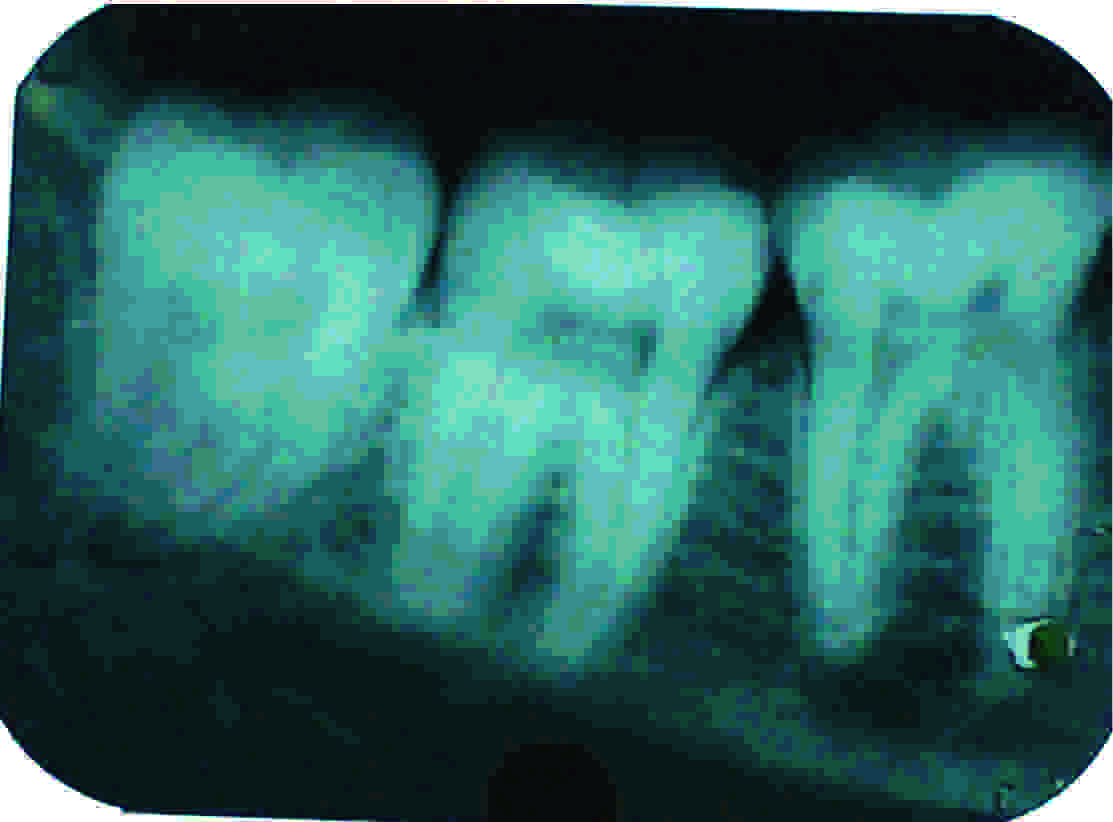
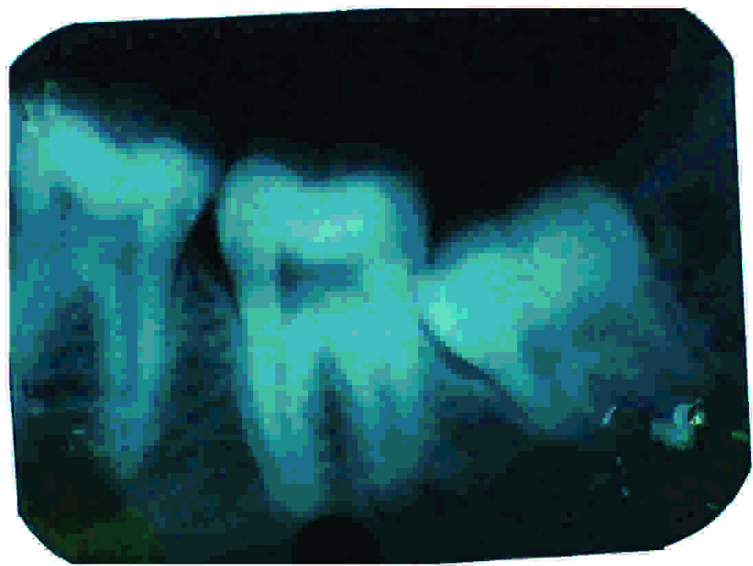
A total of 112 haemodialysis patients who were referred from the Nephrology Department of the Jodhpur Dental College General Hospital clinic for dental examinations, participated in this study. Permission was obtained from the institutional ethical committee to undertake this study. A total number of 2928 radiographs of the posterior and the anterior teeth were evaluated. The radiographs with poor angulations, improper exposures or faulty processing, which could lead to scoring difficulties and the radiographs with carious and restored teeth were excluded from the study. Finally, a total number of 2682 teeth were evaluated. To ensure the accuracy of the diagnosis, all the radiographs were interpreted by two examiners (oral radiologists) in a dark room by using a standard viewing box under the 2X magnification and with the peripheral light being blocked out. Those teeth about which both our examiners were unsure were re-evaluated by a senior dental radiologist. Narrowing was defined as a notable reduction in the size of the pulp chamber and the pulp canals [Table/Fig-1,2]. The definite radiopaque masses inside the pulp chambers were identified as pulp stones. They were scored as present or absent. The OPGs were evaluated for the presence or absence of CAC. The OPGs with low diagnostic qualities were excluded. The radiopaque nodular mass which was adjacent to the cervical vertebrae, inside or below the C3-C4 intervertebral disc level, or to the retromandibular area, generally at an angle of 45° from the angle of the mandible which was independent of the hyoid bone, was considered as a CAC [16].
Intraoral radiograph of the mandibular right quadrant exhibiting narrowing of the dental pulp chamber
Intraoral radiograph of the mandibular left quadrant exhibiting narrowing of the dental pulp chambe
RESULTS
In this study, a total number of 5982 teeth from 112 patients were evaluated. Among the patients, 74 (66.07%) were males and 38 (33.92%) were females. The ages of the patients ranged from 18 to 51 years and the mean age was 24.74 years.
None of the patients who had undergone the haemodialysis showed CAC findings. 84 patients in the haemodialysis patients group had pulp calcification. There was no statistical correlation (p<0.05) between the pulp narrowing and CAC in the haemodialysis patients group [Table/Fig-3].
Distribution of carotid artery calcification (CAC) and pulp narrowing in the haemodialysis group
| No pulp narrowing | Pulp narrowing | Total |
|---|
| No. of patients without CAC (%) | 28 (24.99) | 84 (74.99) | 112 (100) |
| No. of patients with CAC (%) | 0 (00) | 0 (00) | 0 (00) |
| Total no. of patients (%) | 28 (24.99) | 84 (74.99) | 112 (100) |
Five haemodialysis patients had pulp stones. There was no statistical correlation between CAC and the pulp stones (p<0.05) [Table/Fig-4] or between the pulp narrowing and the pulp stones in the haemodialysis patients group (p<0.05).
Distribution of carotid artery calcification (CAC) and pulp stones in the haemodialysis group
| No. of patients without pulp stones | No. of patients with pulp stones | Total |
|---|
| No. of patients without CAC (%) | 74 (66.07) | 38 (33.92) | 112 (100) |
| No. of patients with CAC (%) | 0 (00) | 0 (00) | 0 (00) |
| Total no. of patients (%) | 74 (66.07) | 38 (33.92) | 112 (100) |
DISCUSSION
The atherosclerotic disease in the region of the carotid bifurcation is known to cause 80% of the strokes, which is reported to be the third leading reason for death in most of the countries, after cardiovascular diseases and cancer [17,18]. The vascular injury in ESRD patients is known to be secondary to the metastatic calcification which occurs due to the decreased calcification inhibitors and the increased calcification activators [11,19]. A vascular calcification is an important manifestation of atherosclerosis. The presence and the extent of these vascular calcifications are valuable predictors of the cardiovascular events and the mortality in patients with ESRD [13]. In the ESRD patients, the vascular calcifications are associated with an increased stiffness of the large, elastic-type arteries, such as the aorta and the common carotid artery [15]. The identification of CAC on oral panoramic radiographs has been studied by dental practitioners and researchers. The carotid artery calcifications on the panoramic radiographs are considered as a major sign and a marker for the subsequent vascular risk. So, it has been recommended that all the patients with an evidence of carotid artery calcification in the panoramic radiographs should be referred for a vascular examination and treatment [20].
The dental panoramic radiographs have been found to be valuable for detecting atherosclerosis. The presence of Carotid Artery Calcifications (CACs) on Panoramic Radiographs (PRs) was first shown by Friedlander and Lande. They suggested that these radiographs were able to play an important role in an early diagnosis of CACs, which could result in the more serious conditions of heart disease and stroke [21]. The effectiveness of this type of radiography has been proven by various studies that were done to assess the capabilities of PRs in the diagnosis of CACs in the patients without symptoms, but with high risk of stroke. These studies showed that PRs were not only more commonly used, but that they were also cheaper and easier in comparison to the previous diagnostic methods and that they were also much less invasive [22,23].
It has been suggested that advanced diagnostic examinations such as ultrasonography, MRI and angiography however, may be required to confirm the presence and the extent of the vascular calcification [24,25]. Kansu et al., in their study, used ultrasonography to confirm the carotid calcification in ESRD patients and renal transplant recipients who showed radiographic signs, whereas it was not required in the present study, as none of the patients showed radiographic evidences of a calcification [26]. Pulp calcifications are clinically indiscernible. These are seen as radiopaque structures within the pulp chamber and the root canal on intraoral radiographs. Since so many calcifications are not of sufficient sizes to be discerned on radiographs, a radiographic examination based incidence would be inaccurate. However, radiographs are the only non invasive means of evaluating the pulp calcifications in clinical studies [8]. In general, calcifications of the pulp can be demonstrated more easily by the use of bitewing and periapical radiographs. Previous studies had revealed no significant difference between the periapical and the bitewing radiographs in the diagnosis of pulp calcification [27]. Therefore, in the present study, periapical radiographs were used to determine the pulp calcification.
There is conflicting literature on the association of the presence of pulp calcification and the systemic conditions. Both the narrowing of the dental pulp and CAC have been reported in ESRD patients, but the association between the 2 in ESRD patients and in renal transplant recipients still remains unanswered. Coronary atherosclerosis patients showed pulpal calcifications on radiographic examinations in the studies which were done by Moura and Paiva [28]. Apart from human studies, there are reports on the absence of the relationship between Cardiovascular Disease (CVD) and pulp stones in animals [29,30].
In the present study, no relationship was noted between the presence of pulpal calcification and CAC in ESRD patients. Our findings contradict the findings of Edds et al., [31] who noted a significant relationship between a pre-existing CVD and pulp stones in their pilot study. Differences were also noted between the results of the present study and the results of the only similar study of Kansu et al., when they were compared, where they noted CAC in 6 of the 29 HD patients, whereas CAC was detected in none of the patients of the present study [26]. In the present study, 38 (33.92%) patients had pulp stones, which was higher as compared to that in Kansu et al., study, who noted pulp stones in only 5 (21.7%) patients.
The findings of the present study contradict the findings of some authors. These differences are most likely due to the differences in the composition of the study samples. The aetiology of pulp calcification is still unknown. The diversity and the complexity of the aetiological factors make it very difficult to determine the specific aetiology in any particular subject; however, the aetiology of pulp calcifications has been attributed to long-standing local irritants, such as caries, wasting diseases, pulp-capping procedures, healed fractures, tooth injury restorations, periodontal conditions and orthodontic tooth movements [8,26]. The other reported causes include aging, idiopathic factors, fluoride supplements, hypervitaminosis D or a possible genetic predisposition, as in dentinogenesis imperfecta and dentinal dysplasia, which may be seen even in unerupted teeth [26]. Owing to the diverse and complex aetiological factors, it becomes very difficult to identify the specific underlying cause in a given subject. The limitations of the panoramic radiographs must also be considered when CACs are identified, as it has been shown that panoramic radiographs are unreliable for the detection of CAC as compared to ultrasonography (sensitivity 31.1%) [23].
In conclusion, although intraoral radiographs are easy screening methods that frequently reveal the presence of calcified structures in the pulp, pulp calcification cannot provide any information about CAC. Therefore, the presence of pulp calcification does not seem to serve as a diagnostic marker for carotid atherosclerosis. However, dental practitioners must play their role and contribute to the early detection of the patients who are at a risk of CVD and differentiate them from the anatomic and pathologic forms. Careful evaluation of the dental panoramic radiographs by the dental practitioners might be instrumental in saving the patients’ lives.
[1]. Goga R, Chandler NP, Oginni AO, Pulp stones: a reviewInt Endod J 2008 41:457-68. [Google Scholar]
[2]. Kantaputra PN, Sumitsawan Y, Schutte BC, Tochraeontanaphol C, Vander Woude syndrome with sensorineural hearing loss, large craniofacial sinuses, dental pulp stones and minor limb anomalies: Reports of four-generation Thi familyAm J Med Genet 2002 108:275-80. [Google Scholar]
[3]. Johnson PL, Bevelander G, Histogenesis and histochemistry of pulpal calcificationJ Dent Res 1956 35:714-22. [Google Scholar]
[4]. Parekh S, Kyriazidou A, Bloch-Zupan A, Roberts G, “Multiple pulp stones and shortened roots of unknown etiology,”Oral Surg Oral Med Oral Pathol Oral Radiol Endod 2006 101(6):e139-42. [Google Scholar]
[5]. Sayegh FS, Reed AJ, Calcification in the dental pulpOral Surg Oral Med Oral Pathol 1968 25:873-82. [Google Scholar]
[6]. Bernick S, Nedelman C, Effect of ageing on dental pulpJ Endod 1975 1:88-94. [Google Scholar]
[7]. Philippas GG, Influence of occlusal wear and age on formation of dentin and size of pulp chamberJ Dent Res 1961 40:1186-98. [Google Scholar]
[8]. Ranjitkar S, Taylor JA, Townsend GC, A radiographic assessment of the prevalence of pulp stones in AustraliansAust Dent J 2002 47:36-40. [Google Scholar]
[9]. Hamasha AA-H, Darwazeh A, Prevalence of pulp stones in Jordanian adultsOral Surg Oral Med Oral Pathol Oral Radiol Endod 1998 86:730-32. [Google Scholar]
[10]. Galili D, Berger E, Kaufman E, Pulp narrowing in renal end stage and transplanted patientsJ Endod 1991 17:442-43. [Google Scholar]
[11]. London GM, Drueke TB, Atherosclerosis and arteriosclerosis in chronic renal failureKidney Int 1997 51:1678-95. [Google Scholar]
[12]. London GM, Marty C, Marchais SJ, Guérin AP, Métivier F, de Vernejoul MC, Arterial calcifications and bone histomorphometry in end-stage renal diseaseJ Am Soc Nephrol 2004 15:1943-51. [Google Scholar]
[13]. Blacher J, Guerin AP, Pannier B, Marchais SJ, Safar ME, London GM, Arterial calcifications, arterial stiffness, and cardiovascular risk in end-stage renal diseaseHypertension 2001 38:938-42. [Google Scholar]
[14]. Schiffrin EL, Lipman ML, Mann JF, Chronic kidney disease: effects on the cardiovascular systemCirculation 2007 116:85-97. [Google Scholar]
[15]. Guerin AP, London GM, Marchais SJ, Métivier F, Arterial stiffening and vascular calcifications in end-stage renal diseaseNephrol Dial Transplant 2000 15:1014-21. [Google Scholar]
[16]. Friedlander AH, Panoramic radiography: the differential diagnosis of carotid artery atheromasSpec Care Dentist 1995 15:223-27. [Google Scholar]
[17]. Anderson CS, Jamrozik KD, Burvill PW, Chakera TM, Johnson GA, Stewart-Wynne EG, Determining the incidence of different subtypes of stroke: results from the Perth Community Stroke Study, 1989-1990Med J Aust 1993 158:85-89. [Google Scholar]
[18]. Fatahzadeh M, Glick M, Stroke: epidemiology, classification, risk factors, complication, diagnosis, prevention and medical and dental managementOral Surg Oral Med Oral Pathol Oral Radiol Endod 2006 102:180-91. [Google Scholar]
[19]. Kansu O, Ozbek M, Avcu N, Gençtoy G, Kansu H, Turgan C, The prevalence of carotid artery calcification on the panoramic radio-graphs of patients with renal diseaseDentomaxillofacial Radiol 2005 34:16-19. [Google Scholar]
[20]. Cohen SN, Friedlander AH, Jolly DA, Date L, Carotid calcification on panoramic radiographs: an important marker for vascular riskOral Surg Oral Med Oral Pathol Oral Radiol Endod 2002 94:510-14. [Google Scholar]
[21]. Friedlander AH, Lande A, Panoramic radiographic identification of carotid arterial plaquesOral Surg Oral Med Oral Pathol Oral Radiol Endod 1981 52:20-24. [Google Scholar]
[22]. Chicano RR, Sanchez REO, Castano FL, Merino CC, Panoramic radiograph as a method for detecting calcified atheroma plaques. Review of literatureMed Oral Patol Oral Cir Bucal 2006 11:261-66. [Google Scholar]
[23]. Madden RP, Hodges JS, Salmen CH, Rindal DB, Tunio J, Michalowicz BS, Utility of panoramic radiographs detecting cervical calcified carotid atheromaOral Surg Oral Med Oral Pathol Oral Radiol Endod 2007 103:543-48. [Google Scholar]
[24]. Almog DM, Horev T, Illig KA, Green RM, Carter LC, Correlating carotid artery stenosis detected by panoramic radiography with clinically relevant carotid artery stenosis determined by duplex ultrasoundOral Surg Oral Med Oral Pathol Oral Radiol Endod 2002 94:768-73. [Google Scholar]
[25]. Ohba T, Takata Y, Ansai T, Morimoto Y, Tanaka T, Kito S, Evaluation of calcified carotid artery atheromas detected by panoramic radiograph among 80-year-oldsOral Surg Oral Med Oral Pathol Oral Radiol Endod 2003 96:647-50. [Google Scholar]
[26]. Kansu O, Ozbek M, Avcu N, Aslan U, Kansu H, Gençtoy G, Can dental pulp calcification serve as a diagnostic marker for carotid artery calcification in patients with renal diseases?Dentomaxillofac Radiol 2009 Dec 38(8):542-45. [Google Scholar]
[27]. Tamse A, Kaffe I, Littner MM, Shani R, Statistical evaluation of radiologic survey of pulp stonesJ Endod 1982 8:455-58. [Google Scholar]
[28]. Moura AAM, Paiva JG, Pulpal calcifications in patients with coronary atherosclerosisEndod Dent Traumatol 1987 3:307-09. [Google Scholar]
[29]. Krell KV, McMurtrey LG, Walton RE, Vasculature of the dental pulp of atherosclerotic monkeys: light and electron microscopic findingsJ Endod 1994 20:469-73. [Google Scholar]
[30]. Oguntebi BR, Stafford DS, Cerda J, Robbins F, Vascular changes in the dental pulp in the hypercholesterolemic miniature swineOral Surg Oral Med Oral Pathol 1992 74:351-56. [Google Scholar]
[31]. Edds AC, Walden JE, Scheetz JP, Goldsmith LJ, Drisko CL, Eleazer PD, Pilot study of correlation of pulp stones with cardiovascular diseaseJ Endod 2005 31:504-06. [Google Scholar]