Primary Melanoma of the Spinal Cord: A Case Report
Ruchi Sinha1, Tauseef Husain Rizvi2, Shrijeet Chakraborti3, Chandra Kumar Ballal4, Anup Kumar5
1 Associate Professor, Department of Pathology, Kasturba Medical College, Mangalore, Karnataka, India.
2 Post Graduate, Department of Pathology, Kasturba Medical College, Mangalore, Karnataka, India.
3 Associate Professor, Department of Pathology, Kasturba Medical College, Mangalore, Karnataka, India.
4 Professor, Department of Neurosurgery, Kasturba Medical College, Mangalore, Karnataka, India.
5 Associate Professor, Kasturba Medical College, Mangalore, Karnataka, India.
NAME, ADRESS, E-MAIL ID OF THE CORRESPONDING AUTHOR: Dr. Ruchi Sinha, Department of Pathology, Kasturba Medical College, Lighthouse Hill Road, Mangalore-575001, Karnataka, India.
Phone: +919449991179
E-mail: ruchidoctor@gmail.com
Primary melanoma is an extremely rare tumour of the spinal cord. We are reporting a case of primary melanoma of the spinal cord in a 55-years-old male patient. Magnetic resonance imaging showed an extradural intraspinal lesion opposite the L4 vertebral body. The lesion was completely resected and a microscopic diagnosis of melanoma was made. Thirty eight months later, the patient is alive, with no evidence of any tumour recurrence.
Melanoma, spinal cord, lumbar
INTRODUCTION
Primary melanoma of the Central Nervous System (CNS) accounts for only 1% of all the melanoma cases [1]. Primary malignant melanomas of the spinal cord are extremely rare melanocytic tumours of the CNS and only very few of such cases have been reported in the literature [1]. The first case of primary spinal melanoma was reported by Hirschberg in 1906. Most of the primary spinal melanomas are intradural with or without extradural components; primary extradural spinal melanomas are very rare lesions [2]. We are reporting a case of a primary lumbar spinal extradural melanoma.
CASE REPORT
A 55-year-old male patient with a history of back pain, a weakness in the lower extremities and with urinary and anal incontinences, was admitted. His symptoms had progressively worsened to paraplegia. The neurological signs were consistent with lower spinal cord compression. A careful general physical examination did not reveal any lymphadenopathy, organomegaly or cutaneous lesions. A Magnetic Resonance Imaging (MRI) scan revealed an extradural intraspinal lesion opposite the L4 vertebral body, which was widening the intervertebral foramen. A provisional diagnosis of a neurofibroma was made. Intraoperatively, a bluish, engorged, extradural, intraspinal lesion which measured 1 cm was seen to arise in relation to the right L4 root. The lesion was completely resected and it was sent for a histopathological examination.
Pathological Findings
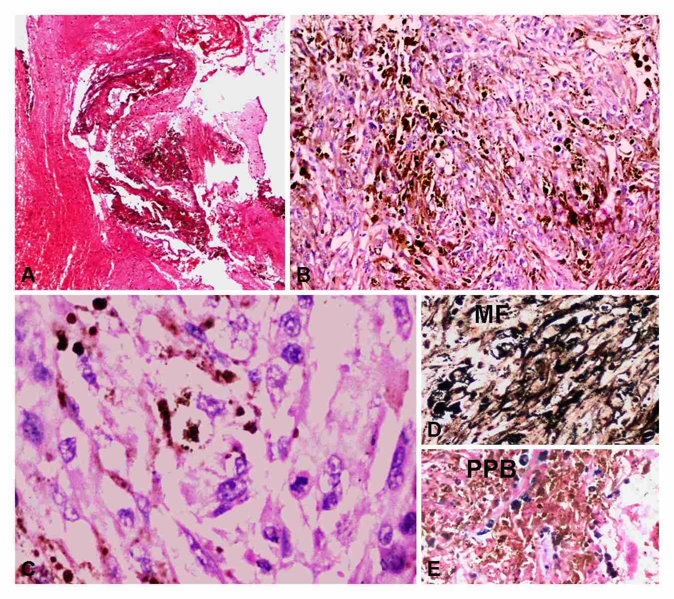
Grossly, multiple black friable tissue bits were received. The histological examination showed a spinal cord tissue with a tumour which was composed of proliferating polygonal to spindle shaped cells which had an abundant intra-cytoplasmic brown to black pigment [Table/Fig-1A, B]. The tumour cells had vesicular nuclei with prominent nucleoli [Table/Fig-1C]. The cells were arranged in tight clusters and sheets. Numerous mitotic figures along with an atypical mitosis were found [Table/Fig-1C]. Bizarre multinucleated giant cells were seen focally. Areas of haemorrhage and necrosis were also present. Masson’s Fontana stain for the melanin pigment was positive [Table/Fig-1D]. A melanin bleach confirmed that the intracytoplasmic pigment was melanin and it highlighted the nuclear features. The Perl’s Prussian Blue stain was negative and it excluded the haemosiderin pigment [Table/Fig-1E].
A: Pigmented mass lesion is seen adjacent to spinal cord (H&E x 40). B: The tumour is composed of spindle to polygonal cell having abundant intra- and extracellular melanin pigment (H&E x 100). C: The tumour cells have vesicular chromatin, prominent nucleoli and mitotic figures (H&E x 100). The melanin pigment stains positively with Masson’s Fontana (MF) stain (D; H&E x 100) and is negative for Perl’s Prussian Blue (PPB) stain (E; H&E x 100) but highlights intra-tumoural haemosiderin
DISCUSSION
The present World Health Organiztion classification classifies primary melanocytic lesions of the CNS into diffuse melanocytosis, melanocytoma, meningeal melanomatosis and malignant melanoma. Among these lesions, primary malignant melanoma of the CNS accounts for only 1% of all melanoma cases [1]. The incidence of primary spinal melanoma is even rarer. According to the Hayward classification [3], the diagnosis of primary spinal cord melanoma is based on the absence of a melanoma outside the CNS, the absence of this lesion at any other site in the CNS and a histologic confirmation of melanoma.
Primary spinal melanoma presents as solitary pigmented lesions. The tumour has an equal sex ratio and it occurs commonly in the fifth decade of life, with an age range of 20 to 80 years [4].
It occurs most frequently in the thoracic segment (42.3%), followed by its occurrence in the cervical (34.6%), thoracolumbar (11.5%), cervicothoracic (7.7%) and the lumbar (3.8%) segments [4]. Till date, only six extradural spinal melanomas have been reported [5,6].
Melanoma is a malignant neoplasm which arises from melanocytes, which are melanin producing cells that arise from the neural crest during embryogenesis and migrate to the skin, mucus membrane, and the CNS [7]. It has been hypothesized that primary melanomas of the spinal cord arise either from the leptomeningeal melanoblasts or from the neuroectodermal rest cells [1,8].
MRI is the radiological method of choice in the diagnosis of spinal cord melanoma. However, it is unable to distinguish it from other spinal cord tumours [7]. The MRI findings of melanomas vary considerably, depending upon the number of melanin-containing cells and the presence or absence of haemorrhages [1,4]. The characteristic signs of a melanoma on MRI is a hyperintense signal in the T1 images and a hypointense signal in the T2 images.
The primary extradural spinal melanomas are extremely uncommon tumours and they have to be differentiated from other extradural lesions like meningiomas, schwannomas, meningeal melanocytomas and vascular malformations such as cavernous hemangiomas [5].
Although the survival rate for CNS melanomas are not exactly known, they have better prognoses than those of cutaneous melanomas [9,10]. Complete surgical excision is the treatment of choice and radiation therapy has been advocated in case of subtotal resection [11].
[1]. Farrokh D, Fransen P, Faverly D, MR findings of a primary intramedullary malignant melanoma: case report and literature reviewAm J Neuroradiol 2001 22:1864-66. [Google Scholar]
[2]. Naing A, Messina JL, Vrionis FR, Daud AI, Uncommon manifestations of common malignancies: case 3. Malignant melanoma arising from a spinal nerve rootJ Clin Oncol 2004 22:194-95. [Google Scholar]
[3]. Hayward RD, Malignant melanoma and the central nervous system. A guide for classification based on the clinical findingsJ Neurol Neurosurg Psychiatr 1976 39:526-30. [Google Scholar]
[4]. Kim MS, Yoon DH, Shin DA, Primary spinal cord melanomaJ Korean Neurosurg Soc 2010 48:157-61. [Google Scholar]
[5]. Jo KW, Kim SR, Kim SD, Park IS, Primary thoracic epidural melanoma: a case reportAsian Spine Journal 2010 4:48-51. [Google Scholar]
[6]. Unal B, Catillo M, MRI features of a primary thoracic epidural melanoma: a case reportClin Imaging 2007 31:273-75. [Google Scholar]
[7]. Kwon SC, Rhim SC, Lee DH, Roh SW, Kang SK, Primary malignant melanoma of the cervical spinal nerve rootYonsei Med J 2004 45:345-48. [Google Scholar]
[8]. Yamasaki T, Kikuchi H, Yamashita J, Aasto R, Fujita M, Primary spinal intramedullary malignant melanoma: case reportNeurosurgery 1989 25:117-21. [Google Scholar]
[9]. Brat DJ, Giannini C, Scheithauer BW, Burger PC, Primary melanocytic neoplasms of the central nervous systemsAm J Surg Pathol 1999 23:745-54. [Google Scholar]
[10]. Larson TC 3rd, Houser OW, Onofrio BM, Piepgras DG, Primary spinal melanomaJ Neurosurg 1987 66:47-49. [Google Scholar]
[11]. François P, Lioret E, Jan M, Primary spinal melanoma: case reportBr J Neurosurg 1998 12:179-82. [Google Scholar]