Metaplastic Ossification in a Juvenile Rectal Polyp: A Rare Histological Finding
Monika Garg1, Jasveen Kaur2, Samridhi Bindroo3, Anuradha Sharma4, Nanak Chand Mahajan5
1 Associate Professor, Department of Pathology,
2 Resident, Department of Pathology,
3 Resident, Department of Pathology,
4 Resident, Department of Pathology,
5 Professor and Head, Department of Pathology, M.M Institute of Medical Sciences and Research, Mullana, Ambala, India.
NAME, ADRES, E-MAIL ID OF THE CORESPONDING AUTHOR: Dr. Monika Garg, Associate Professor, Department of pathology, MMIMSR, Mullana, Ambala, Haryana-133207, India.
Phone: 9417378514
E-mail: monikakash7@yahoo.co.in
An osseous metaplasia is a phenomenon which has been described in a wide variety of tissue types with respect to both neoplastic and non-neoplastic conditions. However, an osseous metaplasia is exceedingly rare in colonic polyps. We are herein representing a case of osseous metaplasia in a juvenile rectal polyp in a six year old boy, with review of the literature on the suggested mechanisms of its aetiology.
Osseous metaplasia, Rectal polyp, Juvenile polyp, Rectum
INTRODUCTION
Osseous metaplasia i.e. a heterotopic bone formation, is rarely detected in the gastrointestinal tract. It is a phenomenon which has been described in a wide variety of tissue types with respect to mainly neoplastic and rarely non - neoplastic conditions. In colonic polyps, its occurrence is extremely rare. The first description of the phenomenon of bone formation within a rectal polyp was given by Sperling in 1981 [1]. It is definitely a striking morphological feature, but it has not been found to have any significance clinically and prognostically [2]. The mechanism which is responsible for this metaplastic change has not been completely understood. We are herein representing a unique case of an osseous metaplasia in a juvenile rectal polyp in a six year old boy and review of the literature on the suggested mechanisms of its aetiology.
CASE REPORT
A six years old boy visited the surgery department of our hospital with the chief complaints of a mass protruding out of the anus and blood in the stools on and off, since 5 months. The local examination of the rectum revealed a mass which protruded out of the anus, on trying to defaecate. There was no bleeding, discharge or ulcer formation, which were associated with it and the mass would go back automatically after the defaecation. A colonoscopic examination revealed a solitary polypoidal growth in the rectum, which measured approximately 1.5x1x0.5 cm. It had a slightly irregular surface and it was reddish in colour. The rest of the colonoscopic examination was unremarkable. Thereafter, a polypectomy with sclerotherapy was performed under general anaesthesia and the polypoidal mass was sent to us for a histopathological examination.
Biopsy
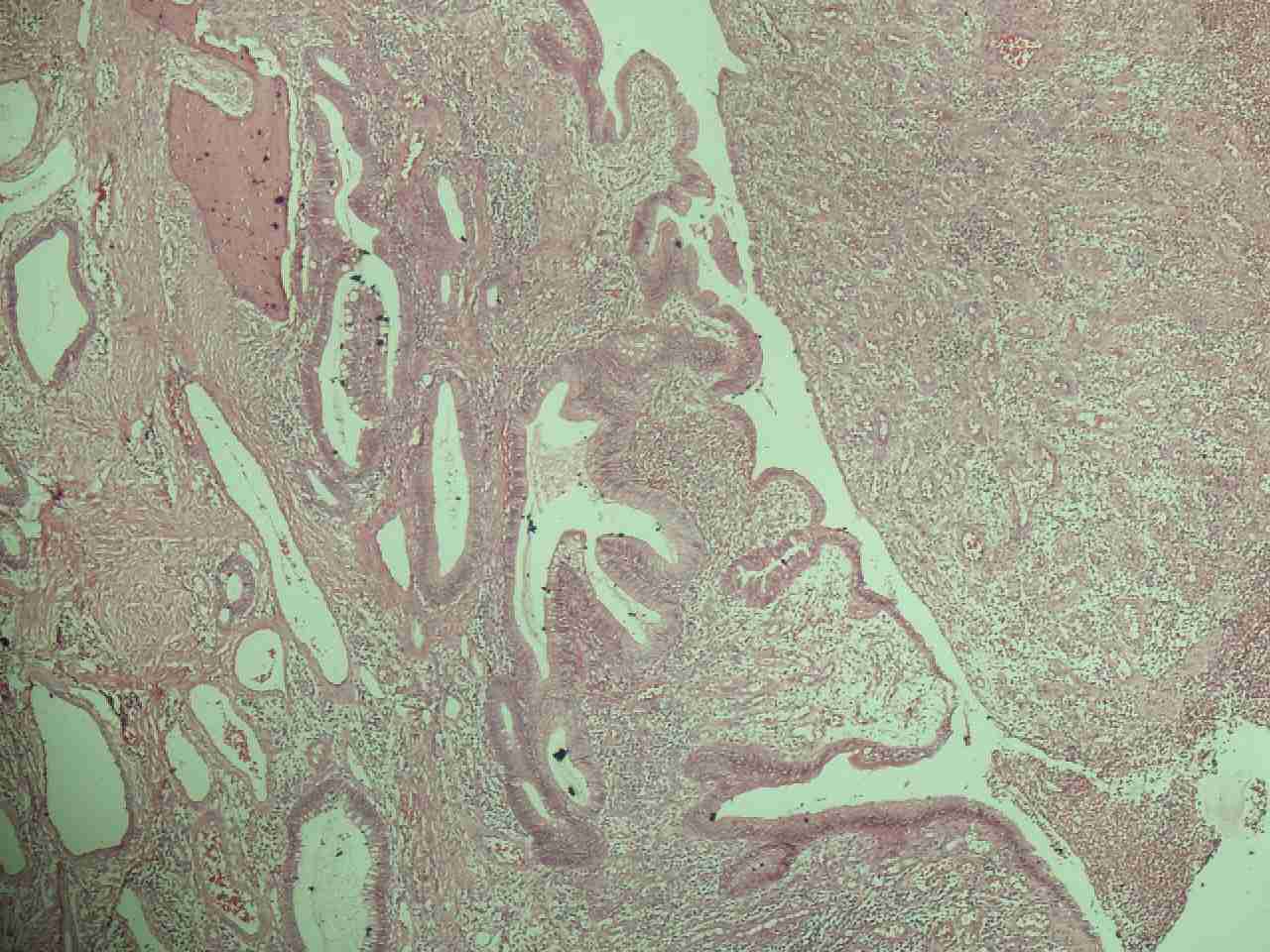
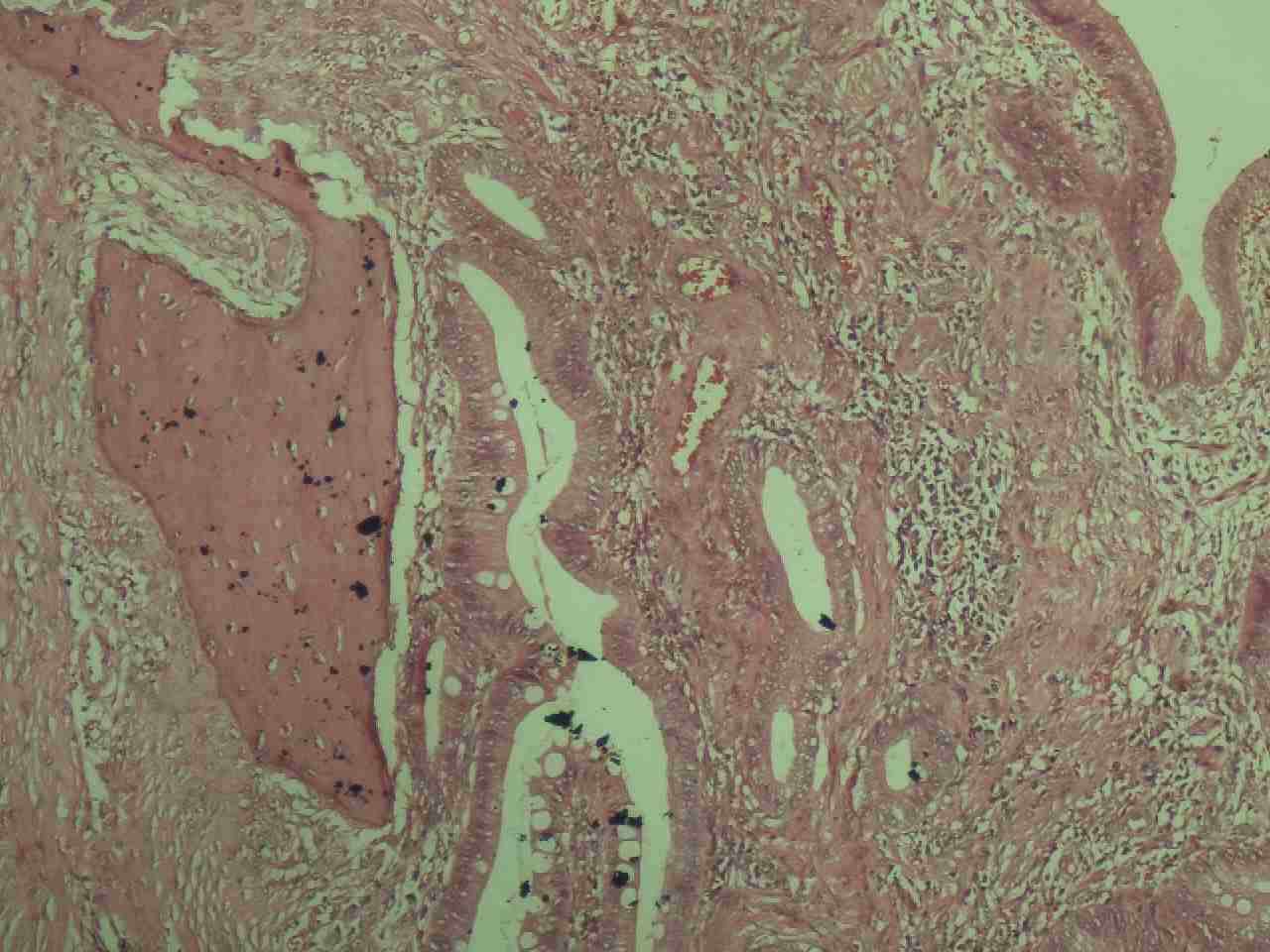
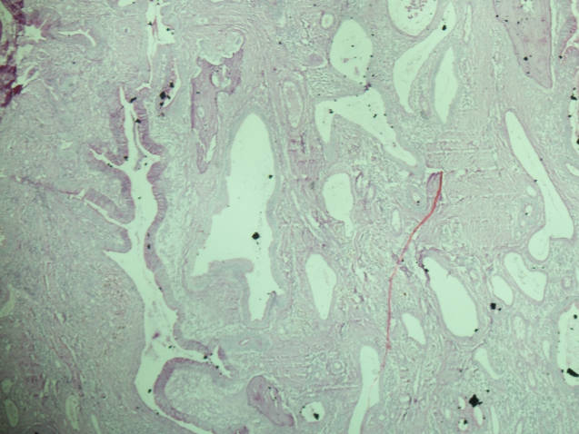
We received a greyish white to a greyish brown polypoidal mass along with the stalk, which measured 1.3x1x0.5 cm. On cut section, the greyish white areas, along with few haemorrhagic areas, were seen. The microscopic examination revealed a polypoidal tissue which was lined by cystically dilated, well-formed, mucin secreting mucosal glands. The foci of the osseous metaplasia were found to be composed of mature bony trabeculae. The lamina propria was infiltrated by a mixed acute and chronic inflammatory cell infiltrate and a granulation tissue. The granulation tissue was comprised of fibrous tissue and prominent small blood vessels [Table/Fig-1,2&3]. This case was reported as a case of an inflammatory juvenile polyp with foci of the osseous metaplasia.
Sections show cystically dilated crypts filled with mucinous material and a bony trabeculae (4x)
Sections show cystically dilated crypts filled with mucinous material and a bony trabeculae (10x)
Sections show cystically dilated crypts filled with mucinous material and a bony trabeculae (4x, PAS stain)
DISCUSSION
Osseous metaplasia which occurs in colonic polyps, is extremely rare [3]. Osseous metaplasia has been described in a wide variety of tissue types with respect to both neoplastic and non-neoplastic conditions. To the best of our knowledge, six cases of juvenile polyps with osseous metaplasia, although around 15 cases of osseous metaplasia in various benign colorectal polyps have been described [Table/Fig-4]. Amongst the cases with juvenile polyps, a majority of the patients were males, with a mean age of 7.4 years (range 3 -15 years). The polyp size vary from 5mm to 20mm, with a mean size of 9mm. A majority of these are located in the rectum.
Summary of reported cases of osseous metaplasia in benign colorectal polyps
| No. | Author | Year | Site | Sex | Age | Size (mm) |
|---|
| Hamartomatous Juvenile |
| 1 | Mark [6] | 1964 | Rectum | Male | 10 | unknown |
| 2 | Drut [7] | 1992 | Rectosigmoid | Male | 5 | 10 |
| 3 | Drut [7] | 1992 | Rectum | Male | 4 | 5 |
| 4 | Groisman [8] | 1994 | Rectum | Female | 3 | 20 |
| 5 | Ahmed [9] | 2009 | Rectum | Male | 15 | 10 |
| 6 | Bhat [10] | 2012 | Rectum | Female | 5 | 10 |
| Inflammatory |
| 7 | Sperling [1] | 1981 | Rectum | Male | 25 | 10 |
| 8 | Castelli [11] | 1992 | Rectum | Female | 22 | 10 |
| 9 | Oono [2] | 2009 | Rectum | Male | 39 | 12 |
| 10 | Odum [4] | 2012 | Rectum | Male | 74 | 10 |
| Dysplastic |
| 11 | Groisman [8] | 1994 | Rectum | Male | 67 | 18 |
| 12 | Cavazza [12] | 1996 | NI | NI | NI | NI |
| 13 | McPherson [13] | 1999 | Cecum | Male | 73 | 20 |
| 14 | Rothstein [14] | 2000 | Sigmoid colon | NI | NI | 25 |
| 15 | Al-Daraji [15] | 2005 | Sigmoid colon | Female | 85 | 15 |
| 16 | White [16] | 2008 | Transverse colon | Female | 63 | NI |
Osseous metaplasia is a well described phenomenon in the setting of intestinal neoplasia, both benign and malignant. Various mechanisms have been suggested, although the pathogenesis of osseous metaplasia still remains unknown. The production of the Bone Morphogenetic Protein (BMP) by the tumour may be a factor [4]. A case series which was published by Dukes in 1939 of osseous metaplasia in rectal carcinoma, suggested that the long duration of the symptoms, the histological picture of the low grade tumour with little potential for metastasis and the presence of areas of necrosis, may be contributory factors [5]. He also suggested an osseous formation, followed by a dystrophic calcification of the necrotic tissue [3, 5]. Juvenile polyps are hamartomatous lesions which may show the occurrence of osseous metaplasia. A study which was done by Marks and Atkinson in 1964 suggested that osseous metaplasia may result from the ability of the fibroblasts to transform into other types of mesodermal tissues, especially osteoblasts [2, 6]. In inflammatory polyps, osseous metaplasia is seen with a histological background of an active chronic inflammation and/or ulceration. A persistent inflammation may result in an osteogenic stimulation. Odum reported a case of osseous metaplasia in an inflammatory polyp of the rectum [4]. No details have been received of osseous metaplasia in hyperplastic polyps. Clearly, the exact mechanism of an osseous metaplasia is still a topic of a significant debate.
An osseous metaplasia is clinically and prognostically insignificant and most of the times, it is an incidental finding. In our case of the juvenile polyps, the pathogenesis could be the result of a metaplastic transformation of the fibroblasts to osteoblasts. Clinically, the presence of this metaplastic bone seems to be innocent. In conclusion, we presented an extremely rare case report of a heterotopic bone formation in a rectal juvenile polyp. The phenomenon was found to be striking, on a histological examination.
[1]. Sperling MH, Friedman CJ, Osseous metaplasia in a benign colon polypGastrointest Endosc 1981 27(3):198-99. [Google Scholar]
[2]. Oono Y, Fu K-L, Nakamura H, Iriguchi Y, Oda J, Mizutani M, Bone formation in a rectal inflammatory polypWorld J Gastrointest Endosc 2010 2(3):104-06. [Google Scholar]
[3]. Hjelkrem M, Fair K, Pabby A, Osseous Metaplasia in a Colon PolypPract Gastroenterol 2012 36(11):58-62. [Google Scholar]
[4]. Odum BR, Bechtold ML, Diaz-Arias A, Osseous Metaplasia in an Inflammatory Polyp of the Rectum: A Case Report and Review of the LiteratureGastroenterol Res 2012 5(2):74-78. [Google Scholar]
[5]. Dukes CE, Ossification in Rectal Cancer: (Section of Surgery: Sub-Section of Proctology)Proc R Soc Med 1939 32(11):1489-94. [Google Scholar]
[6]. Marks MM, Atkinson KG, Heterotopic bone in a juvenile rectal polyp: case reportDis Colon Rectum 1964 7:345-47. [Google Scholar]
[7]. Drut R, Cueto Rúa E, Metaplasia ósea en pólipos juveniles: presentación de dos casosPatol Rev Latinoam 1992 30(1):33-36. [Google Scholar]
[8]. Groisman GM, Benkov KJ, Adsay V, Dische MR, Osseous metaplasia in benign colorectal polypsArch Pathol Lab Med 1994 118(1):64-65. [Google Scholar]
[9]. Ahmed R, Ahmad Z, Qureshi A, Osseous metaplasia in a juvenile retention polyp: a case reportBMJ Case Rep 2009 2009(?) [Google Scholar]
[10]. Bhat V, Roopa A, Shariff S, Juvenile Rectal Polyp with Osseous Metaplasia - A Rare Case with Review of LiteratureInt J Health Sci Res 2012 2(8):114-16. [Google Scholar]
[11]. Castelli MF, Roberts J, Ossification in a benign rectal polypAm J Gastroenterol 1992 87(4):543-44. [Google Scholar]
[12]. Cavazza A, Sassatelli R, De Marco L, [Bone metaplasia in adenomatous intestinal polyp. Report of a case and review of the literature]Pathologica 1996 88(6):511-13. [Google Scholar]
[13]. McPherson F, Maldonado M, Truitt CA, Mamel JJ, Morgan MB, Metaplastic ossification of a benign colonic polyp: case reportGastrointest Endosc 1999 49(5):654-56. [Google Scholar]
[14]. Rothstein RD, LiVolsi VA, Metaplastic ossification of a benign colonic polypGastrointest Endosc 2000 51(2):254 [Google Scholar]
[15]. Al-Daraji WI, Abdellaoui A, Salman WD, Osseous metaplasia in a tubular adenoma of the colonJ Clin Pathol 2005 58(2):220-21. [Google Scholar]
[16]. White V, Shaw AG, Tierney GM, Lund JN, Semeraro D, Osseous metaplasia in an ulcerating tubular adenoma of the colon: a case reportJ Med Case Rep 2008 2:130 [Google Scholar]