The Flexor Indicis Profundus - Its Morphology and Clinical Significance
Prathap Kumar J1, Padmalatha K2, Prakash B S3, Radhika P M4, Ramesh B R5
1 Assistant Professor, Department of Anatomy, M. S. Ramaiah Medical College, Bangalore, Karnataka-560054, India.
2 Assistant Professor, Department of Anatomy, ESIC Medical College & PGIMSR, Bangalore, Karnataka ,India.
3 Professor, Department of Anatomy, Dr. B.R. Ambedkar Medical College, Bangalore, Karnataka- 560045, India.
4 Assistant Professor, Department of Anatomy, M. S. Ramaiah Medical College, Bangalore, Karnataka-560054, India.
5 Professor and Head, Department of Anatomy, Dr. B.R. Ambedkar Medical College, Bangalore, Karnataka- 560045, India.
NAME, ADDRESS, E-MAIL ID OF THE CORRESPONDING AUTHOR: Dr. Prathap Kumar J, Assistant professor, Department of Anatomy, M. S. Ramaiah Medical College, MSRIT Post, Bangalore-560054, Karnataka, India.
Phone: 9035433303, 8880744879, 9035648318
E-mail: dr.prathapkumar@gmail.com
The knowledge on the anatomical variations of the deep flexor muscles is important due to its evolutionary significance. The flexor digitorum profundus is the deep flexor muscle of the forearm. It is a composite muscle with a dual nerve supply. The medial half of muscle is supplied by the ulnar nerve, and lateral half of the muscle is supplied by the anterior interosseous nerve, a branch of the median nerve. It flexes the distal phalanges of the medial four digits.
In the present case, we observed the presence of extensive cleavage of belly and tendon of flexor digitorum profundus to form flexor indicis profundus on the right side in an adult male cadaver. Flexor indicis profundus muscle is an example of progressive type of variation and it is usually asymptomatic, but it may cause compression of the anterior interosseous nerve, which can lead to compression neuropathy. If it is enlarged, it may simulate a ganglion.
Flexor digitorum profundus muscle, Flexor indicis profundus muscle, Anterior interosseous, Nerve, Compression Neuropathy, Ganglion
CASE REPORT
During the routine dissection of the upper limb of a male cadaver which was aged about 50 years, in the Department of Anatomy, Dr. B.R. Ambedkar Medical College, Bangalore, India, a variation in the flexor digitorum profundus muscle in the right forearm was observed. The muscles of the flexor compartment of the right forearm were carefully dissected and photographs were taken.
OBSERVATION
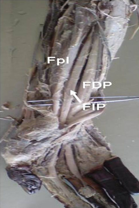
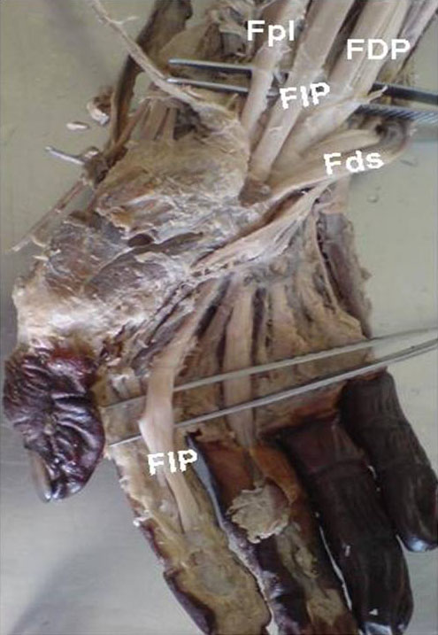
We observed an extensive cleavage of the tendon for the index finger, from the rest of the tendons of the flexor digitorum profundus muscle. The flexor indicis profundus is a part of the flexor digitorum profundus. The flexor indicis profundus originated from the anterior surface of shaft of the ulna and the adjoining interosseous membrane, along with the flexor digitorum profundus. In the upper 3cms, the flexor indicis profundus muscle belly was fused with the flexor digitorum profundus, while in the lower part, the muscle belly was completely separated from the flexor digitorum profundus [Table/Fig-1]. On further dissection, the tendon of the flexor indicis porofundus was found to be present between the flexor digitorum profundus and the flexor policis longus. The tendon was inserted to the palmar surface of the base of the distal phalanx of the index finger [Table/Fig-2].
Extensive cleavage of belly & tendon of Flexor Digitorum Profundus to form Flexor Indicis Profundus. FDP-Flexor Digitorum Profundus, FIP- Flexor Indicis Profundus, Fpl- Flexor pollicis longus.
Tendon of Flexor Indicis Profundus. FDP-Flexor Digitorum Profundus, FIP- Flexor Indicis Profundus, Fpl- Flexor pollicis longus, Fds- Flexor digitorum superficialis.
The anterior interosseous nerve which passed between the flexor digitorum indicis and the flexor policis longus, on a deeper plane, in the right forearm of an adult male cadaver, was observed. The medial half of the flexor digitorum profundus was supplied by the ulnar nerve and the flexor indicis profundus, which is the lateral half of the flexor digitorum profundus, was supplied by anterior interosseous nerve, a branch of the median nerve.
The flexor digitorum profundus muscle on the left side was normal, without any extensive cleavage. No other muscular variations were found in the body.
DISCUSSION
The flexor digitorum profundus is a deep flexor muscle of the forearm. It arises from the upper 3/4th of the anterior and the medial surfaces of the ulna, the adjacent interosseous membrane and the upper 3/4th of the posterior border of the ulna. The muscle ends in four tendons for the medial four digits and it is inserted on to the palmar surfaces of the bases of the distal phalanges. It flexes the distal interphalangeal joint. The medial half of the muscle is innervated by the ulnar nerve and the lateral half is innervated by the anterior interosseous branch of the median nerve, C8 and T1 [1]. Variations in the flexor digitorum profundus and the flexor pollicis longus muscles of the forearm have been reported in the literature, because these muscles have a common phylogenetic development from the pronatoflexor group of Humpry [2].
Muscles are highly variable and are often found in the course of routine dissections of the human body. The three categories of variations may be progressive, retrogressive and atavistic. The muscles which have a tendency to become increasingly complex, represent the progressive type of muscles. The deep flexor muscles of the forearm belong to the progressive group of variations. The muscles which undergo degeneration with a subsequent loss of functions represent the retrogressive type of muscles. Examples of this type are the palmaris longus and the plantaris muscles. The atavistic muscles are the muscular elements which have been lost completely, during the course of evolution and they make an abrupt appearance again. The axillary arch muscle, a remnant of the panniculus carnosus, is an example of the atavistic type of muscles [3]. Anomalous muscles are quite common in the flexor compartment. The flexor digitorum superficialis is the most common muscle, followed by the flexor digitorum profundus, to show anatomical variations [4].
The flexor digitorum profundus is a deep flexor muscle of the forearm, it is a flexor of the wrist and the metacarpophalangeal and the interphalangeal joints. The separation of the tendon for the index finger from the rest of the tendons of the flexor digitorum profundus muscle is a characteristic feature in humans and it can be correlated with the specialization of the functions of the index finger. The degree of the muscle freedom varies. The tendon for the index finger may be quite independent, forming a flexor indicis profundus [5].
The most common variation of the flexor digitorum profundus is at the level at which its tendons become independent. The median nerve always innervates the tendon of the index finger [6].
The variations in the flexor group of muscles of the forearm can be:
Connections between the flexor digitorum superficialis and the flexor digitorum profundus in the form of muscular slips,
Connections between the flexor pollicis longus and the flexor digitorum profundus,
The number of bellies of the muscle may be increased,
The tendon to the middle finger may be absent and,
The tendon to the index finger gets separated from the rest of the muscle to form the Flexor Indicis Profundus [7]. The present case belonged to the 5th type of variation, as has been described above.
An accessory bundle which arises from the coronoid process to form a tendon that joins the tendon of the flexor digitorum profundus and which goes to the index finger, is said to be found in about 20% of the cadavers and it is called the accessory tendon of the flexor digitorum profundus [6].
The flexor muscles of the forearm develop from the flexor mass, which subsequently divides into 2 layers, the superficial and the deep. The deep layer gives rise to the flexor digitorum superficialis, the lexor digitorum profundus and the flexor pollicis longus. The embryological basis of the present variation can be explained as being caused by the additional cleavage of the deep layer of the forearm flexor mass during development [8].
It was reported that these variations have a clinical relevance with respect to the development of the anterior interosseous nerve syndrome and entrapment neuropathy of the median nerve [8]. A cadaveric study on the flexor pollicis longus was done, in which an incidental finding of the flexor indicis profundus was found in two out of seventy six forearms (2.6%) [2].
These muscular variations are usually asymptomatic, but they are of academic interest and are often discovered incidentally during routine anatomical dissections or surgical procedures. However, they may become symptomatic and if they get enlarged, they may present as tumour like masses, simulate ganglions or cause compression neuropathies [3]. These variations should be kept in mind to prevent complications during surgical interventions of these regions. Knowledge on these variations is of importance, not only to the anatomists but also to the surgeons and orthopaedicians.
CONCLUSION
A case of extensive cleavage of both the muscle belly and the tendon of the flexor digitorum profundus muscle which was going to the index finger from the rest of the muscle belly of the right forearm, was observed. Hence, the tendon which was going to the index finger was called the flexor indicis profundus.
The flexor indicis profundus muscle belongs to the progressive type of variation. Such variations are usually asymptomatic. Sometimes, it may cause compression of the anterior interosseous nerve, which can lead to compression neuropathy. If it is enlarged, it may simulate a ganglion.
[1]. Johnson D, Ellis H, Pectoral girdle and upper limbIn: Susan standring, editor. Gray’s anatomy 2005 39th ednEdinburghElsevier Churchill Livingstone:877 [Google Scholar]
[2]. Mangini U, Flexor pollicus longus muscle: its morphology and clinical signifcanceJ Bone Joint Surg Am 1960 42:467-70.:559 [Google Scholar]
[3]. Le Gros Clark WE, The tissues of the body 1980 6th ednOxfordELBS:139-40. [Google Scholar]
[4]. Kopuz C, Fidan B, Islam A, An unusually distal and complete additional flexor profundus muscle to the index fingerJ Anat. 1997 191:465-67. [Google Scholar]
[5]. Bergman RA, Afifi AK, Miyauchi R Opus I: Muscular System: Alphabetical Listing of Muscles: F, Flexor Digitorum Profundus. In: Bergman RA, Afifi AF, Miyauchi R, editors. Illustrated Encyclopedia of Human Anatomic Variation. Available from: URL:http://www.anatomyatlases.org/AnatomicVariants/MuscularSystem/Text/F/16Flexor.shtml [Google Scholar]
[6]. Hollinshead WH, Anatomy for Surgeons: vol-3The back and the limbs 1982 3rd ednPhiladelphiaHarper and Row:400 [Google Scholar]
[7]. Bergman RA, Thompson SA, Afifi AK, Saadeh FA, Compendium of human anatomic variation 1988 Baltimore-MunichUrban and Schwarzenberg:14 [Google Scholar]
[8]. Kumar V, Naveen NS, Murlimanju BV, D’souza PS, A rare muscular variation in the flexor compartment of the forearmInternational Journal of Anatomical Variations 2011 4:115-16. [Google Scholar]