The major structural abnormalities occur in 2–3% of the foetuses, which accounts for 20–30% of the perinatal mortality which is seen in the developed world. A vast majority occur in low-risk women, they are sporadic and therefore, they cannot be anticipated. With the rapid advances in the molecular diagnostic techniques and the recent developments in the in-utero therapy, an early diagnosis of the foetal problems is imperative. The identification of foetal anomalies in utero is important. The prenatal detection of these abnormalities often aids in planning the appropriate obstetrical management and it optimizes the care of the foetuses and the new borns.
To evaluate the antenatal prevalence of the major congenital anomalies in our hospital population and to study the malformation patterns.
This was a cross-sectional and an observational study.
It extended from June 2012 To Aug 2012.
It was done on 250 patients.
MATERIALS AND METHODS
All the pregnant women who attended the Antenatal Clinic of the Obstetrics Department of Saveetha Medical College from June 2012 to August 2012 were examined by transabdominal sonography between 18 -20 weeks by a sonologist of the Radiology Department of Saveetha Medical College. A detailed anatomical survey was done. The inclusion criteria were all the patients without any risk.
The exclusion criterion was pregnancy which was associated with risk factors. The details regarding the parity, age distribution and the gestational age during which the USG was done, were analyzed. When lethal anomalies were detected, after getting the consents of the patients, autopsies were done and the findings were correlated. The foetal chromosome analyses were done in two cases. For the parents with foetal anomalies, counselling was given in a supportive environment and the appropriate management was offered.
RESULTS
In our study, among the 250 cases, 7 anomalies were detected, which gave an incidence 2.97%.
[Table/Fig-1] shows the analysis of the age distribution, which showed that the mean age in our study was 24 years.
Analysis of Age distribution
| Age | Numbers | Percentage |
|---|
| 15-20 | 22 | 8.8 |
| 21-25 | 112 | 44.8 |
| 26-30 | 94 | 37.6 |
| 31- 35 | 18 | 7.2 |
| 36-40 | 4 | 1.6 |
| 40 and above | Nil | Nil |
[Table/Fig-2] shows the distribution of the parity. A majority of the patients were gravida two 53.2%.
| Parity | Numbers | Percentage |
|---|
| Primi | 104 | 41.6 |
| Gravida-2 | 133 | 53.2 |
| Gravida-3 | 12 | 4.8 |
| Gravida-4 and above | 1 | 0.4 |
[Table/Fig-3] shows the gestational age during which the target USG was done and it reveals that in a majority of the cases (48.8%), it was done within 20 weeks of gestation.
Shows Gestational age during which target USG done
| Gestational Age | Numbers | Percentage |
|---|
| 18 weeks 6 days | 18 | 7.2 |
| 19 weeks 6 days | 98 | 39.2 |
| 20 weeks 6 days | 122 | 48.8 |
| 21 weeks 6 days | 12 | 4.8 |
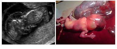
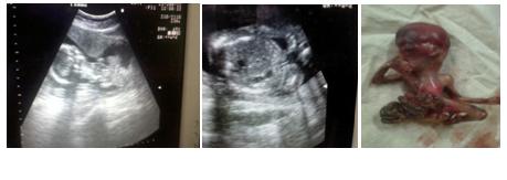
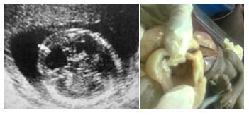
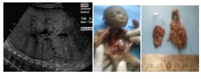
[Table/Fig-4] shows that a variety of anomalies were detected. Five turned out to be lethal, namely, cystic hygroma [Table/Fig-4], body stalk anomaly [Table/Fig-5], non immune foetal hydrops [Table/Fig-6] lost to follow up, the Dandy Walker syndrome [Table/Fig-7] and multicystic dysplastic kidneys [Table/Fig-8]. Two were non lethal, namely, isolated single umbilical arteries and duplication cyst [Table/Fig-9].
Multicystic dysplastic kidney
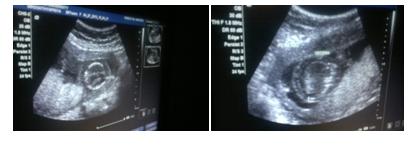
Isolated single umbilical Artery
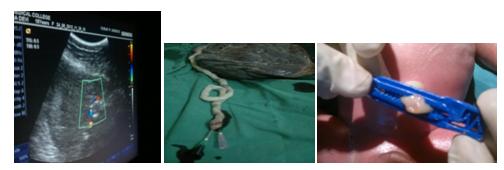
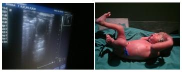
At 32 weeks, the patient with single umbilical artery developed oligohydramnios and abnormal Doppler patterns. An elective LSCS was done and baby was normal duplication cyst [Table/Fig-10], which is a congenital malformation of the alimentary tract. In our case, the cyst was near [1]. the ileum and at term, the baby was delivered by LSCs for obstetric indication and it was normal.
Post natally, the paediatric surgeon’s opinion was obtained and he advised a follow up. For all the cases with anomalies, counselling was given and the parents of the babies with lethal anomalies opted for termination of their pregnancies. Autopsies were carried out in two cases and both showed a very good sonopatholgy correlation. In two cases, the foetal blood was sent for karyotyping, which showed normal karyotypes. The karyotyping results helped us in counselling the parents for their future pregnancies. It was interesting to note that all the lethal anomalies were found in women with no risk and that all of them were primi gravida.
DISCUSSION
Target Scan is an integral part of the antenatal care [2] and it is universally accepted, as was quoted by Geirsson RT and by using which, most of the lethal foetuses can be detected.
In our study, when foetal anomalies were identified in the low risk group, they were given full information, in privacy and the necessary support was provided, as was also reported [3] by Gagnon et al., In our study, foetal autopsies and foetal karyotyping were done when the parents gave their consents and it was easy for us to counsel them regarding the diagnosis and the recurrent risk [1], as was also reported by Desilets V. et al., When the parents did not give their consent to do autopsies of their foetuses, they were explained about the external features [1]. When non lethal anomalies were detected, the parents were well prepared and the handling of the newborns was done appropriately by the paediatricians [3]. In our study, after the lethal anomalies were detected, the accurate information and counselling were given, which helped the couples and their families to make informed decisions. Our study is to assess value of ultrasonography in detecting structural anomalies of the fetus in low risk population which was similar to study conducted by Taipale P et al., [4] Since study was small it did not had impact on Perinatal outcome and it was similar to Ewigman BG et al., [5].
CONCLUSIONS
Target scan has become an indispensable tool because a prenatal detection has several practical benefits, which include a parental preparation, delivery planning and an optimal paediatric care. A prenatal detection can dramatically alter the obstetrical management.
Future Scope: In our study, all non compatible with life anomalies were seen in the young primi without any risk factors.
Hence, the environmental cause has to be studied.