Introduction: Few studies regarding foot changes and health of professional coconut tree climbers of south India are reported. Medical emergencies are very common, especially due to accidental fall from coconut trees, while on job. Objective of the present study is to analyze the altered biomechanics of lower limb joints used by the coconut tree climbers.
Method: Videographs of tree climbing each from a total of 30 male volunteers, all between 30-55 years, engaged in coconut tree climbing profession were collected.
Results: The data revealed the coconut tree climbers are using abnormal rages of foot and lower limb joint motions.
Conclusion: This study establishes an occupationally induced form of altered biomechanics, which leads to professional health hazards.
INTRODUCTION
The ankle-foot complex meets its diverse requirements through its 28 bones that form 25 component joints. The joints include, the proximal and distal tibiofibular joints, the ankle joint, subtalar joint, talonavicular and the calcaneocuboid joints, the five tarso metatarsal joints, five metatarsophalangeal joints and the nine interphalangeal joints. This complexity of the foot structure and the need to perform functions that has to undergo weight-bearing stresses will increase the frequency of ankle or foot problems. The approximate ranges of motion for adults in ankle joint are 20 degrees of dorsiflexion and 30 degrees of plantar flexion, from neutral. The range of motion in subtalar joint is around 15 degrees of eversion and 35 degrees of inversion [1–4].
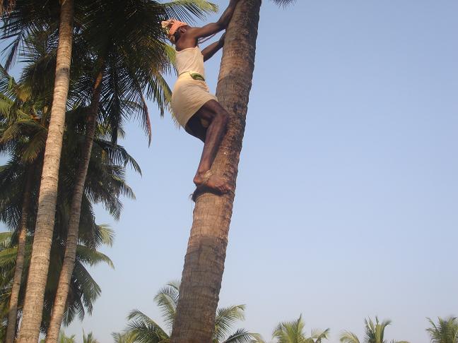
The ankle foot complex must meet both the stability demands and mobility demands in human body. The body requires a flexible foot to accommodate the variations in the external environment, a semi rigid foot that can act as a spring and lever arm for the push off during gait and a rigid foot to enable body weight to be carried with adequate stability [5–8].The alterations of these functions in occupational demands like tree climbing can only be understood when studied in relation to the foot dynamics in gait cycle (the period of time for two steps in walking).In this paper we are presenting an unusual biomechanics of lower limb joints and its implications in professional coconut tree climbers of southern India [Table/Fig-1].This study is of importance, as these professionals are using this altered biomechanics 5-6 hours daily while doing their job, which lead to major anatomical and functional changes in their foot, which over a period of time lead to hazard of falling from trees [9,10].
Lateral view of a professional climber while climbing coconut tree.
The gait cycle is divided into swing phase and stance phase. In swing phase, the foot swings forward to take another step. During the stance phase, the foot is in contact with the ground and Sectioncan be divided into 5 steps in which the stages are heel strike, foot flat, midstance, heel off and toe off [11]. The biomechanics of the foot are best explained by describing what happens to the foot during stance phase of the gait cycle. During heel strike to midstance the foot undergoes pronation. This movement allows the foot to adapt to uneven surfaces and it is during this phase that the foot acts as a shock absorber [11]. As the body weight shifts forward, the feet begin to return to a neutral position in preparation to the heel off. During heel off, the foot begins to supinate. The foot bones, muscles and the plantar fascia act together to supinate, to form a rigid lever. Abnormal amounts of pronation and supination leads to a variety of foot and leg problems. As in walking or climbing, the entire body acting as a closed kinematical chain, these abnormal biomechanics can create lower back, hip, knee, ankle and foot problems [12–17].
OBJECTIVES
To study the functional morphology of lower limb joints of coconut tree climbers. To compare the range of motion of ankle joint required in tree climbing activity in human tree climbers (in the present study) and chimpanzees with that of human terrestrial locomotion (existing data).
i. Subjects
Observations of the coconut tree climbers of different communities in Udupi and Dakshina Kannada districts of southern India were made. The research was approved by the University research committee.
a. Kinematics of the coconut tree climbing
The vertical tree climbing was filmed from the posterior (Back) and lateral(side) aspect as the tree climber ascends from ground to the top of the coconut tree using Sony DCR-SR47E 60GB hand held digital video recorder. Video graphs of tree climbing each from a total of 30 male volunteers, all between 30-55 years, engaged in coconut tree climbing profession were collected. Great care was taken as not to film the face of coconut tree climbers and the movie film was shown to each climbers. The distance between the video camera and the climbing subject varied because the climbing episodes were filmed from the floor. Frames of vertical tree climbing were viewed individually with the help of Motion View software (Version 7.2).
b. Video sequence analysis
The videos selected were opened in the video sequence analysis software (Motion View 7.2) and analyzed. The individual climbing step duration, the cadence per unit time, the sequence of movements in all the joints of the climbers, “stance” phase duration, “swing” phase duration and range of motion in lower limb joints, especially the foot and ankle complex are analyzed and tabulated [Table/Fig-2 & 3].
Summary of video sequence analysis of coconut tree climbing process in relation to gait cycle and accompanying motions of ankle and foot
| Time Duration (Sec) | Ankle movement (Degree) |
|---|
| Upward hops (n=73) | To climb down/step (n=59) | Ascend and descend (n=26) | Cadence/10sec (n=26) | Dorsiflexion (n=24) | Plantarflexion(n=24) |
|---|
| Swing Phase | Stance phace | Total time | Late swing | Early stance | Mid stance to take off | Early stance in downward hops |
|---|
| 0.71 | 1.17 | 1.88 | 1.53 | 57.29 | 6.35 | 33.3 | 24.5 | 24.38 | 37.6 |
| ± | ± | ± | ± | ± | ± | ± | ± | ± | ± |
| 0.05 | 0.05 | 0.07 | 0.7 | 5.9 | 0.2 | 3.63 | 1.92 | 3.6 | 3.02 |
Summary of phases of coconut tree climbing in relation to gait cycle and accompanying motions of the lower limb joints
| % | Hip joint | Knee joint | Ankle joint | Subtalar joint (qualitative) |
|---|
| Government | 0 | Flexion(31°-64°) | Flexion (34°-59°) | Dorsi flexion(20°-36°) | Inversion |
| 35 | Flexion(84°-90°) | Flexion(90°-102°) | Plantar flexion(16°-44°) | Inversion |
| 100 | Flexion(5o)- full extension | Flexion(5o)- full extension | Plantar flexion | Neutral(slight inversion) |
| Private | 10 | Flexion(5°-7°) | Flexion(4°-6°) | Plantar flexion | Neutral | (slight inversion) |
| 100 | Flexion(29°-49°) | Flexion(21°-84°) | Dorsi flexion (34°-47°) | Inversion |
To get the reasonable accurate range of ankle dorsiflexion and plantarflexion, several criteria were applied to identify video that could be used. First, only the lateral view of the tree climber was selected to estimate dorsiflexion at the ankle joint. The ankle nearer to the observer only used for measuring dorsiflexion. Only the video in which the opposite hip and shoulder were obscured by the near hip and shoulder were considered as lateral. Second, videos of lateral views had to be captured within 2-5 meters from the ground to minimize the angular errors that can be introduced by height [18]. Of the 30 videos obtained from coconut tree climbers 24 met with the above criteria and were measured.
The angle of dorsiflexion at the ankle joint was measured by drawing a straight line from the knee to the heel and another straight line from the heel through the metatarsophalangeal joint of the fifth metatarsal. The angle was then subtracted from 900 to make it comparable to the results from the literature (The ankle is 900 in neutral position). Dorsiflexion angles of the same video stills measured a week apart to test the reliability, and suggest that a maximum measurement error is ±3degrees.
Foot inversion (qualitative, as quantitative value does not give accurate results) associated with vertical climbing was assessed from both lateral and posterior views of climbing from all video captured.
RESULTS
The cadence /minute was found to be much less than normal walking(36-39). Increased range of ankle dorsiflexion was seen in late swing to early stance [Table/Fig-2]. Feet of coconut climbers were always inverted except late stance and early swing. The lower limb range of movements were found typical in coconut tree climbing activity [Table/Fig-2 & 3].
DISCUSSION
Some researchers have argued that the last ancestor of human being was probably quite chimpanzee like including in its locomotion [19]. But whether early hominins were adept tree climbers are not very clear [18]. Kinematic data on vertical climbing in wild chimpanzees proves that they use large range of dorsiflexion (45.5°±7.1°) at the ankle joint while climbing trees [18]. Most of their bodyweight is likely to be supported on a single highly dorsiflexed ankle at the stance phase during climbing, while the opposite foot is pushing off the substrate.
Although the coconut tree climbers use the similar vertical upward hops, most of them use ankle belt and grip both feet together while climbing. The maximum dorsiflexion during normal walking in modern humans is only 15°-20° [11,20–22]. In our study, the coconut tree climbers had values of 33.3°±3.63° dorsiflexion while doing their routine occupation which is very high compared to the normal walking function of the human ankle joint.
Studies indicate that dorsiflexing the human ankle to 45° results in soft-tissue failure and severe injury, but our study reported no such soft tissue injuries in coconut tree climbers. But there is clear evidence that the increased range of dorsiflexion used for tree climbing will exert much pressure on the soft tissues around the ankle and foot which make them change or adapt to the occupation [23].
The anterior aspect of the distal tibia in climbing hominoids is predicted to be mediolaterally expanded as the contact area between the tibia and the talus shifts anteriorly during dorsiflexion [18]. As invasive parameters are not included in our study, whether the tibia or any other skeleton of the foot complex adapted itself for this extreme dorsiflexion is unclear.
Literature indicates that from heel strike to the first 20% of stance phase the foot is in slight plantar flexion during normal walking. The foot begins to dorsiflex at this time and by 70% of stance phase the ankle dorsiflexion reaches from 7°to 14°.
In the final 30% of stance phase the foot plantar flexes again to prepare for the push off phase of walking and reaches a range of 14°-20°at push off [24–26]. But, the vertical climbing in coconut tree requires initial dorsiflexion in stance phase (20°-36°) followed by plantar flexion (16°-51°),by push off. This extreme range also will affect the muscles supporting the osteoarchitecture of the foot [27].
Foot inversion at the subtalar and transverse tarsal joint are very important motion for positioning the sole of the foot against the vertical substrate during climbing bouts in arboreal primates. They have a bowed femur and an obliquely oriented tibia relative to the horizontal plane of the ankle joint.
This particular anatomy positions the knees lateral to the centre of gravity and places the free foot in an inverted set [18]. But in contrast, the lower limbs are adapted for terrestrial bipedalism in humans. Humans have an obliquely oriented femur with a bicondylar angle and a tibia which is perpendicularly oriented relative to the plane of the ankle joint. Thus the sole of the human foot is directed plantar rather than medially when in neutral position.
This particular relationship among the geometrics of the femur, tibia and foot in human maladapted the human skeleton for arboreal locomotion. Even though the human foot is maladapted in its anatomy for dorsiflexion and inversion together in climbing as in arboreal primates, the coconut tree climbers of south India climb coconut trees with inversion and plantar flexion rather than dorsiflexion.
CONCLUSIONS
Arboreal primates safely and effectively climb trees as they are capable of extreme dorsiflexion at the ankle and inversion at the subtalar joints. The anatomical orientations of their lowerlimbs are suitable for arboreal habitat. Studies suggest that early hominion skeleton did not have adapted foot with extreme dorsiflexion and inversion so was not significant parts of their locomotor habits. A coconut tree climber who climbs more than 30 trees a day in 7925-6 hours and is capable of extreme dorsiflexion, inversion and plantar flexion, similar to arboreal primates suggest that there is a probability for the skeletal adaptations in the foot. The occupational hazard; falling from coconut tree while on job is seen in more experienced climbers. The altered biomechanics used by them in their profession might be the reason for it. As there is no published data regarding the biomechanics of coconut tree climbing, their occupational habits and probable osteological and functional atavism, we hope that this study provides the preliminary data for further studies.
[1]. Procter P, Paul JPL, Ankle joint biomechanicsJ Biomech 1982 15:627-634. [Google Scholar]
[2]. Katoh Y, Chao EYS, Laughman RK, Biomechanical analysis of foot function during gait and clinical applicationsClin Orthop 1983 177:23-33. [Google Scholar]
[3]. Nigg BM, Biomechanical analysis of ankle andfoot movementMed Sci Sports Exerc 1987 23:22-29. [Google Scholar]
[4]. Jenkyn TR, Anas K, Nichol A, Foot segment kinematics during normal walking using a multisegment model of the foot and ankle complexJ Biomech Eng 2009 131(3):034504 [Google Scholar]
[5]. Kayano J, Dynamic function of medial foot archJournal of the Japanese Orthopaedic Association 1986 60:1147-56. [Google Scholar]
[6]. Chan YY, Fong DT, Yung PS, Fung KY, Chan KM, A mechanical supination sprain simulator for studying ankle supination sprain kinematicsJ Biomech 2008; Aug 7 41(11):2571-4. [Google Scholar]
[7]. Chang R, Van Emmerik R, Hamill J, Quantifying rear foot-forefoot coordination in human walkingJ Biomech 2008 20(41)(14):3101-5. [Google Scholar]
[8]. Glaister BC, Schoen JA, Orendurff MS, Klute GK, A mechanical model of the human ankle in the transverse plane during straight walking: implications for prosthetic designJ Biomech Eng 2009 131(3):034501 [Google Scholar]
[9]. Bhat PS, Kumar A, The medial longitudinal arch of foot in Tree climbing professionalsScientific Medicine 2009 1(2) [Google Scholar]
[10]. George B M, Rao M S, Kumar A, Suvarna N, D’Souza J S, Health of Coconut Tree Climbers of Rural South India – Medical Emergencies, Body Mass Index and Occupational Marks: A Quantitative and Survey StudyJournal of Clinical and Diagnostic Research 2012 February 6:57-60. [Google Scholar]
[11]. Levangies PK, Norkin CC, Joint structure and Function 2001 3rd editionjaypee brothers:392-4. [Google Scholar]
[12]. Tiberio D, The effect of excessive subtalar joint pronation on patellofemoral mechanics: A theoretical modelJ Orthop Sports Phys Ther 1987 9:160-5. [Google Scholar]
[13]. Donatelli RA, Normal anatomy and biomechanics. In: Donatelli RA, ed.The Biomechanics of the Foot and Ankle 1996 2nd edPhiladelphia. PAFA Davis:3-31. [Google Scholar]
[14]. McClay I, Manal K, Coupling parameters in runners with normal and excessive pronationJ Appl Biomech 1997 13:109-24. [Google Scholar]
[15]. Smith J, Szczerba JE, Arnold BL, Martin DE, Perrin DH, Role of hyperpronation as a possible risk factor for anterior cruciate ligament injuriesJathl Train 1997 32:25-28. [Google Scholar]
[16]. Borton DC, Saxby TS, Tear of the plantar calcaneonavicular (spring) ligament causing flatfoot. A case reportJ Bone Joint Surg Br 1997 79(4):641-3. [Google Scholar]
[17]. Van Boerum DH, Sangeorzan BJ, Biomechanics and pathophysiology of flat footFoot Ankle Clin 2003; Sep 8(3):419-30. [Google Scholar]
[18]. DeSilva J M, Functional morphology of the ankle and the likelihood of climbing in early homininsProc Natl Acad Sci U S A 2009 21;106(16):6567-72. [Google Scholar]
[19]. Gebo DL, Climbing, brachiation and terrestrial quadrupedalism: Historical precursors of hominin bipedalismAm J Phys Anthropol 1996 101:55-92. [Google Scholar]
[20]. Siegler S, Chen J, Schneck CD, The three-dimensional kinematics and flexibility characteristics of the human ankle and subtalar joints—Part I: KinematicsJ Biomech Eng 1988 110:364-73. [Google Scholar]
[21]. Lundberg A, Goldie I, Kalin B, Selvik G, Kinematics of the ankle/foot complex: Plantarflexion and dorsiflexionFoot Ankle 1989 9:194-200. [Google Scholar]
[22]. Rome K, Ankle joint dorsiflexion measurement studies. A review of the literatureJ Am Podiatr Med Assoc 1996 86:205-11. [Google Scholar]
[23]. Parenteau CS, Viano DC, Petit PY, Biomechanical properties of human cadaveric ankle-subtalar joints in quasi-static loadingJ Biomech Eng 1998 120:105-11. [Google Scholar]
[24]. Czerniecki JM, (Foot and ankle biomechanics in walking and running. A reviewAm J Phys Med Rehabil 1988 67:246-52. [Google Scholar]
[25]. Rodgers M M, Dynamic Biomechanics of the Normal Foot and Ankle During Walking and RunningPhysical Therapy 1988 68(12):1822-30. [Google Scholar]
[26]. Kojima S, Nakajima Y, Takada J, Kinematics of the compensatory step by the trailing leg following an unexpected forward slip while walkingJ Physiol Anthropol 2008 27(6):309-15. [Google Scholar]
[27]. Kannus VPA, Evaluation of abnormal biomechanics of the foot and ankle in athletes- ReviewBr J Sp Med 1992 26(2):83-89. [Google Scholar]