Unusual ‘Tick Mark’ Calcification on Chest Radiograph in Rheumatic Heart Disease - CT Imaging Revealing Pericardial Calcification
Peter George1, Archana Kaveri Badekila2, Narasimha Hegde3, Hadihally Suresh Byregowda4
1 Associate Professor, Department of Medicine,
2 Resident, Department of Medicine,
3 Professor, Department of Medicine,
4 Professor, Department of Radiodiagnosis & Imaging, Father Muller Medical College, Mangalore, Karnataka, 575002- India.
NAME, ADDRESS, E-MAIL ID OF THE CORESPONDING AUTHOR: Dr. Peter George, Associate Professor, Department of Medicine, Father Muller Medical College, Father Muller Road, Mangalore, Karnataka, S -575002, India.
Phone: +91 9845177660, +91 824 2238000 Fax: +91 824 2436352
E-mail: drpetergeorge2002@yahoo.com
The identification and the interpretation of subtle opacities on chest radiographs are challenging for clinicians. At times, especially when they are found incidentally, some opacities may be considered as artefacts or insignificant and they are neglected. In the present case, an unusual ‘tick mark’ - shaped dense opacity was incidentally found over the cardiac shadow, and CT imaging revealed pericardial calcification. Pericardial, valvular and atrial calcifications in rheumatic heart disease have been described in the literature.
Pericardial calcification, Rheumatic heart disease, Mitral stenosis, ‘Tick mark- shaped’ Calcification, Chest radiograph, CT imaging
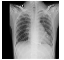
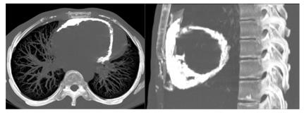
Many a times, the identification and the interpretation of subtle opacities on chest radiographs are challenging for clinicians. Especially, when they are found incidentally, some opacities may be considered as artefacts or insignificant and they are neglected. In this report, the ‘tick mark’ pattern of calcification over the heart shadow on the chest radiograph [Table/Fig-1] was difficult to be identified as a pericardial calcification. The opacities had appeared like an image artefact at the very first instance. The presence of these calcified lesions, with clinical evidence of mitral valvular disease, prompted further cardiac CT imaging leading to the diagnosis [Table/Fig-2]. The patterns of the calcifications which occur in rheumatic heart disease have been described in the literature [1,2]. These calcifications in rheumatic heart disease, usually occur in the valves, the left atrium and the pericardium [1]. In this patient, a rheumatic aetiology was considered for the association of the pericardial calcifications with valvular heart disease.
Chest radiograph showing ‘tick mark - shaped’ calcified opacity over the heart shadow and straightening of left heart border
CT images showing hyperdense opacities like ‘egg shell’ in the pericardium suggestive of pericardial calcification
Even in this era of rapid advances in imaging techniques, the age old chest radiographs can give remarkable insights for identifying the underlying disease in the routine clinical practice of medicine.
[1]. Ferguson EC, Berkowitz EA, Cardiac and pericardial calcifications on chest radiographsClin Radiol 2010 65(9):685-94. [Google Scholar]
[2]. Bouwman RA, van Schijndel RJ, Images in clinical medicine. Pericardial calcification noted long after Sydenham’s choreaN Engl J Med 2009 361(14):1386 [Google Scholar]