Syringocystadenoma Papilliferum of the Scalp in an Adult Male – A Case Report
Mima Maychet B Sangma1, Simon David Dasiah2, Ramachandra Bhat V3
1 Assistant Professor, Department of General Surgery, Indira Gandhi Medical College & Research Institute, Pondicherry-605009, India.
2 Professor & Head, Department of General Surgery, Indira Gandhi Medical College & Research Institute, Pondicherry-605009, India.
3 Professor & Head, Department of Pathology, Indira Gandhi Medical College & Research Institute, Pondicherry-605009, India.
NAME, ADDRESS, E-MAIL ID OF THE CORRESPONDING AUTHOR: Dr. Mima Maychet B. Sangma, Assistant Professor, Department of General Surgery, IGMC & RI, Vazhudavur Road, Kathirkamam, Pondicherry-605009, India.
E-mail: mimamaychet@gmail.com
Syringocystadenoma papilliferum is a benign adnexal skin tumour of the apocrine or the eccrine type with characteristic histological features and varied and non-distinct clinical findings. It is relatively a rare neoplasm, which is called as a childhood tumour, since it usually appears at birth or during puberty. A case of syringocystadenoma papilliferum of the scalp in an adult male has been presented, which was clinically diagnosed at first as keratocanthoma of the scalp but was later histologically confirmed as syringocystadenoma papilliferum.
Skin hamartoma, Apocrine sweat glands, Sebaceous nevus, Basal cell carcinoma
INTRODUCTION
Syringocystadenoma papilliferum is a rare benign hamartomatous adnexal tumour which originates from the apocrine or the eccrine sweat glands. It is relatively a rare neoplasm, predominantly a childhood tumour. In about 50% of those who are affected, it is present at birth, and in a further 15%-30%, the tumour develops before puberty [1]. The lesion of syringocystadenoma papilliferum usually measures between 1 to 3 cm and <4cms in diameter. The tumour has varied clinical presentations. The plaque type which presents as a hairless area on the scalp, is commonly associated with a sebaceous nevus of Jadassohn. Appearance of the lesion in the face and neck region is seen in the linear type and the solitary nodular type shows predilection for the trunk [2]. A presentation with multiple lesions is rare, and in those lesions which arise outside the head and neck region, it is even more uncommon. Syringocystadenoma papilliferum may occur de-novo or within a nevus sebaceous. It occasionally co-exists with other tumours such as basal cell carcinomas and verrucous carcinomas [3]. We are presenting a case which was clinically diagnosed as keratocanthoma of the scalp but was later histologically diagnosed as syringocystadenoma papilliferum.
CASE REPORT
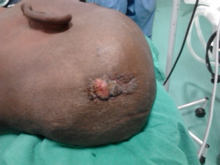
A 38 years old male presented with the complaint of a swelling on the scalp, which was there for the past 12 years [Table/Fig-1]. Initially, the patient had a nodule of around 1 cm over the right side of the parietal region of the scalp, which did not increase in size. There was no growth of hair over the swelling. The nodule turned into a 1 cm ulcer in the past 2 years, which gradually increased in size, with a serous discharge in the past 8 months. The lesion attained the size of 3.5cm in length with 1.5cms in breadth. The lesion consisted of multiple pink to red to brown, firm nodules that occurred in groups. The base was not indurated and the lesion was not fixed to the deeper structures. There was no regional lymphadenopathy. No other skin lesions were noted elsewhere.
Scalp lesion of syringocystadenoma papilliferum of the scalp

The Operative Procedures: The patient was operated under local anaesthesia. The lesion was excised completely with a normal margin of around 1 cm and with a depth upto the subcutaneous plane. He was followed up for 10 months, he showed no recurrence and he had a good cosmetic recovery.
Histopathological Examination: Gross findings; the skin showed a nodular ulcerated lesion with everted margins, which measured 3.5x2x1.5cm above the surface. The cut surface showed that the lesion appeared to sit over the dermis.
Microscopy Findings: The HPE report showed an epidermis with papillomatosis, with several cystic invaginations into the dermis. These invaginations showed many papillae which were lined by two rows of cuboidal to columnar epithelial cells, with oval nuclei and a pale eosinophilic cytoplasm. Some of these cells showed a decapitation secretion. The deep dermis showed few tubular glands with lumina. The stroma contained a dense mononuclear cell infiltrate, which comprised predominantly of plasma cells. The sebaceous glands appeared to be normal. The histopathological features were diagnostic of syringocystadenoma papilliferum [Table/Fig-2].
HP Section showing papillary invagination with plasma cells in stroma (HPE, x 100)

DISCUSSION
Syringocystadenoma papilliferum is a rare benign tumour which is believed to be derived from the apocrine or the eccrine sweat glands. It usually appears at birth or during infancy, around the time of puberty, but in this case, it started appearing at the age of 26 years. The nodular variety has its predilection for the trunk, but here, it presented on the scalp. Its association with a hamartomas lesion of a follicular or a sebaceous origin is common [4]. In about one-third of the case, syringocystadenoma papilliferum is associated with a nevus sebaceous. Multiple tumours of adnexal origin (such as trichoblastomas, aporine adenomas, hidradenoma papilliferum, porona follicular, trichilemmoma etc.) have been reported to arise on a sebaceous nevus, among which Syringocystadenoma papilliferum may be included [5]. Basal Cell Carcinoma (BCC) development has been reported in upto 10% of the cases [6]. In the majority of the cases, there is a coexistent nevus sebaceous. Squamous cell carcinoma (SCC) may also develop, but much less frequently. Till now, only two cases of verrucous carcinoma in conjunction with Syringocystadenoma papilliferum, have been published [7]. Ductal carcinomas which arise from Syringocystadenoma papilliferum have been reported as well [8]. An ulceration or a rapid enlargement is indicative of a malignant transformation. Syringocystadenocarcinoma papilliferum is a malignant counterpart of syringocystadenoma papilliferum [9]. The diagnosis is clinically suspected and histologically confirmed. Due to the risk of a malignant change, a prophylactic surgical excision, followed by a detailed histological examination, is the treatment of choice. Considering the size and the ulceration, an excision was done with a 1cm normal margin, but it proved to be histologically benign and a malignant transformation of the tumour was not seen, even after a follow up for a period of ten months.
In conclusion, syringocystadenoma papilliferum is a rare neoplasm, and even though it is called as a childhood tumour as it usually appears at birth, during infancy or around the time of puberty, it rarely appears in adults. The nodular variety has a predilection for the trunk, but here, it presented on the scalp. In the present case, it was clinically diagnosed at first as keratocanthoma of the scalp, but later, it was histologically confirmed as syringocystadenoma papilliferum. Such a presentation of this tumour may generate multiple differential diagnoses and it must be sent for a histopathological examination.
[1]. Karg E, Koram I, Varga E, Ban G, Turi S, Congenital syringocystadenoma papilliferumPediatrics Dermatol 2008 25:132-33. [Google Scholar]
[2]. Katoulis AC, Bozi E, Syringocystadenoma papilliferumOrphanet Encyclopedia April. 2004 [Google Scholar]
[3]. Yorukoglu A, Demirkan N, Tasli L, Kolluksez U, P86 syringocystadenoma papilliferumMelanoma research June 2010 20:e79-e80. [Google Scholar]
[4]. Pinkus H, Life history of naevussyringocystadenomatouspapilliferusArch Dermatol Syphil 1954 69:305-22. [Google Scholar]
[5]. Stavrianeas NG, Katoulis AC, Stratigeas NP, Development of multiple tumours in a sebaceous nevus of JadassohnDermatology 1997 195:155-58. [Google Scholar]
[6]. Helwig EB, Hackney VC, Syringocystadenoma papilliferum: Lesion with and without naevus sebaceous and basal carcinomaArch Dermatol 1955 71:361-72. [Google Scholar]
[7]. Monticciolo NL, Schmidt JD, Morgan MB, Verrucous carcinoma papilliferumAnn Clin Lab Sci 2002 32:434-37. [Google Scholar]
[8]. Hu Gel H, Reguena L, Ductal carcinoma arising from a syringocystadenoma papilliferum in a nevus sebaceous of JadassohnAm J Dermatopathol 2003 23:490-93. [Google Scholar]
[9]. Ishida-Yamamoto A, Sato K, Wada T, Syringocystadenoma papilliferum: A case report and immunohistochemical comparison with its benign counterpartJ Am Acad Dermato 2001 45:755-59. [Google Scholar]