Pleomorphic Adenoma of the Lacrimal Gland in an Eleven Years Old Girl
Ananthalakshmi Vijayakumar1
1 Assistant Professor, Pathology, Sree Balaji Medical College and Hospital, Bharath University, India.
NAME, ADDRESS, E-MAIL ID OF THE CORRESPONDING AUTHOR: Ananthalakshmi Vijayakumar, 2/8, 5th Cross Street, New Colony, Chromepet, Chennai, India - 600044.
Phone: 9500043179 E-mail : drananthalakshmi@gmail.com
We are presenting a case of pleomorphic adenoma of the lacrimal gland in a 11 years old girl. This is a rare presentation. Pleomorphic adenoma is the most common epithelial tumour of the lacrimal gland, which represents 12% of all the lacrimal fossa lesions. It typically presents in middle age and is rare in children who are under the age of sixteen years, with only a few previously reported cases.
Pleomorphic adenoma, Lacrimal gland, Children
BACKGROUND
Pleomorphic adenoma of the lacrimal gland is a rare presentation in children.Pleomorphic adenoma is the most common epithelial tumour of the lacrimal gland, which represents 12% of all the lacrimal fossa lesions [1]. It typically presents in middle age and is rare in children who are under the age of 16 years, with only a few previously reported cases [2–7]. We are reporting a case of a lacrimal gland pleomorphic adenoma in a 11 year old girl.
CASE PRESENTATION
A 11 years old girl child presented with a history of a slowly progressing
painless swelling in the lateral aspect of the right eye, of 6 months duration. The examination of her right eye revealed a firm, nodular, non tender, non pulsatile and irreducible swelling in the lateral aspect of the right orbit. The conjunctiva over the swelling was freely movable. There was a mild conjunctival congestion over the swelling. The ocular examination was otherwise within normal limits. The left eye examination was within normal limits.
Ultrasonography revealed a tumour which showed a medium reflectivity with a moderate sound attenuation, with a well defined contour. Computerized tomography scan of the right orbit revealed a homogenous soft tissue mass in the lateral aspect of the right orbit, which was not infiltrating the surrounding tissues, which was suggestive of a benign tumour.
Tumour resection was performed under general anaesthesia, by doing a modified lateral orbitotomy. The periorbita was elevated from the lateral wall and the roof of the orbit. The periosteum was incised inferiorly to the lower pole of the orbital part of the gland and the incision extended anteriorly and posteriorly, to beyond the palpable limits of the neoplasm. The incision was continued superiorly at either end of this incision, and the superior periorbita was incised medially to the upper pole of the gland, thereby mobilizing the gland on an intact ‘island’ of the free periorbita. Peroperatively, a firm slightly lobulated swelling which was free from other structures, was seen. The tumour was removed in toto by doing a blunt dissection and an intact capsule was left behind. This tumour had no connection with any other structures in the orbit. The post operative period was uneventful.
METHODS
The surgical specimen was formalin fixed and paraffin embedded. The sections were stained with the routine haematoxylin and eosin stains. A written consent was obtained from the patient for the publication of the patient’s details.
RESULTS
Gross Appearance
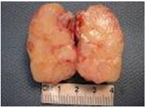
Grossly, the specimen weighed 36.8 g and it measured 4 × 3 × 2 cm [Table/Fig-1].
The external surface appeared grey white and nodular.
The cut section showed a glistening appearance [Table/Fig-1].
Gross specimen measuring 4×3×2 cm

Microscopic Appearance
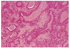
The histological evaluation revealed a tumour tissue which was composed of epithelial elements which were arranged in a trabecular pattern, with glands and cysts. Foci of squamous metaplasia were present. The epithelioid cells were cuboidal to columnar. The stroma was chondromyxoid. No malignant component was present [Table/Fig-2].
Cellular lesion with biphasic patternrecurrence

The clinical presentation and the macroscopic aspects, together with the histological pattern and the cytological characteristics, addressed the diagnosis towards that of a lacrimal gland pleomorphic adenoma.
DISCUSSION
A pleormorphic adenoma is the most common benign epithelial lacrimal gland tumour. The incidence of pleomorphic adenoma in the literature has been reported to be 9–27% of all the lacrimal gland tumours [1, 2].
The ectopic lacrimal gland tissue may be found in the caruncle, the bulbar conjunctiva, the outer canthus and the lower lid intraorbital and intraocular regions and in any location in and around the eye. Pleomorphic adenomas of the orbit can be found in the lacrimal gland, in the palpebral lobe of the lacrimal gland which is associated with a raised intraocular pressure, in the upper lid, in the lower lid developing in Krause’s gland, in the lacrimal sac and in the orbit [3].
Pleomorphic adenoma of the lacrimal gland usually manifests as a slowly progressive, painless superolateral orbital mass. The chronically expanding tumour typically produces a well-corticated fossa in the orbital rim, but a true bone destruction is rare. The chronicity, the absence of pain, and the radiological evidence of a bony fossa, suggest that a lacrimal gland mass is benign rather than malignant [2].
Pleomorphic adenoma presents an excellent prognosis, particularly, following a complete excision, with the pseudocapsule intact [8]. If the tumour is incompletely removed, a recurrence may occur, which may usually takes many years to become clinically apparent. Although the recurrent tumours are usually as benign as the original ones, some may become frankly malignant many years (mean- 17.4 years) after the initial excision of the pleomorphic adenomas [8–13]. The malignant components of pleomorphic adenomas are usually carcinomatous, which show the features of either an adenocarcinoma or a poorly differentiated carcinoma, sometimes sarcomatous ones. Furthermore, a malignant evolution in a tumour patternrecurrence may be associated with an intracranial invasion to the cavernous sinus, although most of the recurrences involve only the intraorbital structures. An intracranial infiltration following a malignant transformation in recurrent lacrimal gland tumours, have previously been reported [8–11].
The recommended treatment for suspected pleomorphic adenomas of the lacrimal gland is a complete surgical resection or an en block excision. However, an incomplete or a piecemeal removal of the lesion or a rupture of its pseudocapsule, inevitably results in a recurrence, which may evolve malignant changes in the original tumour [9, 11]. A biopsy of the lesion may cause seeding of the tumour cells into the orbit and a clinical recurrence [14]. Thus, it has been emphasized that a complete excision of the lesion within the pseudocapsule is essential to prevent the potential malignant evolution in recurrent pleomorphic adenomas [11, 12].
CONCLUSION
Given an appropriate clinical and a radiological presentation, and without any regard to the patient’s age and the duration of the symptoms, the diagnoses of lacrimal gland pleomorphic adenomas should be considered, even in the paediatric age group.
[1]. Shields CL, Shields JA, Eagle RC, Clinicopathologic review of 142 cases of lacrimal gland lesionsOphthalmology 1989 96:431-35. [Google Scholar]
[2]. Font RL, Gamel JW, Epithelial tumors of the lacrimal gland: an analysis of 265 cases. In: Jakobiec FA, edOcular and adnexal tumors 1978 BirminghamAesculapius:787-805. [Google Scholar]
[3]. Patyal S, Banarji A, Bhandauria M, Gurunadh VS, Pleomorphic adenoma of a subconjunctival ectopic lacrimal glandIndian J Ophthalmol 2010 58(3):245-47. [Google Scholar]
[4]. Sanders TE, Mixed tumor of the lacrimal glandArch Ophthalmol 1939 21:239-60. [Google Scholar]
[5]. Tsunoda S, Yabuno T, Sakaki T, Pleomorphic adenoma of the lacrimal gland manifesting as exophthalmos in adolescenceNeurol Med Chir 1994 34:814-16. [Google Scholar]
[6]. Faktorovich EG, Crawford JB, Char DH, Benign mixed tumor (pleomorphic adenoma) of the lacrimal gland in a 6-year-old boyAm J Ophthalmol 1996 122:446-47. [Google Scholar]
[7]. Mercado GJV, Gündüz K, Shields CL, Pleomorphic adenoma of the lacrimal gland in a teenagerArch Ophthalmol 1998 116:962-63. [Google Scholar]
[8]. Zimmerman LE, Sanders TE, Ackerman LV, Epithelial tumors of the lacrimal gland: prognostic and therapeutic significance of histologic typesInt Ophthalmol Clin 1962 2:337-67. [Google Scholar]
[9]. Ni C, Cheng SC, Dryja TP, Cheng TY, Lacrimal gland tumoursInt Ophthalmol Clin 1982 22:99-120. [Google Scholar]
[10]. Perzin KH, Jakobiec FA, Livolsi V, Desjardins L, Lacrimal gland malignant mixed tumors (carcinomas arising in benign mixed tumors): a clinico-pathologic studyCancer 1980 45:2593-606. [Google Scholar]
[11]. Riley FC, Henderson JW, Report of a case of malignant transformation in benign mixed tumor of the lacrimal glandAm J Ophthalmol 1970 70:767-71. [Google Scholar]
[12]. Yamasaki T, Kikuchi H, Yamabe H, Yamashita J, Multiple intracranial metastases following malignant evolution in recurrent pleomorphic adenoma of the lacrimal glandNeurol Med Chir (Tokyo) 1990 30:1038-42. [Google Scholar]
[13]. Henderson JW, Farrow GM, Primary malignant mixed tumors of the lacrimal glandOphthalmology 1980 87:466-75. [Google Scholar]
[14]. Lloyd GA, Lacrimal gland tumours: the role of CT and conventional radiologyBr J Radiol 1981 54:1034-38. [Google Scholar]