A Rare Finding of the Superficial Palmar Arch-Developmental and Clinical Significance
Alok Saxena1, Kishore Kumar Agarwal2, Biswabina Ray3, Samuel Pyrtuh4
1 Senior Resident, Veer Chandra Singh Garhwali Government Medical Sciences and Research Institute, Srinagar, Uttarakhand, India.
2 Assistant Professor, Veer Chandra Singh Garhwali Government Medical Sciences and Research Institute, Srinagar, Uttarakhand, India.
3 Additional Professor, AIIMS, Bhopal, India.
4 Demonstrator, Ross University School of Medicine, Caribbean.
NAME, ADDRESS, E-MAIL ID OF THE CORESPONDING AUTHOR: Mr. Alok Saxena, H.No. 171/2, Saket Colony Roorkee, Uttarakhand, India.
Phone: 9897699599 Email: alok.sxna@gmail.com
The ulnar artery provides a major blood supply to the hand in the form of the superficial palmar arch, with the assistance of the radial artery. A rare pattern of the superficial palmar arch was observed in a formalin fixed, male cadaveric left hand. The ulnar artery was only involved in the formation of this arch, which provided three common palmar digital arteries which ran into the second, third and the fourth spaces between the corresponding digits and one proper palmar digital artery which ran along the ulnar side of the little finger. The main trunk of the ulnar artery bifurcated to supply the thumb and the index finger. The superficial branch of the radial artery did not participate in the arch formation. The arch was completed by the radial artery proper on the dorsolateral surface of the hand, after joining the point of bifuracation of the ulnar artery.
Collateral circulation, Radial artery, Ulnar artery
INTRODUCTION
The acceptance of the term, ‘complete superficial palmar arch’ (SPA) is based on the anastomosis of the Ulnar Artery (UA) and the superficial branch of the Radial Artery (RA) [1]. Al Turk et al., considered that the major vessel arose from the SPA and fed the thumb and the index finger as the first common digital artery [2]. There are very few schools of thought which explain the development of the arterial pattern in humans. As suggested by De Vriese, the arteries arise from the initial capillary reticulum in association with each of the principal nerves [3]. Singer opined the development of the arterial system by a bud formation from the axial arterial trunk [4]. A study which was conducted by Rodriguez- Niedenfuhr on human embryos, described the commencement of the vascular system at stage 12 (3-5mm Crown-rump length; 26th day) and differentiation of the arterial wall into all the segments, which included the radial artery and the palmar arch in stage 23 (27-31mm Crown-rump length; 54th day). According to this theory, a capillary plexus is formed, which enters the limb bud and grows along with it distally. Thereafter, only the axial artery supplies the limb and the capillary plexus, giving rise to the arteries of the antebrachial region through a sprouting mechanism [5].
CASE REPORT
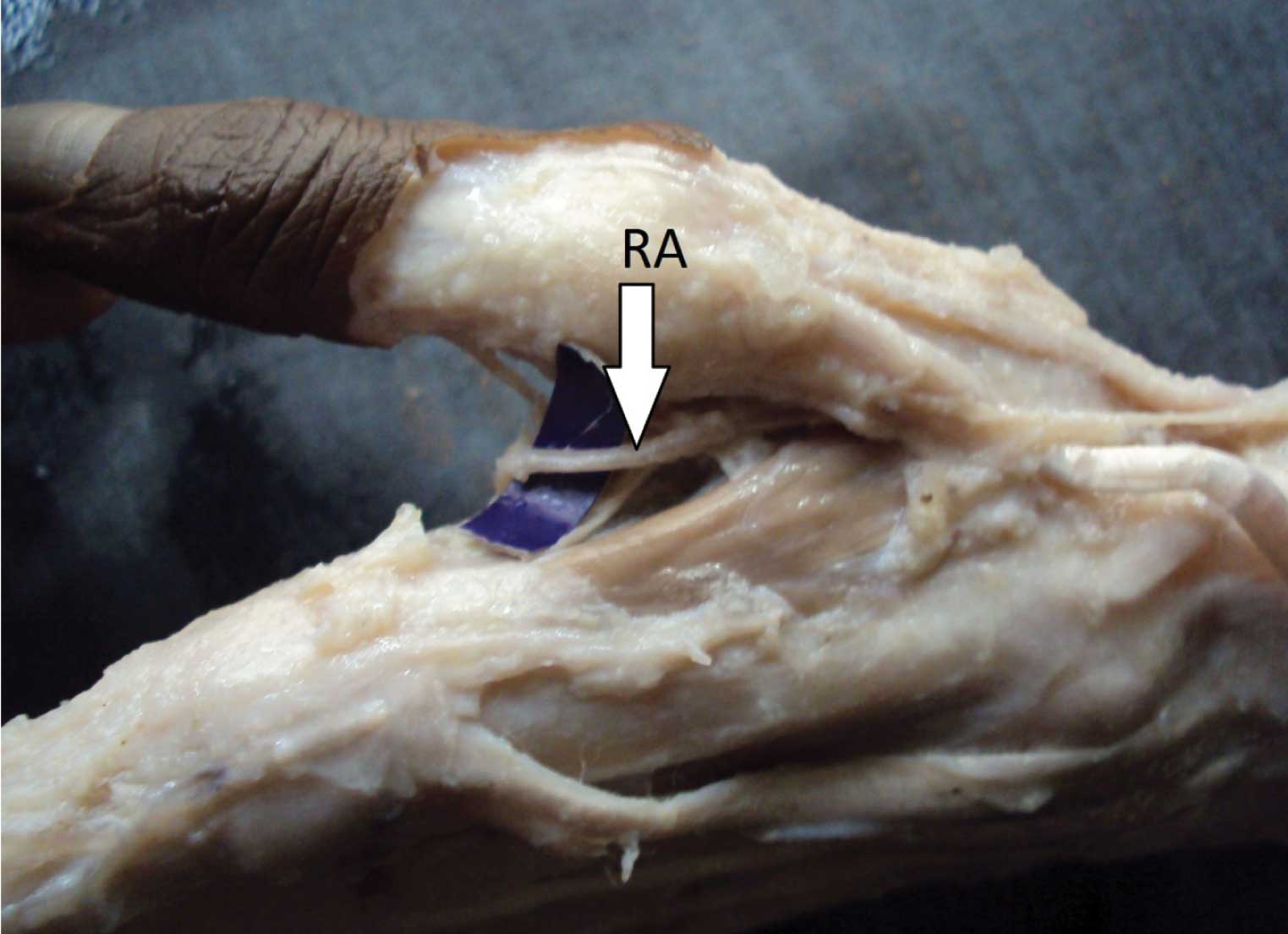
During the routine dissection of a formalin fixed male cadaver, we came across an unusual pattern of the SPA. The vessels which contributed to the SPA were identified and a digital photographic record was documented. The arch was devoid of the superficial branch of the RA. It was solely formed by the UA on the palmar region and it provided three common palmar digital arteries which ran into the second, third and the fourth spaces between the corresponding digits and one proper palmar digital artery which ran along the ulnar side of the little finger. The main trunk of the UA bifurcated and fed the thumb and the index finger [Table/Fig-1]. The ulnar artery completed the arch after joining the RA proper on the dorsolateral surface of the hand [Table/Fig-2] instead of joining the superficial palmar branch of the RA, since this branch was completely in absentia.
Picture depicting the ulnar type superficial palmar arch (SPA) and its branching pattern in left hand. Note the bifurcation of ulnar artery (UA) nourishing thumb and index finger

Dissected specimen showing Radial Artery (RA) on dorsolateral
aspect of the same hand
DISCUSSION
Quain described in his book, that the RA is said to be “usual”, when it originates proximal to the elbow joint: provided a recurrent branch runs along the forearm, close to the supinator longus muscle: turns at the dorsum of the hand beneath the extensor tendons of the thumb: enters in the palm by passing between the first two metacarpal bones, and between the heads of the first dorsal interosseous muscle: and, finally, lies in the palm beneath the tendons of the flexor muscles. The “usual” UA commences from the close proximity of the elbow joint: it gives the interosseous artery and two branches: it approaches the flexor carpi ulnaris and the ulnar nerve, above the middle of the forearm: it enters the palm and lies between the palmar fascia and the flexor tendons and divided into branches to supply the digits [6].
Previous literatures have defined the variations in the construction of a complete SPA and their prevalence [Table/Fig-3]. A higher proportion of the ulnar type arch was recorded in a cadaveric study which was conducted on 650 dissections [7]. Karlsson also found a greater incidence of the presence of the ulnar type arch [8]. On the contrary, no ulnar arch was noticed in a study which was done on 25 white Caucasian subjects by using a Doppler ultrasonic flowmeter [2]. A study which was based on fifty dissected hands showed a very minimal prevalence of the ulnar arch [9]. The data which was recorded by Loukas et al., revealed lesser values in comparison to the classical radio ulnar arch and greater values than the median ulnar and the radio median ulnar arches [10]. Ottone et al’s results revealed a 20.9% ulnar arch and a 43% classical radioulnar arch [11].
Prevalence of various patterns of superficial palmar arches (SPA)
| Authors | Year | Prevalence of types of complete SPA (%) |
|---|
| Classical radio ulnar (%) | Ulnar type (%) | Median-ulnar (%) | Radio-median-ulnar (%) |
|---|
| Coleman and Anson | 1961 | 34.5 | 37 | 3.8 | 1.2 |
| Karlsson et al., | 1982 | 32 | 64 | 4 | - |
| Al Turk et al., | 1984 | 78 | - | 4 | 2 |
| Patnaik et al., | 2002 | 76 | 2 | - | - |
| Loukas et al., | 2009 | 40 | 35 | 15 | 6 |
| Ottone et al., | 2010 | 43 | 20.9 | - | - |
In the present study, we found an exclusive vascular pattern in the cadaveric hand, which did not show absolute similarity with the findings of the above mentioned studies. We observed the origin of the princeps pollicis and the radialis indicis from the SPA. These results were consistent with those of the study which was conducted by Loukas, except the existence of the RA. The artery was present in our findings but it was absent in the study of Loukas [10].
Various diagnostic procedures have been implemented to assess the viable collateral circulation in the hand. Gull published a case of pseudoaneurysm in an eighty years old male which was diagnosed by using contrast enhanced computerized tomography angiography and Doppler, which were used to delineate the details of the aneurysm and the collateral circulation [12]. The modified Allen’s test is considered as a primary screening method due to its feasibility and Doppler ultrasonography can be used as an alternative, in case unsatisfactory results are obtained by using the former test [13]. A collateral circulation is the remedy for diseases of the palm, which can be established by an anastomosis between the radial and the ulnar arteries [14]. The radial and the ulnar arteries are the main sources to the palm and the further supply can be added by the median and the interosseous arterial systems. The collateral circulation can be assessed by using the modified Allen’s test, Doppler ultrasound, pulse oximetry and arterial angiography [15].
Our finding was rare, as it showed an anastomosis between the RA and the UA, since the former can be cropped from the dorsolateral surface of the hand without affecting the collateral circulation in the hand, for grafting purposes. It should be noted that the patients with coronary disease must be screened before the RA harvesting. Knowledge on the variations in the arterial patterns may be a useful guide for the radiologists and the vascular surgeons when they perform grafting procedures and fistula arterial surgeries. The variations are equally important for the anatomists, to help in enhancing the knowledge of the medical students, so that they can deal with such type of cases in their clinical practice.
[1]. Fazan VPS, Borges CT, Da silva JH, Caetano AG, Filho OAR, Superficial palmar arch: An arterial diameter studyJ Anat 2004 204:307-11. [Google Scholar]
[2]. Al Turk M, Metcalf WK, A study of superficial palmar arteries using the Doppler ultrasonic flowmeterJ Anat 1984 138:27-32. [Google Scholar]
[3]. De Vriese B, Recherchessur L’, Evolution des vaisseaux sanguins des members chez l’ homeArchiv Biol 1902 18:665-730. [Google Scholar]
[4]. Singer E, Embryological pattern persisting in the arteries of the armAnatomical Record 1933 55:403-09. [Google Scholar]
[5]. Rodriguez-Niedenfuhr M, Burton GJ, Deu Sanudo JR, Development of the arterial pattern in the upper limb of staged human embryos: normal development and anatomic variationsJ Anat 2001 199:407-17. [Google Scholar]
[6]. Quain R, Anatomy of the Arteries of the Human Body 1844 LondonTaylor and Walton [Google Scholar]
[7]. Coleman SS, Anson BJ, Arterial pattern in the hand based upon a study of 650 specimensSurgery, Gynaecology, Obstetrics 1961 113:409-24. [Google Scholar]
[8]. Niechajev Karlsson S, Arterial anatomy of the upper extremityActa Radiologica Diagnosis 1982 23:115-21. [Google Scholar]
[9]. Patnaik VVG, Kalsey G, Singla RK, Palmar arterial arches- A morphological studyJ AnatSoc India 2002 51:187-93. [Google Scholar]
[10]. Loukas M, Tubbs S, Louis Jr RG, Apaydin N, Princeps pollicis artery arising from the superficial palmar archSingapore Med J 2009 50:391-92. [Google Scholar]
[11]. Ottone NE, Prum N, Dominguez M, Blasi E, Medan C, Shinzato S, Analysis and clinical importance of superficial arterial palmar irrigation and its variants over 86 casesInt J Morphol 2010 28:157-64. [Google Scholar]
[12]. Gull S, Spence AJ, Loan W, Superficial palmar arch aneurysm after carpal tunnel decompression, a rare complication: A case reportCase Reports in Medicine 2011 2011:3Article ID 595120, .doi: 10.1155/2011/595120 [Google Scholar]
[13]. Ruengsakulrach P, Eizenberg N, Fahrer C, Fahrer M, Buxton BF, Surgical implications of variations in hand collateral circulation: anatomy revisitedJ Thorac Cardiovasc Surg 2001 122:682-86. [Google Scholar]
[14]. Vollala VR, Nagabhooshana S, Rao M, Potu BK, Pamidi N, Bolla SR, Unorthodox superficial palmar arch observed in South Indian cadaver: A case reportCases Journal[doi: 10.4076/1757-1626-2-6362] 2009 July [cited 2011 Aug 04] [Approximately 3 p.] http://casesjournal.com/casesjournal/article/view/6362 [Google Scholar]
[15]. Bataineh ZM, Moqattash A complex variation on superficial palmar archFolia Morphol 2006 65:406-09. [Google Scholar]