The brachial plexus is a major and a complicated plexus at the root of the neck. It is formed by the ventral primary rami of the C5, C6, C7, C8 and the T1 spinal nerves. During the routine under graduate dissection of the right upper limb of an adult female cadaver, a variant pattern of a two trunked brachial plexus was encountered. The upper trunk was formed by the fusion of the C5 and the C6 roots. The C7 root, instead of continuing as the middle trunk, joined with the roots of C8 and T1 to form the lower trunk.
On the left side, the usual pattern of the brachial plexus was seen. The knowledge on such variations are of interest to anatomists, clinicians, anesthesiologists and especially, to surgeons. These are of immense importance during surgical explorations of the axilla and the arm region and also during nerve blocks. It also helps the clinicians in getting a proper understanding of some previously unexplained clinical symptoms. Further, the ontogeny and the phylogeny of this entity have been discussed in detail.
Brachial plexus, Roots, Trunks, Cords
INTRODUCTION
The brachial plexus is formed by the ventral primary rami of the C5, C6, C7, C8 and the T1 spinal nerves. The upper trunk of the plexus is formed by the union of the C5 and the C6 roots; the middle trunk is the continuation of the C7 root and the lower trunk is formed by the union of the C8 and the T1 roots. Each trunk divides into the ventral and dorsal divisions. The ventral divisions of the upper and the middle trunk join to form the lateral cord; the ventral division of the lower trunk continues as the medial cord and the posterior divisions of all the three trunks join to form the posterior cord of the brachial plexus [1].
The brachial plexus may be damaged in open, closed or obstetrical injuries, it may be pressed upon by the cervical rib or it may be involved in a tumour [2]. The knowledge on its variations is important for explaining incomprehensible clinical signs [3]. Apart from the clinicians, these variations are also of interest to the radiologists, anaesthesiologists, neurosurgeons, neurologists, vascular surgeons and the orthopaedic surgeons [4]. One such case which had two trunks of the brachial plexus (with absence of the middle trunk) on the right upper limb of a female cadaver, was encountered in our department, which has been reported here.
CASE REPORT
During a routine under graduate dissection of the upper limb of an adult female cadaver in the Department of Anatomy, Govt. Medical College, Amritsar (Punjab), India, the following variant of the brachial plexus was encountered.
On the right side, there were two trunks instead of the usual three. The upper trunk was formed by the fusion of the C5 and the C6 roots. The C7 root, instead of continuing as the middle trunk, joined with the roots of C8 and T1 to form a single trunk (which has been termed as the lower trunk in this case). The lateral cord was formed as a continuation of the anterior division of the upper trunk (C5, C6). The medial cord was formed as a continuation of the Shreeanterior division of the lower trunk (C7, 8, T1). Thus, the fibres of C7 were going into the medial cord instead of the usual lateral cord. The root value for the lateral cord was C5, 6 instead of the usual C5-7 and that of the medial cord was C7,8, T1 instead of the usual C8, T1. The posterior cord was formed by the fusion of the posterior divisions of the upper (C5, 6) and lower (C7, 8, T1) trunks. The further branchings of the three cords were normal [Table/Fig-1].
Showing a two trunked brachial plexus.
AA: Axillary artery; C5- T1: Roots of brachial plexus; MC: Medial cord
MCA: Medial cutaneous nerve of arm; MCN: Musculocutaneous nerve
MN: Median nerve; LC: Lateral cord; LT: Lower trunk
PC: Posterior cord; UN: Ulnar nerve; UT: Upper trunk
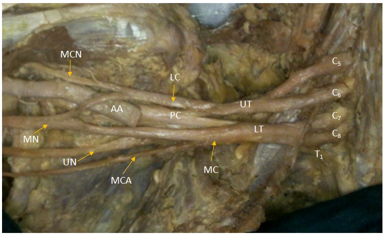
On the left limb, the usual pattern of the brachial plexus was seen.
DISSCUSSION
The common variations in the formation of the brachial plexus i.e. the prefixed and postfixed plexuses, have been well documented [5].
Formation of the lower trunk of the brachial plexus by the C7,C8 and T1 roots is very rare. Only Nayak et al., [6] had earlier encountered an absence of the middle trunk, with the C7 root joining C5 and C6 to form the upper trunk; the lower trunk being formed by C8 and T1. In our case, the two trunks were formed, but the middle trunk (C7 root) had joined C8 and T1 instead of joining C5 and C6. So, it was different from the one which was reported by them.
Ontogeny
Rao and Chaudary [7] are of the opinion that developmentally, the human brachial plexus appears as a single radicular cone in the upper limb bud. Initially, a plexus is formed by the anastomosis between the spinal nerves and then it develops into a solid plate that finally divides into separate trunks and then, divisions. The posterior division supplies the extensor muscles and their surfaces and the anterior division supplies the flexor muscles and their surfaces [8].
The guidance of the developing axons is regulated by the expression of chemoattractants and chemorepulsants in a highly co-ordinated site specific fashion.
Tropic substances such as the brain-derived neurotropic growth factor, neutrin-1, neutrin-2, the c-kit ligand, etc. attract the correct growth cones that happen to take the right path [9]. Any alterations in the signalling between the mesenchymal cells and the neuronal growth cones or the circulatory factors at the time of fission of the brachial plexus cords, can lead to significant variations [10].
Ontogenically, the present variation may be due to a failure on the part of the radicular cone of the nerves of the upper limb to divide into different trunks. This may be attributed to the disproportionate display of the chemoattractants and the chemorepulsants.
Phylogeny
If we trace the phylogeny, no trunk formation is seen in amphibians, reptiles and dogs. Two trunks are formed in marsupials and Lemurs. However, in them, the two lowest roots form an inferior trunk and the others form a superior trunk. Similarly, in gorilla, two trunks are formed, with a root value of C4,5, 6 for the first trunk and of C7,8, T1 for the second trunk [11]. Thus, the present case was more close to that of a gorilla, with the upper trunk being formed from the C5, C6 roots (C4 not contributing) and the lower trunk being formed from C7,8,T1.
Clinical implications
The knowledge on the variations which are involved in the formation of the brachial plexus is important, not only for anatomists, but also for radiologists, anaesthesiologists, neurosurgeons and orthopaedic surgeons. It will be of great use in the surgical treatment of tumours of the nerve sheaths, such as schwannomas and neurofibromas. The awareness on the variations might also help in treating non-neural tumours like lipomas. Orthopaedic procedures of the cervical spine also need a thorough knowledge on the normal and the abnormal formation of the brachial plexus [12].
Such a variant brachial plexus with two trunks, with the lower trunk having a root value of C7,8, T1, may give a confusing clinical picture if it is affected by Klumpke’s paralysis. In such cases, the injury may not be restricted to T1 or C8 only, but rather, it may extend to C7 as well. So, clinicians must be familiar with such an anomaly when they encounter an extended Klumpke’s paralysis case.
[1]. Shetty SD, Nayak BS, Madhav V, Braganza CS, Somayaji SN, A study on the variations in the formation of the trunks of brachial plexusInt J Morphol 2011 29(2):555-58. [Google Scholar]
[2]. Ellis H, Clinical Anatomy: Applied anatomy for students and junior doctors 2006 11thOxfordBlackwell Publishing Ltd:189-97. [Google Scholar]
[3]. Gumusburn E, Adiguzel E, A variation of the brachial plexus characterised by the absence of musculocutaneous nerve: A case reportSurg Radiol Anat 2000 22(1):63-65. [Google Scholar]
[4]. Collins JD, Shaver ML, Disher AC, Miller TQ, Compromising abnormalities of the brachial plexus as displayed by magnetic resonance imagingClin Anat 1995 8:1-16. [Google Scholar]
[5]. Matejcik V, Variations of nerve roots of brachial plexusBratisl Lek Listy 2005 106(1):34-36. [Google Scholar]
[6]. Nayak BS, Nagabhooshana S, Venkatramanal V, A rare variation in the formation of the upper trunk of brachial plexus: a case reportJ Neuro Anat 2005 4:37-38. [Google Scholar]
[7]. Rao PVVP, Chaudary SC, Communication of the musculocutaneous nerve with the median nerveEast African Med J 2000 77(9):498-503. [Google Scholar]
[8]. Hamilton WJ, Boyd JD, Mossman HW, Peripheral nervous system. InHuman Embryology (pre-natal development of form and function) 1962 3rdCambridge, EnglandW. Heffer and Sons Limited:358-60. [Google Scholar]
[9]. Larson WJ, Development of peripheral nervous system. InHuman Embryology 2001 3rdPennsylvaniaChurchill Livingstone:115-16. [Google Scholar]
[10]. Sannes HD, Reh TA, Harris WA, Axon growth and guidance. InDevelopment of nervous system 2000 New YorkAcademic Press:189-97. [Google Scholar]
[11]. Miller RA, Comparative studies upon the morphology and distribution of the brachial plexusAm J. Anat 1932 :143-66. [Google Scholar]
[12]. Royse CE, Sha S, Soeding PF, Royse AG, Anatomical study of the brachial plexus using surface ultrasoundAnaesth Intensive Care 2006 34(2):203-10. [Google Scholar]