Aberrant Adrenal Tissue in Omentum: An Incidental Finding on Ovarian Cystectomy
Meenu Pujani1, Neha Kawatra Madan2, Monisha Choudhury3, Meenakshi Rao4
1 Senior Residents, Department of Pathology, Lady Hardinge Medical College, New Delhi 110001, India.
2 Senior Residents, Department of Pathology, Lady Hardinge Medical College, New Delhi 110001, India.
3 Director Professor, Department of Pathology, Lady Hardinge Medical College, New Delhi 110001, India.
4 Postgraduate Student, Department of Pathology, Lady Hardinge Medical College, New Delhi 110001, India.
NAME, ADDRESS, E-MAIL ID OF THE CORRESPONDING AUTHOR: Dr. Meenu Pujani, Department of Pathology, Lady Hardinge Medical College, New Delhi-110001, India.
Phone: 9818538609
E-mail: drmeenupujani@gmail.com
Sir,
Aaberrant adrenal tissue is a rare occurrence in adults. It is divided into ectopic or heterotopic and accessory adrenal tissue: the former is derived from the adrenal primordium that has migrated to other organs like the kidney and the liver at an embryonic stage, whereas when the medullary tissue migrates to the region of the adrenal cortex and the fragments of the cortical tissue may be split off, forming the accessory adrenal tissue [1]. An accessory adrenal tissue may be found in at least 50% of the neonates and infants if it is meticulously searched for; however, with advancing age, the accessory tissue atrophies and it is present in less than 1% of all the adults [2].
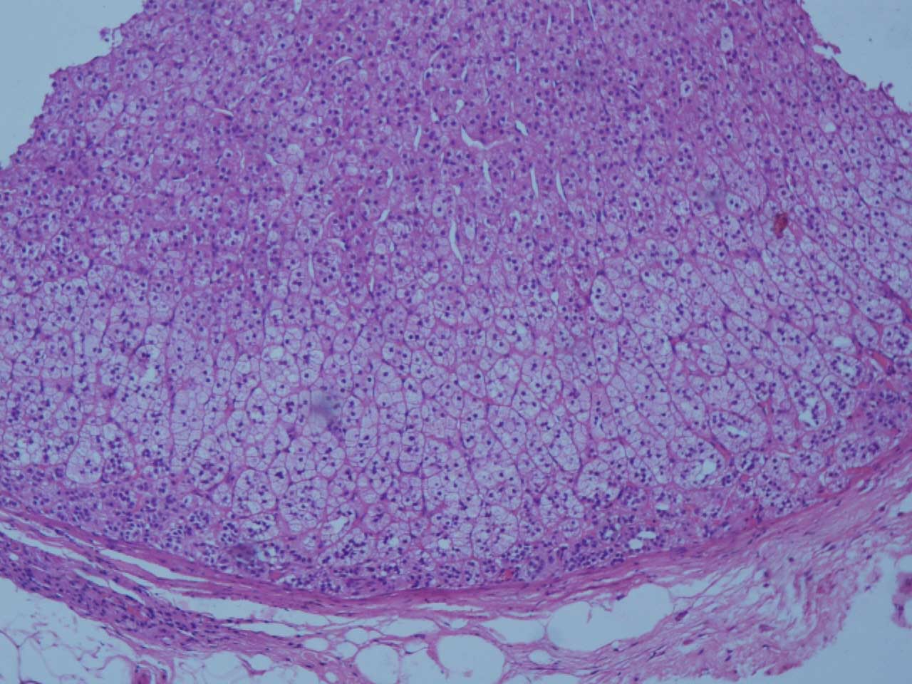
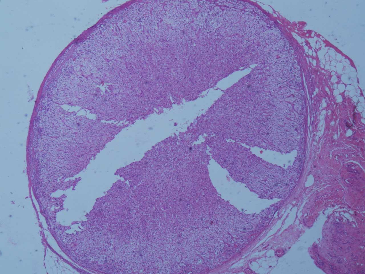
We are reporting a case of a 21 years old unmarried woman who came with the complaint of lower abdominal pain. On ultrasonography, a 6x7 cm anechoic cyst was seen in the left ovary. A laparoscopic cystectomy was performed. A small, yellowish brown nodule which was 0.5 cm in diameter and which was embedded in the omentum was also found, which was clinically suspected to be a caseous lymph node. It was also excised and sent for a histopathological examination. A microscopic examination of the ovarian cyst revealed a serous cystadenoma. The excised nodule was composed of encapsulated adrenal cortical tissue which comprised of the zona glomerulosa and the zona fasciculata which were surrounded by omental adipose tissue [Table/Fig-1 and 2]. A medullary component was not present.
Tissue section showing adrenal cortical tissue: Zona glomerulosa and Zona fasciculata (H&E, 100X)
Tissue section showing adrenal cortical nodule with omental fat (H&E, 40X)
Since the discovery of the yellowish nodules of the adrenal tissue by Morgagini in 1740, several researchers have noticed the presence of the ectopic adrenal tissue in the lung, brain, pancreas, transverse colon and the large intestine [1,3,4]. The unusual location of the ectopic tissue could be related to the misplaced mesothelial cells or the autonomous differentiation of the mesodermal elements [5].
Occasional cases which have described the occurrence of the ectopic adrenal tissue in the omentum, have been reported in the literature [1,6]. Most of the ectopic adrenal nodules are clinically silent. However, the importance of recognition of the ectopic adrenal tissue lies in the fact that it could be the only adrenal tissue in the body, which if accidentally removed surgically, may prove to be fatal. It may become hormonally functional or it may rarely undergo a malignant transformation- Phaeochromocytoma, Leydig’s cell tumour and adrenal adenoma have been documented in the literature [4]. The patients who have undergone bilateral adrenalectomy due to a pathologic ACTH production and compensatory hyperplasia of the aberrant adrenal tissue, could lead to relapse of the symptomatology.
It is important to recognize the existence of such an aberrant tissue at unusual sites like the omentum and to treat them accordingly, in order to avoid any potential complications.
[1]. Shin YM, Hepatic adrenal rest tumor mimicking hepatocellular carcinomaKorean J Hepatology 2010 16:338-41. [Google Scholar]
[2]. Jaffe HL, The suprarenal glandArch Pathol Lab Med 1927 3:414-53. [Google Scholar]
[3]. Schecter DS, Aberrant adrenal tissueAnn Surg 1968 167:421-26. [Google Scholar]
[4]. Bal A, Adhya AK, Mahajan JK, Ectopic adrenocortical rest in the wall of the large intestineIndian J Pathol Micro 2009 52:130-31. [Google Scholar]
[5]. Bozic C, Ectopic fetal adrenal cortex in the lung of newbornVirchows Arch Path Anat 1974 363:371-74. [Google Scholar]
[6]. Macmillan SF, Gilbert JB, Aberrant adrenal tumor of upper part of abdomenArch Surg 1940 40:77-82. [Google Scholar]