Crystallizing Galactocele – An Unusual Diagnosis on Fine Needle Aspiration Cytology
Dhiraj B Nikumbh1, Sushama R Desai2, Pallavi A Shrigondekar3, Ajay Brahmnalkar4, Avinash M Mane5
1 Department of Pathology, Krishna Institute of Medical Sciences Karad, Maharashtra, India.
2 Department of Pathology, Krishna Institute of Medical Sciences Karad, Maharashtra, India.
3 Department of Pathology, Krishna Institute of Medical Sciences Karad, Maharashtra, India.
4 Department of Medicine, Krishna Institute of Medical Sciences Karad, Maharashtra, India.
5 Department of Pathology, Krishna Institute of Medical Sciences Karad, Maharashtra, India.
NAME, ADDRESS, E-MAIL ID OF THE CORESPONDING AUTHOR: Dr. Dhiraj B Nikumbh, Associate Professor, Department Of Pathology, JMF’s Acpm Medical College, Dhule, Maharashtra, India.
E-mail: drdhirajnikumbh@rediffmail.com
A galactocele is a benign cystic lesion that generally occurs during late pregnancy and lactation. Fine Needle Aspiration Cytology (FNAC) reveals a milky fluid, which is diagnostic as well as therapeutic.
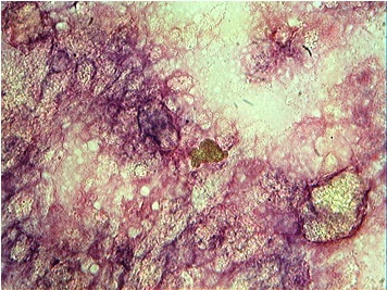
A 27 years old female was referred to our hospital with the chief complaint of a non-tender nodule in her right breast since 2 months. One and a half year back, she had delivered a male baby by a spontaneous, full term, vaginal delivery. She had breast fed her child for one year and thereafter, intermittently to the day of presentation. On physical examination of the breast, a discrete, mobile, nodule measured 1.5 X 1.0 cm was palpable in the upper outer quadrant of the right breast. The clinical impression was of a fibroadenoma. The clinician advised an FNAC of the lesion. We performed 2 needle passes which resulted in the aspiration of a thick, milky material, with a reduction in the size of the lesion. The cytology smears were studied with H and E staining, as well as Leishman’s staining. The smears showed numerous, distinct, compact and semitransparent to dark blue/purple crystals on H and E staining [Table/Fig-1] and refractile crystals with defined borders of variable shapes and sizes on Leishman’s staining [Table/Fig-2]. The background showed granular, amorphous and proteinaceous material which was admixed with frothy appearing lipid micelles and variable sized crystals [Table/Fig-3]. The distinct epithelial fragments, bipolar nuclei and fibrous stroma were not seen. In view of the clinical history of the lactation and the cytology, a diagnosis of a crystallizing galactocele was made.
Cytological features of crystallizing galactocele as mainly numerous distinct, compact semitransparent to dark blue/purple crystals (H & E,x100).
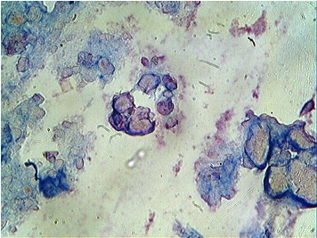
Many refractile blue crystals with defined borders of variable shape and size. (Leishman, x400).
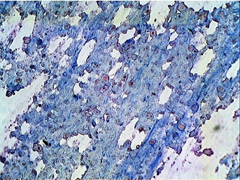
Granular, amorphous, proteinaceous material in the background admixed with distinct, variable sized crystals (H&E, x100).
The physiological changes of the breast during pregnancy and lactation make the clinical, radiological and the pathological evaluation of a breast mass particularly challenging [1].
Although most of the palpable breast lesions in the pregnant and lactating patients are benign, 3% of the breast cancers are diagnosed in this population and an evaluation of a breast mass should not be delayed for any reason [2]. FNAC is a well adapted, cost effective technique for the initial evaluation of clinically suspicious breast masses which are found during pregnancy and in the post-partum period [3]. The cytological features of a galactocele which are considered to be specific to pregnancy and lactation, were assessed by only two small series [4] and by a single case report of Raso DS et al., [5].
A galactocele is an uncommon benign lesion of the breast and it is defined as an encysted collection of milk products that is lined by a flattened cuboidal epithelium [4, 5]. The major features which are required for the development of a galactocele can be initiated by several features: a secretary breast epithelium, the presence of prolactin stimulation, and some form of ductal obstruction [5]. In addition to lactation, several breast lesions which result in a ductal obstruction or a generalized condition such as a breast surgery, the transplacental passage of prolactin, and oral contraceptives can create the factors for the development of a galactocele [4, 5].
Nevertheless, the diagnosis of a galactocele is often difficult to make on sonography alone, and a pathologic diagnosis is often warranted [4, 5].
To conclude, in a pregnant or a lactating female, a mass which is associated with a milky, viscous aspirate on FNAC, it is of paramount importance, to consider a crystallizing galactocele.
We are reporting this case due to the unusual features of the galactocele, which was diagnosed on FNAC as a crystallizing galactocele. To the best of our knowledge, this is the second case report after Raso DS et al’s which was made in 1997.
[1]. Hogge JP, de Paredes ES, Magnant CM, Lage J, Imaging and management of breast mass during pregnancy and lactationBreast J 1995 5:272-83. [Google Scholar]
[2]. Eedarapilli P, Jain S, Breast cancer in pregnancyJ Obstet Gynaecol 2006 26:1-4. [Google Scholar]
[3]. Silverman JF, Philadelphia, WB Saunders, Breast In: Comprehensive cytopathologyM Bibbo(edi) 1991 :703- 70. [Google Scholar]
[4]. Kline TS, Masquerades of malignancy: A review of 4241 aspirates from the breastActa Cytol 1981 25:263-66. [Google Scholar]
[5]. Raso DS, Greene WB, Silverman JF, Crystallizing galactocele: A case reportActa Cytol 1997 41:863-70. [Google Scholar]