The Limb-Abdominal Wall Complex Defects, a form of Amniotic Band Sydrome: A Rare Case Report
Sudhanshu Ku. Das1, Sidharth Sankar Maharana2, Monalisa Subudhi3, P. V. Subha Rao4
1 Assistant Professor, Department of Pediatrics,
2 Tutor, Department of Anatomy,
3 Tutor, Department of Microbiology,
4 Professor & HOD, Department of Pediatrics, Konaseema Institute of Medical Sciences, Amalapuram, Andhra Pradesh-533201, India.
NAME, ADRESS, E-MAIL ID OF THE CORESPONDING AUTHOR: Dr. Sudhanshu ku. Das, Assistant Professor, Department of Pediatics, Konaseema Institute of Medical Sciences, Amalapuram, Andhra Pradesh-533201, India.
Phone: 09441140581
E-mail: swayam.dr007@gmail.com
The limb-body wall complex defects a form of amniotic band syndrome which consists of a polymal formation with a thoracic and /or an abdominal-schisis, eventration of the internal organ and anomalies of the extremities. We are presenting a case of a limb-body wall complex defect with the phenotype of a placenta-abdominal attachment, anomalies of the abdominal wall defect, absence of the right lower limb and genitourinary defects.
Amniotic band syndrome, Limb body wall complex
INTRODUCTION
The amniotic band syndrome (ABS) and/or the Limb Body Wall Complex (LBWC) is a rare syndrome which results from two distinct phenotypic forms, a placenta-cephalic attachment and a placenta-abdominal attachment [1]. In newborns, it has been estimated to occur in 1 in 1300–2000 births [2]. Its pathogenesis includes germ disc disruption, genetic disruption, vascular disruption and amniotic disruption [3].
CASE REPORT
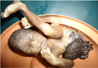
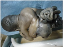
The foetus was a female and it was stillborn. It was the result of a pregnancy which was terminated at 28 weeks, as was revealed by an ultrasound examination. It weighed 498 g and had multiple congenital malformations [Table/Fig-1] with no family history. There was a facial deformity and a right side abdominal wall (abdominoschisis) defect, which were associated with an exteriorized bowel, a bilobed, globular and a congested liver, a right kidney and absence of the labia majora, with an exposed urinary bladder [Table/Fig-1] and [Table/Fig-2].
Facial deformity with an exteriorized bowel, a bilobed, globular, congested liver and absence of right lower limb, with left side clubfoot
exteriorized bowel, right kidney and absence of the labia majora, with exposed urinary bladder
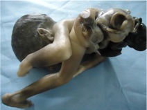
The apex of the heart was turned upwards and it was compressed with both lungs partially and it was severely hypoplastic (ultrasonographic findings).The diaphragm was inverted. The limb defect was limited to absence of the right side lower limb, with a clubfoot on the left side [Table/Fig-1]. The foetal trunk was flexed and scoliotic, with a twist which had a spinal deformity [Table/Fig-3].
Flexed and scoliotic trunk having spinal deformity
The placenta weighed 350 g and it was attached to the left side of the abdomen. The umbilical cord was short and lustreless, but without any knot, twist or constriction.
DISCUSSION
ABS is a group of congenital anomalies which are caused by the bands of the amnion (inner lining of the “bag of water”) that attach to the foetus due to an early rupture of the amnion (which usually occurs between 28 days after conception, to 18 weeks of gestation) [4]. The abnormalities result from an attachment or constriction, leading to webbing of the fingers and/or toes, amputation of the limbs and severe defects of the head and face, the spine, the umbilical cord and/or the body wall.
The ABS includes many congenital anomalies like:
The amniotic band disruption complex,
Amniochorionic mesoblastic fibrous strings,
Aberrant tissue bands,
Amniotic deformities,
The ADAM complex,
The amniotic adhesion malformation syndrome and
Limb and /or body wall defects [4].
In the Limb-Body-Wall Complex (LBWC), there is a constellation of abnormalities which include myelomeningoceles or caudal regression, thoracoabdominoschisis, or abdominoschisis and limb defects [5]. At least two of the three abnormalities which are listed above are necessary to make a diagnosis of LBWC. The presence of bands is unnecessary for the diagnosis of the amniotic band syndrome, in the presence of the characteristic foetal anomalies. However, an ultrasound detection of the bands is helpful in confirming the diagnosis of the amniotic band syndrome as the cause of the foetal deformities which can be made at the end of the first trimester. Colour Doppler is very useful for the detection of the umbilical cord. The indications for a foetal surgery may be either a life-threatening condition if it involves constriction of the umbilical cord, or more commonly, a threatened limb amputation which is caused by the amniotic band syndrome. More often, there is a band-like deformation that requires Z-plasties to surgically correct it. A foetoscopic laser is an option for releasing the amniotic bands that threaten to amputate a hand. It is a successful procedure [6].
[1]. Torpin R, Amniochorionic mesoblastic fibrous strings and amnionic bands: associated constricting fetal malformations or fetal deathAm J Obstet Gynecol 1965 91:65-75. [Google Scholar]
[2]. Ossipoff V, Hall BD, Etiologic factors in the amniotic band syndrome: a study of 24 patientsBirth Defects Orig Artic Ser 1977 13(3D):117-32. [Google Scholar]
[3]. Miller ME, Graham JM Jr, Higginbottom MC, Smith DW, Compression related defects from early amnion rupture: evidence for mechanical teratogenesisJ Pediatr 1981 98:292-97. [Google Scholar]
[4]. Kulkarni ML, Gopal PV, Amniotic band syndromeIndian Pediatr 1990 27:471-76. [Google Scholar]
[5]. Van Allen MI, Curry C, Gallagher L, Limb body wall complex. I. PathogenesisAm J Med Genet 1987 28:529-48. [Google Scholar]
[6]. Torpin R, Amniochorionic mesoblastic fibrous strings and amniotic bandsAm J Obstet Gynecol 1965 91:65-75. [Google Scholar]