Myiasis is the infestation of the tissues and organs of animals or man by fly larvae. Ophthalmic myiasis has been reported from various world regions. In this study, we are presenting the clinical manifestations of external ophthalmomyiasis which was caused by the larvae of the sheep nasal botfly, Oestrus ovis, in 10 patients in the Tirunelveli and the Tuticorin districts of Tamil Nadu state India. All the patients were farmers, who worked in close contact with sheep and goats. All the patients presented with severe conjunctivitis. The larvae were observed in the bulbar conjunctiva and following their removal, the symptom of eye inflammation improved in a few hours.
Myiasis, Oestrus ovis, External ophthalmomyiasis, Conjunctivitis, Tamil Nadu
INTRODUCTION
Myiasis is the infection of the tissues or organs of animals or man by the larvae of a dipteran fly. The common sites are skin wounds. Eyes, nose, nasal sinuses, throat, and the urogenital tract are the less common sites [1–3]. The Ophthalmomyiasis in human beings is caused by the larvae of Oestrus ovis. Oestrus ovis, the common sheep nasal botfly breeds in the nasal cavity and the sinuses of sheep. The flies enter the nostril and deposit larvae. The larvae crawl into the nose and attach to the mucous membrane with the help of oral hooks. When they mature, they fall out from the nasal passage to pupate in the ground. It takes a year to complete its life-cycle feeding on mucous substances. It is very interesting to note that the adult flies do not feed in sheep [4] since they have rudimentary mouth parts. The adult flies dart their larvae in the eyes of human beings, especially in the eyes of sheep and goat farmers/workers. The first stage larvae (L1) do not mature in the unnatural human hosts but they erratically wander the ocular sites and produce ophthalmomyiasis. An ocular involvement occurs in about < 5% of all the cases of human myiasis [5]. Ophthalmomyiasis is classified as ophthalmomyiasis externa if the larvae are present on the conjunctiva and as ophthalmomyiasis interna when there is an intraocular penetration of the larvae [6]. Cases of ophthalmomyiasis externa have been reported from various parts of the world [7–9]. Myiasis appears to be fairly common but it is underestimated in many rural areas. However, this condition is rare in India and there are only a few sporadic reported cases [6, 10–13]. Most of these reports are limited to one case. To the best of our knowledge, no case of ophthalmomyiasis externa which was caused by Oestrus ovis has been reported from this part of India. We are presenting the clinical manifestations in 10 patients with ophthalmic myiasis from the Tirunelveli and the Tuticorin districts of Tamil Nadu, India. The present case therefore highlights the awareness among the ophthalmologists regarding larval conjunctivitis in the spring and summer seasons, with a mention of the timely diagnosis and treatment of this rather rare infestation.
CASE HISTORY
We investigated all of the patients who were referred with an evidence of an ophthalmic inflammation to a tertiary care hospital during the year 2010. Most of those who live in this area were involved in agricultural activities and/or sheep raising animal husbundary activites as shepherds. Each subject underwent a thorough physical examination and was asked in detail about his/her medical history and, in particular, about the symptoms which were related to the development of myiasis. Attempts were made to directly detect any larvae which were present in the eye cases. The diagnosis of myiasis was made by a direct visualization of the larvae, by using a magnifier lens or an ophthalmoscope. After the instillation of tetracaine eye drops, the larvae were removed by an applicator or by washing the eyes with normal saline.
Ten patients (8 males and two females) were proved to have ophthalmic myiasis. The age of the patients ranged between 25 and 65 years. This condition occurred in the early rainy fall season (80%) and in summer (20%) in the year 2010 in the Tirunelveli and the Tuticorin districts of Tamil Nadu. All of the 10 cases lived in rural areas and were in close contact with sheep and goats. The major clinical features have been shown in [Table/Fig-1].
Clinical findings in 10 patients with ophthalmomyiasis
| No. of patient | Gender (age) | Season | No of larva | Complaint and findings |
|---|
| 1 | Male (25) | Early Rain fall | 4 | Rhinorrhea, swelling and chemosis, redness, itching, foreign body sensation, lacrimation |
| 2 | Male (28) | Summer | 3 | Itching, foreign body sensation, lacrimation, pain, swelling and chemosis, redness |
| 3 | Male (42) | Early Rain fall | 2 | Foreign body sensation, lacrimation, pain, swelling and chemosis |
| 4 | Male (47) | Early Rain fall | 5 | Itching, foreign body sensation, lacrimation, swelling and chemosis, redness |
| 5 | Male (35) | Summer | 6 | Itching ,foreign body sensation, lacrimation, swelling and chemosis, redness |
| 6 | Male (25) | Early Rain fall | 10 | Itching, lacrimation, rhinorrhea, swelling and chemosis, redness |
| 7 | Female(30) | Early Rain fall | 3 | Itching, foreign body sensation, lacrimation, swelling and chemosis, redness |
| 8 | Male (65) | Early Rain fall | 4 | Itching, foreign body sensation, lacrimation, pain, swelling and chemosis, redness |
| 9 | Male (35) | Early Rain fall | 6 | Rhinorrhea, swelling and chemosis, redness, itching, foreign body sensation, lacrimation |
| 10 | Female (26) | Early Rain fall | 4 | Itching, foreign body sensation, lacrimation, pain, swelling and chemosis, redness |
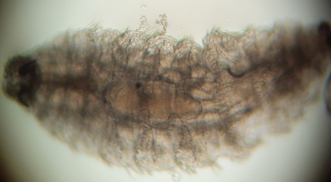
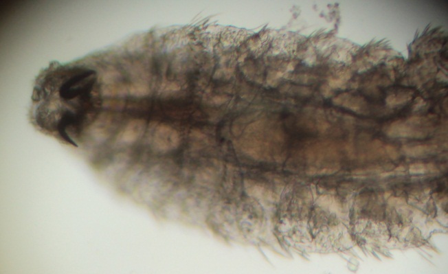
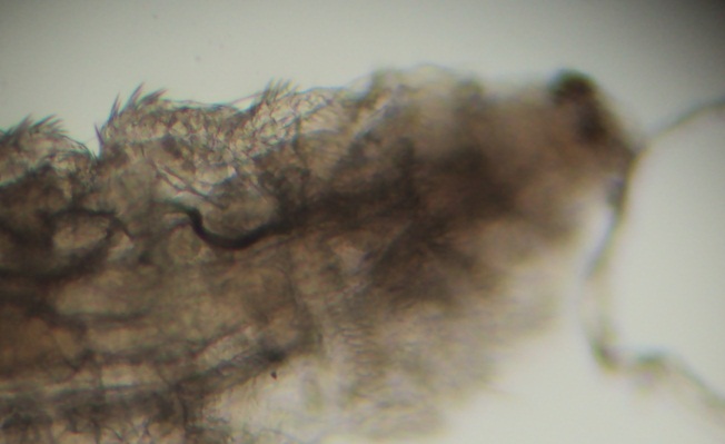
In each case, the symptoms began while the affected individual was working on a farm/grazing or while he/she was washing or cleaning the sheep in a flock, during the days. None of the patients had a history of allergic reactions in the past. The first symptom, sensing the presence of a foreign body in the eye and itching, had always appeared abruptly. In all the cases, the eye involvement was unilateral and extra ocular and motile larvae were present in the bulbar conjunctivae and in the fornices. There was no evidence of a corneal or an intraocular involvement. A slit lamp examination revealed white coloured larvae which varied from one to five and measured 2–5 mm in length. They were mounted on a microscope slide, examined carefully and photographed under a microscope. The larvae were identified as the first instars of Oestrus ovis (Diptera: Oestridae), which is a larviparous dipteran, on the basis of their spindle shape and the pair of sharply curved mouth-hooks [8]. The pattern of the spinules on the dorsal surface consisted of a complete row of denticles on the third segment and a broadly interrupted row on both the fourth and fifth segments, with 22 to 25 terminal hooks being arranged in two scallops [Table/Fig-2,3 and 4].
Entire maggot (L1) of Oestrus ovis recovered from eye (x40)
Anterior end of first instar larva (L1) of Oestrus ovis (x100)
Posterior end of first instar larva (L1) of Oestrus ovis (x100)
Following the removal of all the larvae, the symptoms completely resolved within a few hours. In the patients who had remained without treatment for some time, the swelling of the eye had increased rapidly, and this prevented the eye opening.
DISCUSSION
Ophthalmic myiasis is caused by the deposition of fly larvae in the human eyes. Various species of flies are able to provoke ophthalmomyiasis, which include Oestrus ovis, latrine fly (Fannia), house fly (Musca domestica) and cattle botfly (Hypoderma) [14,15]. Oestrus ovis is by far, the most common cause of ophthalmic myiasis in man [16]. Sheep bot (Oestrus ovis) is a cosmopolitan parasite of sheep and goats. The sheep nose bot is a hairy, yellowish, bee-like fly, which is about the size of a common horsefly. The adult flies are 12-14 mm in length but they are rarely seen [17]. The adult female fly is active during summer and early fall. Its eggs are retained in the body until they hatch. The gravid adult female fly swarms around the head of the animals and ejects the first-instar larvae which have previously hatched from the eggs in its vagina, in a stream of milky fluid on to the nostrils of the host [18]. The flies may deposit as many as 500 larvae in the nostrils of sheep. A direct contact between the fly and its host is not necessary for the infestation [19]. The larvae of Oestrus ovis mature in the mucous membrane of the nasal cavities, where they remain for up to ten months and then are sneezed out of the nostrils [17]. The larvae pupate in the soil, with the pupal period lasting for three or more weeks, depending on the temperature. The adults then emerge from the pupae and may live as long as 28 days [17].
The ophthalmic myiasis in human beings, which is caused by Oestrus ovis, was described for the first time in 1947 by James [12]. More scattered cases were reported since then from the Mediterranean area, like Italy, and also from Russia, Serbia (previous Yugoslavia), Africa, America, and Oman [10,11]. Many cases have been reported recently from various parts of India. Myiasis is more common than what has been indicated by the previously published reports [20,21]. Chandra et al, Reingold et al, Vertsrynge et al, Masoodi and Hosseini from Iran, Greogary et al from Iraq, Senthilvel et al from the Salem district of Tamil Nadu, Sreejith et al., from Hyderabad and Seema et al., from Maharashtra reported few cases of the Oestrus ovis infection in humans [22–29]. This is the first series on ophthalmomyiasis in 10 cases, which was caused by Oestrus ovis from the Tirunelveli and the Tuticorin districts of Tamil Nadu state in India.
Human myiasis mostly occurs in the rural areas, where man lives in close contact with small ruminants. Sheep and goat are the main hosts for myiasis which is caused by Oestrus ovis and humans are infested accidentally. Ocular myiasis which is caused by the infection of the first stage larvae of the sheep botfly, which is associated with mucopurulent conjunctivitis in the human eyes, is a rare occurrence. However, if the sheep botfly, Oestrus ovis abounds, there may be chances of the deposition of its larvae in the conjunctival sac of the human eyes. The incidences of ocular myasis which are associated with mucopurulent conjunctivitis, which has been described in the present cases, may be occupationally contracted. The erosion of the epithelial tissues on the eye ball as well as on the conjunctival cavity, may be prone for the infection of microbes [30]. This was evidenced from the analysis of the cases which were reported in this present study.
Symptoms such as severe eye irritation, redness, foreign body sensation, pain, lacrimation, and swelling of the lids, and also, the predominance of male patients and the warmer climate which have been presented in this study, are similar to those which have been described in other reports [10, 31]. Rhinorrhoea, which has been reported in a few studies [32], may point out to an allergic reaction, in addition to the local irritation which is induced by the fly larvae.
Complications such as corneal ulcers, invasion into the eye globe, and decreased vision are not usual and none of these complications were encountered in our patients. The factors which predispose to ophthalmic myiasis have been reported to be eye infection, young age, alcohol abuse and debility, but none of these factors were present in our patients. The treatment consists of anaesthetizing the larvae and the eye, followed by removal of the larvae. Antihistamine drops and/or topical antibiotics may also be used, as needed.
Myiasis should be considered as an occupational disease among farmers and shepherds. An awareness on larval conjunctivitis in rural areas, especially during the early rainy season and summer, leads to a more prompt diagnosis, and institution of the specific therapy for the disease.
[1]. Al-Dabagh C, Al-Mufti N, Shafiq M, Rawas AL, Al-Saffar S, A second record from Iraq of human myiasis caused by larvae of the sheep botfly Oestrus ovisAnn Trop Med Parasitol 1980 74:73-77. [Google Scholar]
[2]. Amr ZS, Amr BA, Abo-Shehada MN, Ophthalmomyiasis externa caused by Oestrus ovis in the Ajloun area of northern JordanAnn Trop Med Parasitol 1993 87:259-62. [Google Scholar]
[3]. Katz SI, Taylor R, Cutaneous myiasisSouth Med. J. 1971 64:759-60. [Google Scholar]
[4]. Fraser CM, Berveron JA, Asamays DU, Aiello SE, The Merck Veterinary ManualA hand book of diagnosis and therapy for the Veternarian 1991 7thU.S.A.Merck and Co, INC1792 [Google Scholar]
[5]. Keller AP, Keller AP, 3rd Myiasis of middle earLaryngoscope 1970 80:646-50. [Google Scholar]
[6]. Thompson JH, Knutson LV, Culp OS, Larva of Scenopinus sp (Diptera: Scenopinidae) causing human urogenital myiasisMayo Clin Proc 1970 45:597-601. [Google Scholar]
[7]. Minar J, A case of eye myiasis in man caused by first instar larvae of Oestrus ovis L (Diptera: Oestridae) in IranFolia Parasitol. (Praha) 1976 23:283-84. [Google Scholar]
[8]. Janbakhsh B, Pirouz MS, Tirgari S, Agha-Mohammadi A, A case of ophthalmomyiasis in man by Oestrus ovis Linnaeus in Tehran (Diptera: Oestridae)Acta Medica Iranica 1977 20:19-26. [Google Scholar]
[9]. Grammer J, Erb C, Kamin G, Wild MR, Riedinger C, Kosmidis P, Ophthalmomyiasis externa due to the sheep botfly Oestrus ovis in South-West GermanyGev J Ophthalmol 1995 4:188-95. [Google Scholar]
[10]. Victor R, Bhargva K, Ophthalmomyiasis in Oman: a case report and commentsWilderness Environ Med 1998 9:32-35. [Google Scholar]
[11]. Maretic Z, Nadenic LA, Ladavic J, Zekic R, Ophthalmomyiasis due to Oestrus ovisActa Trop 1973 33:369-72. [Google Scholar]
[12]. Patel SJ, Extraocular myiasis due to the larva of Oestrus ovisEast Afr Med J. 1975 52:167-69. [Google Scholar]
[13]. Wolfelschneider P, Wiedemann D, External ophthalmic myiasis caused by Oestrus ovis (sheep and goat botfly)Klin Monatsbl Augenheilkd 1996 209:256-58. [Google Scholar]
[14]. Mandell GL, Douglas RG, Bennett JE, Mandell Dolin R., Douglas Bennett Dolin Principles and Practice of Infectious Disease 2000 5thPhiladelphiaChurchill Livingstone [Google Scholar]
[15]. Vaughan DG, Asharg T, Riardan EP, General Ophthalmology 1999 5thStamfordAppleton and Lange [Google Scholar]
[16]. Grammer J, Erb C, Kamin G, Wild MR, Riedinger C, Kosmidis P, Ophthalmomyiasis externa due to the sheep botfly Oestrus ovis in South-West GermanyGev J Ophthalmol 1995 4:188-95. [Google Scholar]
[17]. Lloyd JL, Brewer MJ, Sheep bot fly, biology and management April 1992 Available from: http://www.ces.uwyo.edu/PUBS/B966.PDF [Google Scholar]
[18]. Verstrynge K, Foets B, External ophthalmomyiasis: A case reportBull Soc Belge Ophthalmol 2004 294:67-71. [Google Scholar]
[19]. Odat TA, Gandhi JS, Ziahosseini K, A case of ophthalmomyiasis externa from Jordan in the Middle EastBr J Ophthalmol 2007 91Video report [Google Scholar]
[20]. Khurana S, Biswal M, Bhatti HS, Pandav SS, Gupta A, Chatterjee SS, Ophthalmomyiasis: Three cases from North IndiaIndian J Med Microbiol 2010 28:257-61. [Google Scholar]
[21]. Pandey A, Madan M, Asthana AK, Das A, Kumar S, Jain K, External ophthalmomyiasis caused by Oestrus ovis: a rare case report from IndiaKorean J Parasitol 2009 47:57-9. [Google Scholar]
[22]. Chandra DB, Agrawal TN, Ocular myiasis caused by Oestrus ovisIndian J Ophthalmol 1981 29:199-200. [Google Scholar]
[23]. Reingold WJ, Robin JB, Leipa D, Kondra L, Schanzlin DJ, Smith RE, Oestrus ovis ophthalmomyiasis externaAm J Ophthalmol 1984 97:7-10. [Google Scholar]
[24]. Masoodi M, Hosseini K, External ophthalmomyiasis caused by sheep botfly (Oestrus ovis) larva: A report of 8 casesArch Iran Med 2004 7:136-39. [Google Scholar]
[25]. Greogary AR, Scott S, Harold L, Ophthalmomyiasis caused by the sheep bot fly Oestrus ovis in northern IraqOptom Vis Sci 2004 81:586-90. [Google Scholar]
[26]. Verstrynge K, Foets B, External ophthalmomyiasis: A case reportBull Soc Belge Ophthalmol 2004 294:67-71. [Google Scholar]
[27]. Sreejith RS, Reddy AK, Ganeshpuri SS, Garg P, Oestrus ovis ophthalmomyiasis with keratitisIndian J Med Microbiol 2010 28:399-402. [Google Scholar]
[28]. Bose Seema, Saini Santosh, Barapatre Rekha, Mathew Stephen, Ophthalmomyiasis External: A Case ReportJournal of Clinical and Diagnostic Research 2012 6:1079-80. [Google Scholar]
[29]. Senthilvel K, Muralidharan J, Thiruvenkadan A.K., Karunanithi K, Human opthalmomyiosis due to Oestrus ovis: A reportJ Vet Parasitol 2008 22:63-64. [Google Scholar]
[30]. Mazzeo V, Ercolani D, Trombetti D, Todeschini R, Gaiba G, External ophthalmomyiasis Report of four cases [in Germany]Int Ophthalmol 1987 11:73-76. [Google Scholar]
[31]. Cameron JA, Shoukrey NM, Al-Garni AA, Conjunctival ophthalmomyiasis caused by the sheep nasal botfly (Oestrus ovis)Am J Ophthalmol 1991 112:331-34. [Google Scholar]
[32]. MacDonald PJ, Chan C, Dickson J, Jean-Louis F, Heath A, Ophthalmomyiasis and nasal myiasis in New Zealand:a case seriesN Z Med J. 1999 112:445-47. [Google Scholar]