On dissection of the abdomen of an adult male cadaver, in addition to the normal inferior vena cava on the right side, an unusual venous channel which connected the left renal vein with the left common iliac vein was found; (probably the left inferior vena cava). The left testicular and the left suprarenal veins were opening into the left renal vein as usual. Other than this, a retrocaval ureter was found on the right side.
The works of previous authors have highlighted the incidence of a venacaval duplication and its surgical implications, but here, we are presenting a unique case of a double inferior vena cava with an anomalous retrocaval ureter. A conglomeration of such vascular malformations is of immense surgical importance, and it is indicative of a grossly defective angiogenesis.
Keeping in mind the clinical relevance of the variations which were observed, an attempt was made to explain them in the light of the embryogenic development.
Retrocaval ureter, Venacaval duplication, Angiogenesis
INTRODUCTION
Embryogenesis of the Inferior Vena Cava (IVC) is a complex process which involves the formation of anastomoses between three paired embryonic veins. Based on the abnormal regression or the persistence of various embryonic veins, numerous vena caval anomalies may be encountered, which include: transposition of the IVC, an azygos continuation of the IVC, a circumaortic left renal vein, a retroaortic left renal vein, duplication of the IVC, a double IVC with a retroaortic right renal vein, a hemiazygos continuation of the IVC, a retrocaval ureter and absence of the infrarenal IVC or the entire IVC [1].
Duplication of the IVC (D-IVC): This is a relatively uncommon congenital anomaly with a reported incidence of 0.2%–3%. A majority of the cases are clinically silent and they are diagnosed incidentally during imaging studies which are done for other reasons [2].
D-IVC can be of following types:
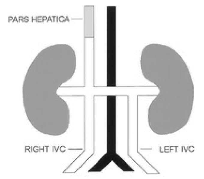
An incomplete D-IVC: The left common iliac vein ascends as a duplicated left IVC and it drains into the Left Renal Vein (LRV). The LRV then crosses the aorta anteriorly and it joins the right IVC in a normal fashion [Table/Fig-1].
A complete D-IVC: In this, the left IVC does not drain into the LRV, but after receiving the LRV, it continues as a preaortic trunk that travels obliquely and empties into the right IVC [2].
Diagram showing duplicated IVC
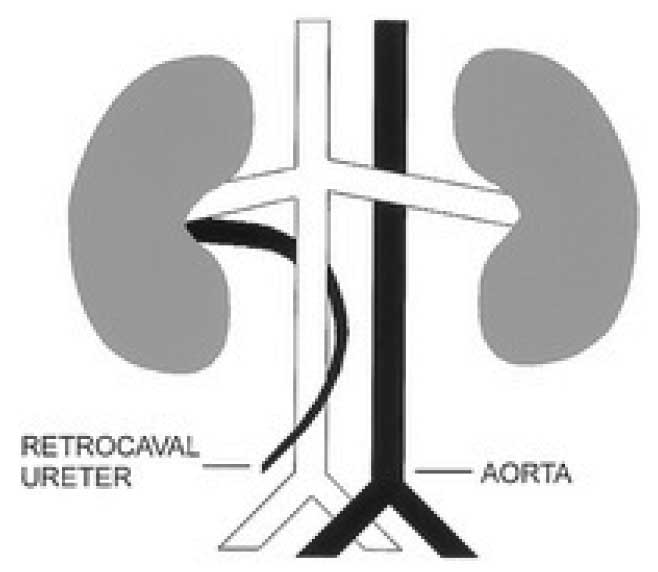
A circumcaval ureter or a retrocaval ureter: This is a rare, congenital venous anomaly. In this, the proximal ureter deviates medially and it courses behind the IVC, thereby partially encircling it. Then it exits anteriorly between the IVC and the aorta and returns to its normal position [Table/Fig-2]. Since its first description which was made by Hochstetter in 1893, approximately 200 cases have Singhbeen reported worldwide. It is reported to be present in 0.06-0.17% of the autopsy materials. The incidence is greater in males than in females, with a ratio of 2.8:1 [3].
Diagram showing Retrocaval ureter
A retrocaval ureter almost invariably involves the right side, but when it is reported on the left side, it is usually associated with either a partial or a complete situs inversus or a D-IVC [3].
The term, ‘retrocaval ureter’ is used interchangeably with a ‘circumcaval ureter’ or a ‘postcaval ureter’, which describes the anatomic relationship of the ureter to the embryologically abnormal IVC. The designation, ‘preureteric vena cava’ more accurately describes the embryologic malformation; however, this is not a practical term because the functional abnormality is only manifest clinically due to the obstruction of the ureter [3].
CASE REPORT
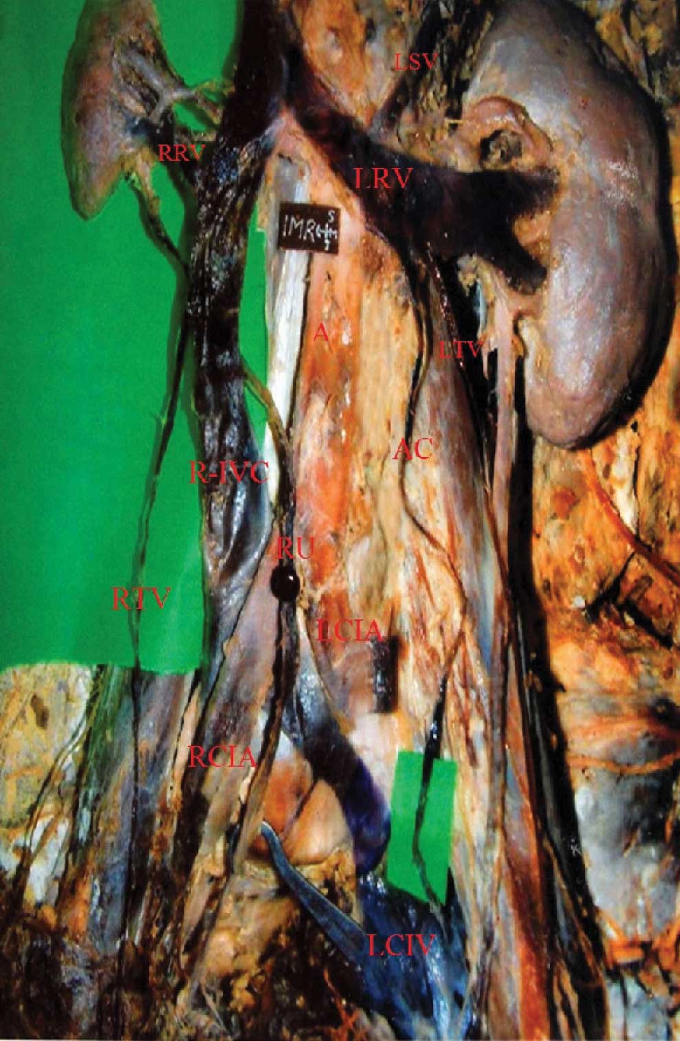
During a routine dissection of the abdomen in a 45 year old adult male cadaver, an abnormal venous channel (21.5 cms long) was found on the left side, which connected the left renal vein with the left common iliac vein. This channel took origin, 7.8 cms distal to the point of union of the two Common Iliac Veins (CIVs). The right and left renal veins terminated into the right sided IVC, at the level of the disc between the L1 and L2 vertebrae and the upper border of the L1 vertebra respectively [Table/Fig-3].
Photograph of dissected abdomen showing: Anomalous channel (AC) connecting left renal vein (LRV) with left common iliac vein (LCIV); the right IVC (R-IVC) being normal.
Retrocaval ureter (RU). (RRV- Right renal vein, LSV- Left suprarenal vein, LTV- Left testicular vein, A- Aorta, RCIA- Right Common iliac artery, LCIA- Left common iliac artery).

The CIVs fused slightly cranial to the level of origin of the inferior mesenteric artery, from the aorta (i.e at the level of the lower border of the body of the L4 vertebra). The resultant complex then continued superiorly as the right-sided IVC. The anomalous channel on the left side ended into the left renal vein. Thereafter, the left renal vein drained into the right IVC [Table/Fig-3].
In addition to this, an anomalous retrocaval ureter was also seen on the right side. The right ureter coursed medially behind the right IVC. Then, it took exit anteriorly between the IVC and the aorta to regain its normal position [Table/Fig-4].
Magnified version of Fig-3. (Focussing on site of origin of left sided anomalous channel from the left common iliac vein).
DISCUSSION
The congenital anomalies of the IVC and its tributaries are a relatively rare pathology, usually with an asymptomatic iter. From the bioptic material, their incidence has been estimated to be 2-3% and the percentage of the intraoperative findings varies in different series, between 0.2-0.6%. Among its various anomalies, a caval duplication and a left positioned IVC are most commonly found [1].
The D-IVC was found unexpectedly during resection of abdominal aortic aneurysms, in patients who were investigated by venography for varicoceles or in CT scans which were done for diagnosing Hodgkin’s lymphoma. Peroperatively, the left IVC was encountered in patients with the Leriche’s syndrome [4].
Minniti et al., reported 2 variants of the IVC: the D-IVC with an azygos continuation of the posterior-medial vein and the D-IVC with hemiazygos and azygos continuations of the left one [5]. Mano et al reported a case of deep venous thrombosis which was accompanied by the D-IVC [6]. Itoh et al., and Surucu independently reported cases of the D-IVC, which were associated with the left suprarenal vein draining into the left vena cava and the right testicular vein draining into the right renal vein [7, 8].
The D-IVC which was recorded in our case resembled the incomplete type of D-IVC of the classification which was given by Ng WT and Ng SSM [2].
Wang et al., reported a case of a right-sided retrocaval ureter in a 21 years old male [9]. Pierro et al reported two cases of left-sided circumcaval ureters without situs inversus or caval duplications, while Brooks reported a similar case which was associated with situs inversus [10, 11]. Numerous cases of retrocaval ureters have also been reported in children and pregnant females [12]. Bilateral postcaval ureters in an acardiac foetus have also been reported [12].
Bateson and Atkinson distinguished the two types of retrocaval ureters according to the radiological appearance and the site of the ureteral narrowing. These are:
Type I: The ureter crosses behind the IVC, at the level of the L3 vertebra and it exhibits an “S-shaped“ deformity.
Type II: The renal pelvis and the upper ureter lie horizontally. The retrocaval segment of the ureter is at the same level as that of the renal pelvis and it exhibits a “sickle shaped” deformity [13].
The retrocaval ureter which was observed in our case apparently fitted into the Type I of the given classification.
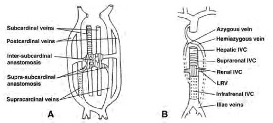
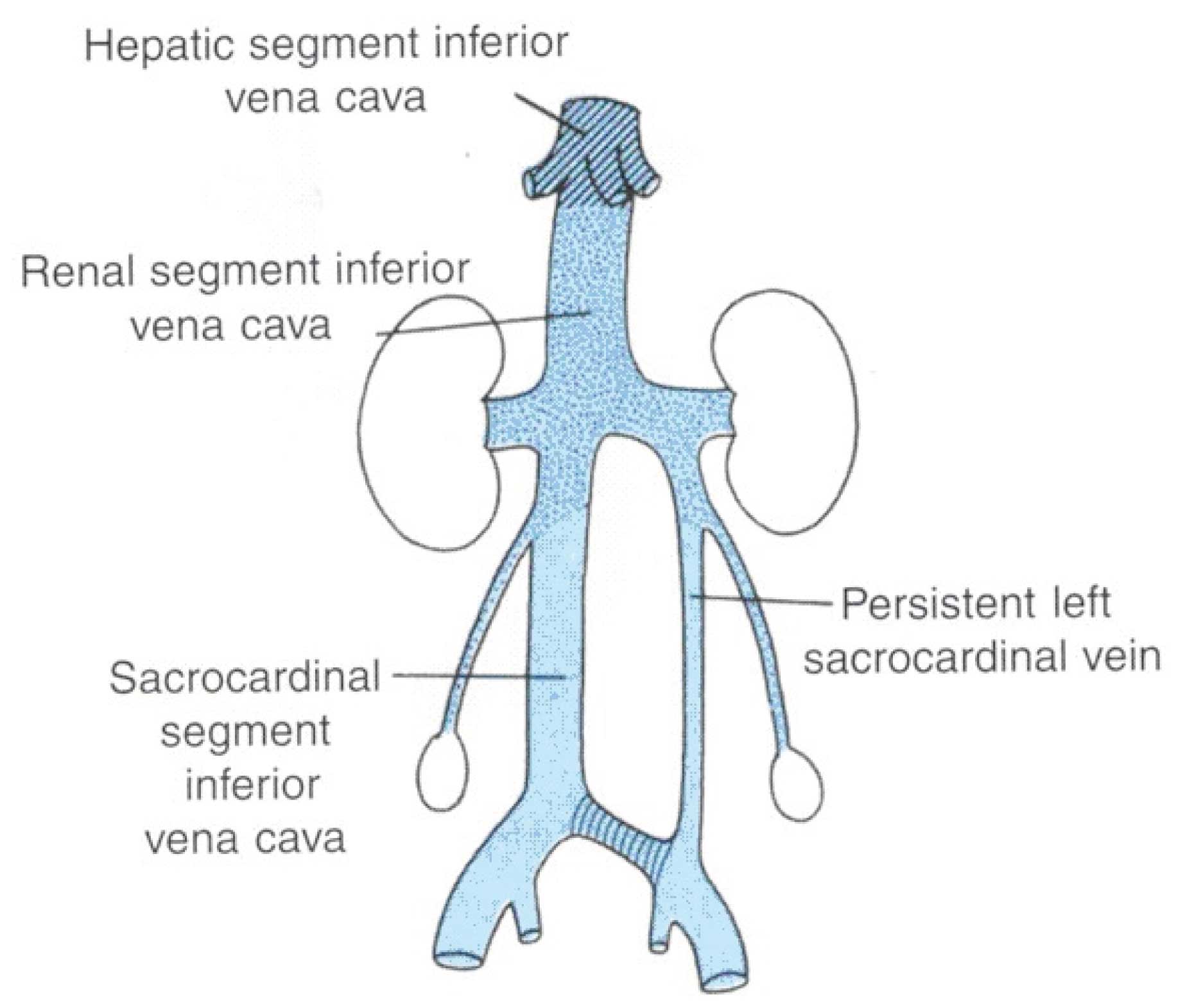
The embryological development of a normal right sided IVC has been shown in [Table/Fig-5]. In this figure, it is evident that the infrarenal segment of the IVC (i.e. under the level of the renal veins) develops from the caudal part of the right supracardinal vein only, since the part of the left supracardinal vein which is caudal to the kidneys, degenerates. However, if the left supracardinal vein persists; along with one of the midline anastomoses, it leads to a variety of postrenal IVC anomalies. These anomalies include renal venous collars, a left-sided IVC and a D-IVC (as the one which was seen in our case) [Table/Fig-6].
Diagram showing Embryogenesis of normal right-sided IVC
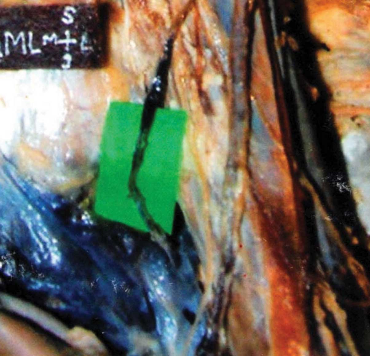
Diagram showing Embryogenesis of D-IVC
A retrocaval ureter is, strictly speaking, a developmental anomaly of the IVC and not of the ureter. The [Table/Fig-7(a) & 7(b)] show that the normally postcardinal veins do not participate in the formation of a definitive right-sided IVC. But, if the right posterior cardinal vein fails to regress in the lumbar portion, it forms a part of the right-sided IVC. Subsequently, it courses anterior to the ureter for a variable distance, the ureter being trapped dorsal to the IVC; resulting in the formation of a retrocaval ureter [Table/Fig-7(c)].
Diagram showing Embryogenesis of Retrocaval ureter
CLINICAL SIGNIFICANCE
Although anomalies of the IVC are uncommon, their recognition is important before vascular procedures which involve the renal pedicle are performed, for a correct interpretation of the cross-sectional images, in radio nuclide venography or in catheterization and opacification of the IVC; so as to avoid making erroneous diagnoses of the retroperitoneal and the mediastinal masses, and to alert the surgeons and the angiographers about the potential sources of the complications preoperatively [14].
[1]. Milloy FJ, Anson BJ, Cauldwell EW, Variations in the inferior vena caval veins and in their renal and lumbar communicationsSurg. Gynaecol. Obstet 1962 115:131-42. [Google Scholar]
[2]. Ng WT, Ng SSM, Double inferior vena cava: a report of three casesSingapore Med. J. 2009 50(6):211-13. [Google Scholar]
[3]. Uthappa MC, Anthony D, Allen C, Retrocaval ureter: MR appearancesBritish Journal of Radiology 2002 75:177-79. [Google Scholar]
[4]. Rispoli P, Conforti M, Cassatella R, Varetto G, Melloni CD, Raso AM, Left sided inferior vena cava in patients submitted to aortoiliac surgery, our experience and review of literatureJournal of Cardiovascular Surgery 2001 42(2):249-55. [Google Scholar]
[5]. Minniti S, Visentini S, Procacci C, Congenital anomalies of the venae cavae: embryological origin, imaging features and report of three new variantsEur. Radiol 2002 12:2040-55. [Google Scholar]
[6]. Mano A, Tatsumi T, Sakai H, Imoto Y, Nomura T, Nishikawa S, A case of deep venous thrombosis with a double inferior vena cava effectively treated by suprarenal filter implantationJpn. Heart. J. 2004 45:1063-69. [Google Scholar]
[7]. Itoh M, Moriyama H, Tokunaga Y, Miyamoto K, Nagata W, Satriotomo I, Embryological consideration of drainage of the left testicular vein into the ipsilateral renal vein: analysis of cases of a double inferior vena cavaInt. J. Androl 2001 24:142-52. [Google Scholar]
[8]. Surucu HS, Erbil KM, Tastan C, Yener N, Anomalous veins of retroperitoneum: clinical considerationsSurg. Radiol. Anat 2001 23:443-45. [Google Scholar]
[9]. Wang LT, Lo HC, Yu DS, Sun GH, Wu CC, Fong CJ, Ureteral obstruction caused by a duplicated anomaly of inferior vena cavaInt. J. Urol 2005 12:842-44. [Google Scholar]
[10]. Pierro J, Soleimanpour M, Bory JL, Left retrocaval ureter associated with left inferior vena cavaAm. J. Roentgenol 1990 155:545-56. [Google Scholar]
[11]. Brooks RJ, Left retrocaval ureter associated with situs inversusJ. Urol 1962 88:484 [Google Scholar]
[12]. Hyams BB, Schneiderman C, Mayman AB, Retrocaval ureterCan. Med. Assoc. J. 1968 98(1):45-49. [Google Scholar]
[13]. Bateson E, Atkinson D, Circumcaval ureter: a new classificationClin. Radiol 1969 20:173-77. [Google Scholar]
[14]. Royal SA, Callen PW, CT evaluation of the anomalies of the inferior vena cava and left renal veinAJR 1979 132:759-63. [Google Scholar]