Ochronotic Spondyloarthropathy
Upasana Ranga1, Senthil Kumar Aiyappan2, Natarajan Shanmugam3, Saveetha Veeraiyan4
1 Assistant Professor, Department of Radiodiagnosis and Imaging, Saveetha Medical College and Hospital, Thandalam, Kanchipuram, Tamilnadu - 602105, India.
2 Assistant Professor, Department of Radiodiagnosis and Imaging, Saveetha Medical College and Hospital, Thandalam, Kanchipuram, Tamilnadu - 602105, India.
3 Assistant Professor, Department of Orthopaedics, Saveetha Medical College and Hospital, Thandalam, Kanchipuram, Tamilnadu - 602105, India.
4 Associate Professor, Department of Radiodiagnosis and Imaging, Saveetha Medical College and Hospital, Thandalam, Kanchipuram, Tamilnadu - 602105, India.
NAME, ADDRESS, E-MAIL ID OF THE CORRESPONDING AUTHOR: Dr. Senthil Kumar Aiyappan, Assistant Professor, Department of Radiodiagnosis and Imaging, Saveetha Medical College and Hospital, Thandalam, Kanchipuram, Tamilnadu – 602105, India.
Phone: 91-44-26811173 senthilkumarpgi@yahoo.co.in
Alkaptonuria, Intervertebral disc calcification, Vacuum phenomenon
CASE REPORT
We are describing here, a case of ochronotic spondyloarthropathy who presented with a gradually increasing backache and stiffness and was diagnosed with the help of plain radiographs which showed intervertebral disc calcification. This was later confirmed by urine tests.
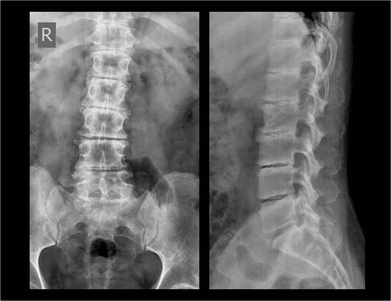
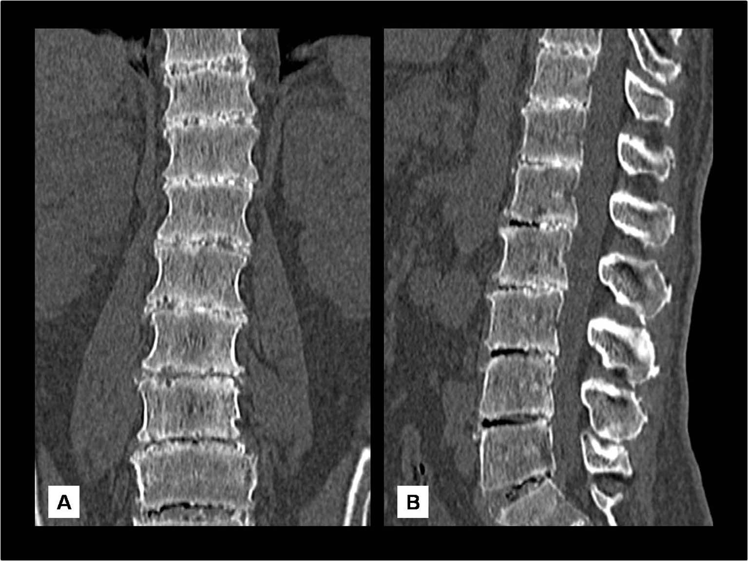
A 40-years-old male patient came to our hospital with the complaints of a gradually progressing backache and mild stiffness in the lower back which were there for the past 2 years. The patient did not give any history of trauma, fever, cough, abdominal pain or constitutional symptoms. After a local physical examination, X-rays of the lumbosacral spine were advised by the orthopaedic surgeon. The X-rays of the lumbosacral spine (anteroposterior and lateral views) showed loss of the lumbar lordosis with minimal scoliosis, reduced disc spaces, calcification of all the visualized intervertebral discs and the vacuum phenomenon at multiple levels, along with endplate changes in the spine [Table/Fig-1]. The patient gave a history of a similar illness in his father. After examining the X-rays, a detailed history was taken, along with a complete general physical examination. The patient revealed that his underwears used to get stained brownish black since childhood. His urine sample turned dark in colour spontaneously, when it was exposed to air and it showed high levels of homogentisic acid. Computed Tomography (CT) of his dorsolumbar spine confirmed the X- ray findings [Table/Fig-2 and 3]. The disc and endplate changes were better appreciated in the CT scan. Based on the urine sample findings and the radiological features, a diagnosis of ochrontic spondyloarthropathy was made.
Plain X-rays of lumbosacral spine (anteroposterior and lateral views) showing loss of lumbar lordosis with minimal scoliosis, intervertebral disc calcification and vacuum phenomenon at multiple levels along with endplate changes.
Coronal (A) and sagittal (B) CT sections of dorsolumbar spine showing intervertebral disc calcification and vacuum phenomenon at multiple levels.
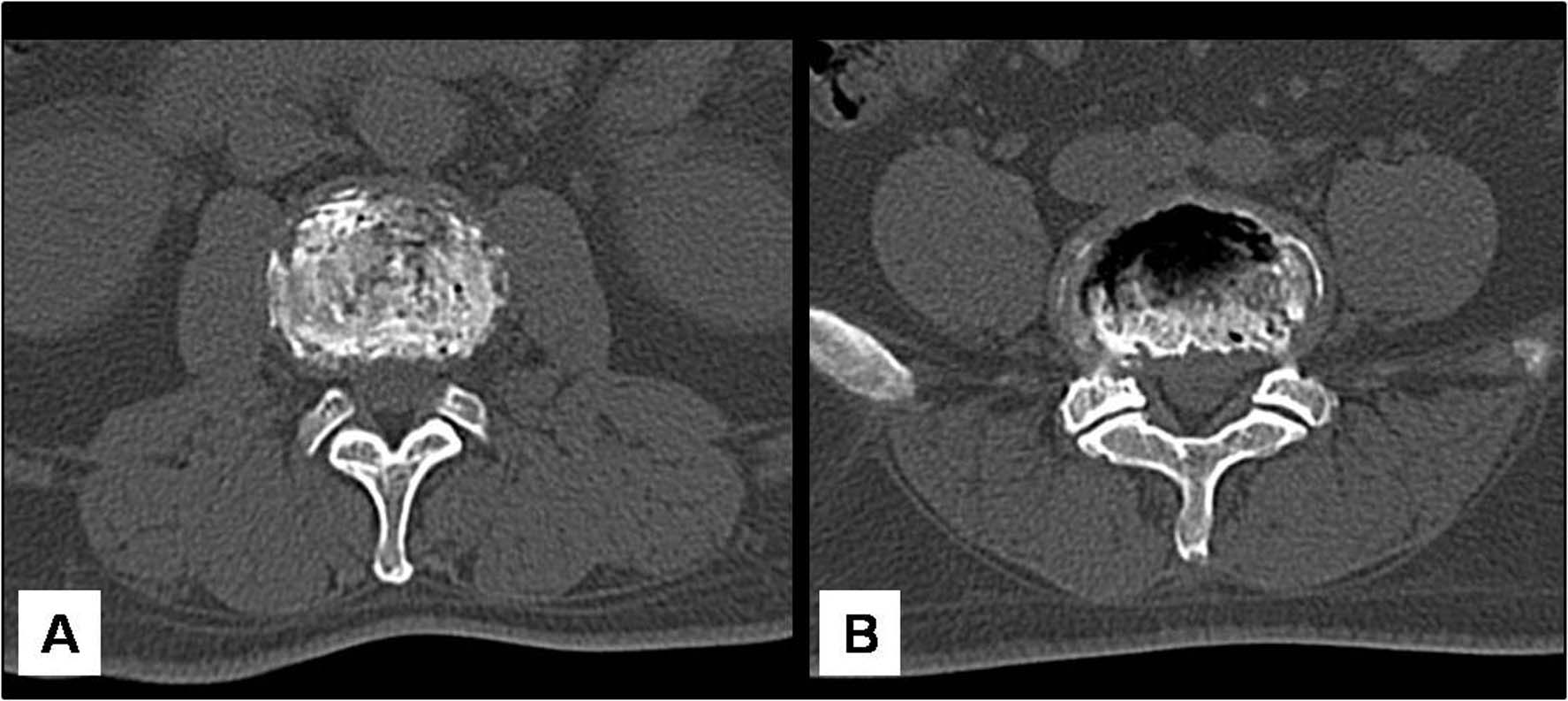
Axial CT sections at two different intervertebral disc levels showing diffuse dense disc calcification (A) and vacuum phenomenon (B).
DISCUSSION
Alkaptonuria is a rare autosomal recessive disorder with an incidence of 1 in 250000 to 1 in 1000000 live births [1]. It results from a deficiency of the enzyme homogentisate, 1,2-dioxygenase which is required for the oxidation of homogentisic acid, an intermediate product which is formed during the metabolism of tyrosine amino acid. Over time, it starts getting deposited in the connective tissues of the body in an oxidized polymerized form, as ochre-coloured pigment granules (ochronosis). A part of it gets excreted in the urine.
Alkaptonuria can be detected early in life as the dark staining of diapers. The standing urine gradually becomes black coloured due to the oxidation of homogentisic acid to a dark, pigment-like, polymeric form. The symptoms which are caused by the pigment deposition in the connective tissues of the body become apparent after adulthood. A bluish black pigmentation can be noticed over the sclera, skin, tympanic membranes, oral mucosa and the nails. Renal calculi, nephrocalcinosis and renal failure can also occur. The cardiac valves may also show pigment deposition.
Ochronotic spondyloarthropathy shows a gradually progressive course. The changes result from the deposition of the ochronotic pigment within the articular cartilages. With the loss of elasticity, the cartilages become brittle, they develop calcification and they later break down, leading to secondary degenerative changes [2]. The dorsolumbar spine is commonly affected and it shows reduced intervertebral disc spaces, a dense disc calcification (a wafer like calcification) and the vacuum phenomenon, with relatively little osteophytic changes [3]. The annulus fibrosis gets calcified first, followed by the nucleus pulposis in the late stages [2]. Osteopaenia is also noted. A spontaneous vertebral fusion with ankylosis can occur in the later stages. The hip and the knee joints show reduced joint spaces with subchondral sclerosis and cystic changes.
Clinically, the diagnosis of alkaptonuria can be made on the basis of the classical triad of arthritis, the ochronotic pigmentation and dark coloured urine. The increasing disc calcification and the associated vacuum phenomenon with minimal osteophytic changes, are the specific imaging findings of alkaptonuria [3]. This condition however, has to be differentiated from the other causes of the intervertebral disc calcification, some of which include calcium pyrophosphate dihydrate deposition disease (relatively faint, peripheral disc calcification), haemochromatosis, hyperparathyroidism (peripheral disc calcification), acromegaly (anterior disc calcification, normal or increased disc spaces and posterior scalloping of the vertebral bodies) and ankylosing spondylitis (central disc calcification, a maintained disc space, syndesmophytes and a ligamentous calcification) [4].
[1]. Phornphutkul C, Introne WJ, Perry MB, Bernardini I, Murphey MD, Fitzpatrick DL, Natural history of alkaptonuriaN Engl J Med. 2002 347:2111-21. [Google Scholar]
[2]. Laskar FH, Sargison KD, Ochronotic arthropathy: A review with four case reportsJ Bone Joint Surg Br. 1970 52(4):653-66. [Google Scholar]
[3]. Orzincolo C, Castaldi G, Scutellari PN, Cicognani P, Bariani L, Feggi L, Ochronotic arthropathy in alkaptonuria- Radiological manifestations and physiological signsRadiol Med. 1988 75:476-81. [Google Scholar]
[4]. Weinberger A, Myers AR, Intervertebral disc calcification in adults a reviewSemin Arthritis Rheum 1975 18:69-75. [Google Scholar]