A Hairy Cell Leukaemia Variant – A Rare Case Report
Pankaj Pande1, Balasaheb Ramling Yelikar2, Mahesh Kumar U3
1 Associate Professor, Department of Hematology,
2 Professor and Head , Department of Pathology,
3 Assistant Professor, Department of Pathology, BLDE University’s, Shri B.M.Patil Medical College, Bijapur, India.
NAME, ADDRESS, E-MAIL ID OF THE CORRESPONDING AUTHOR: Dr. Pankaj Pande, Associate Professor, Department of Hematology, Department of Hematology and Blood Banking.
Phone: 09739317309
E-mail: bldepathology@yahoo.com
The aim of the article is to present a rare case of Hairy cell leukaemia variant (HCl-V) which is a distinct clinico-pathological entity with intermediate features between classical HCl (HCl-C) and B-cell prolymphocytic leukaemia. It is an uncommon disorder accounting for approximately 0.4% of chronic lymphoid malignancies and 10% of all HCl cases. A 58 year old woman presented with pain abdomen and loss of weight. On examination she had massive splenomegaly. Peripheral smear was reported as chronic lymphoproliferative disorder (? Hairy cell leukemia or splenic lymphoma with villous lymphocytes). On Bone marrow examination, differential diagnosis was given as splenic lymphoma with villous lymphocytes (SLVL) and prolymphocytic variant of Hairy cell leukemia. On flow cytometric analysis, these cells were positive for CD11c, CD19, CD20, and CD22. Based on the clinical, peripheral smear, bone marrow and flow cytometry findings, a diagnosis of hairy cell leukaemia variant was confirmed. The differential diagnosis should always include SLVL, HCL-C and Japanese variant HCL because they have different clinical and biological features, particularly regarding their response to purine analogue-based treatment or splenectomy.
Flow cytometry, Hairy cell leukemia variant, Massive splenomegaly
INTRODUCTION
Hairy Cell Leukaemia (HCL) is a rare and an indolent form of a small, mature, B-cell leukaemia which is characterized by oval or indented (bean-shaped) nuclei and abundant “hairy” cytoplasm, which involves the Peripheral Blood (PB), Bone Marrow (BM) and the spleen [1]. As compared to the classical HCL, the hairy cell leukaemia variant (HCL-v) is a more aggressive disease. It is no longer considered to be biologically related to HCL and it has been included in the 2008 World Health Organization (WHO) classification as a provisional entity [2]. HCL is a rare disease which comprises about 2% of the lymphoid leukaemia in the west [1]. Its incidence is lower in Asia and it is even lower in India.
CASE PRESENTATION
A 58 year old woman presented with pain in the abdomen and loss of weight. Her physical examination revealed a moderately pale woman without jaundice and fever. The respiratory, cardiovascular and the nervous systems were within normal limits. Her abdominal examination revealed a firmly enlarged spleen which extended down to the level of the umbilicus. The liver was not palpable and no signs of chronic liver disease or ascites were present. No lymphadenopathy was noted.
PATHOLOGY
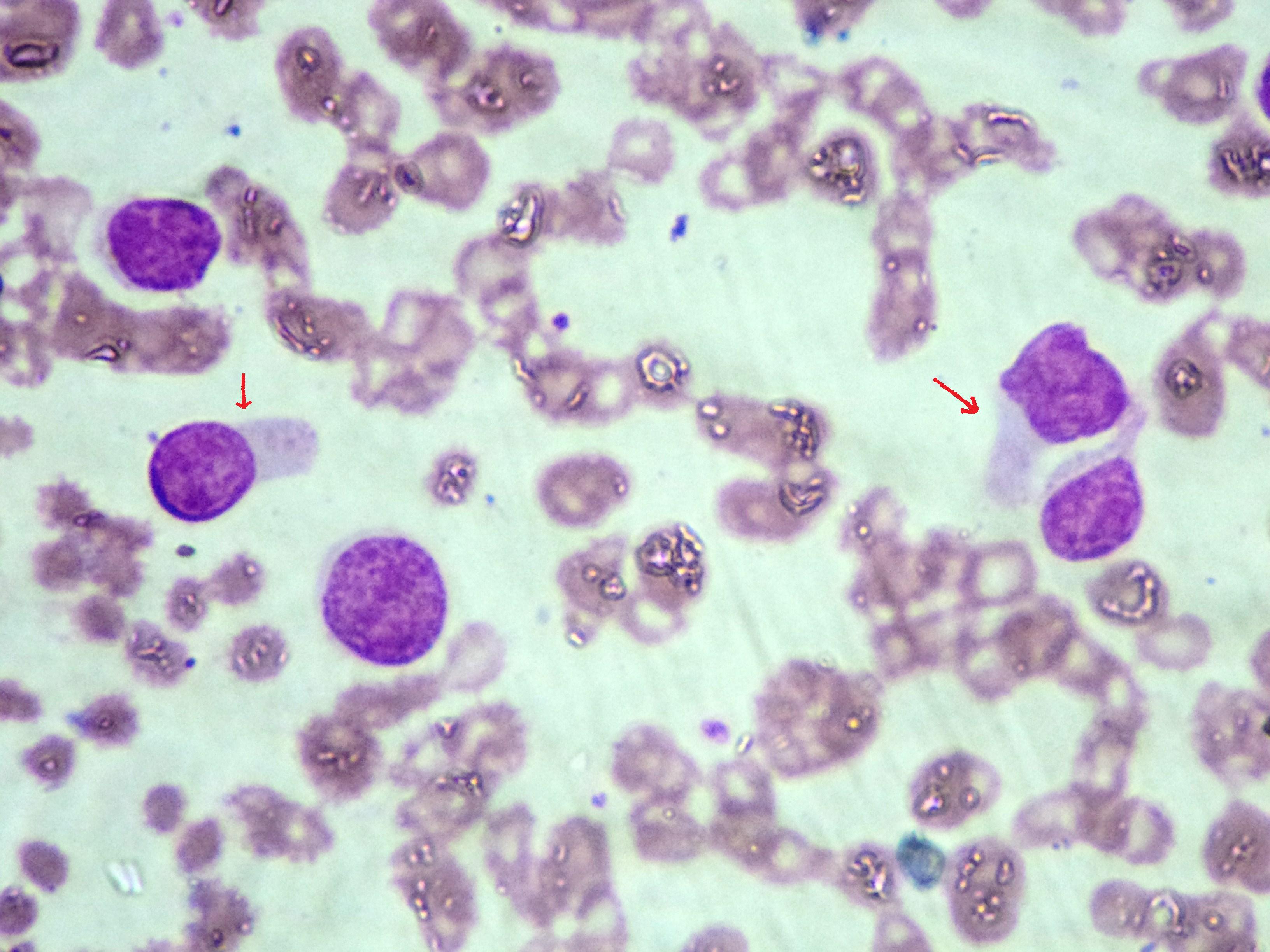
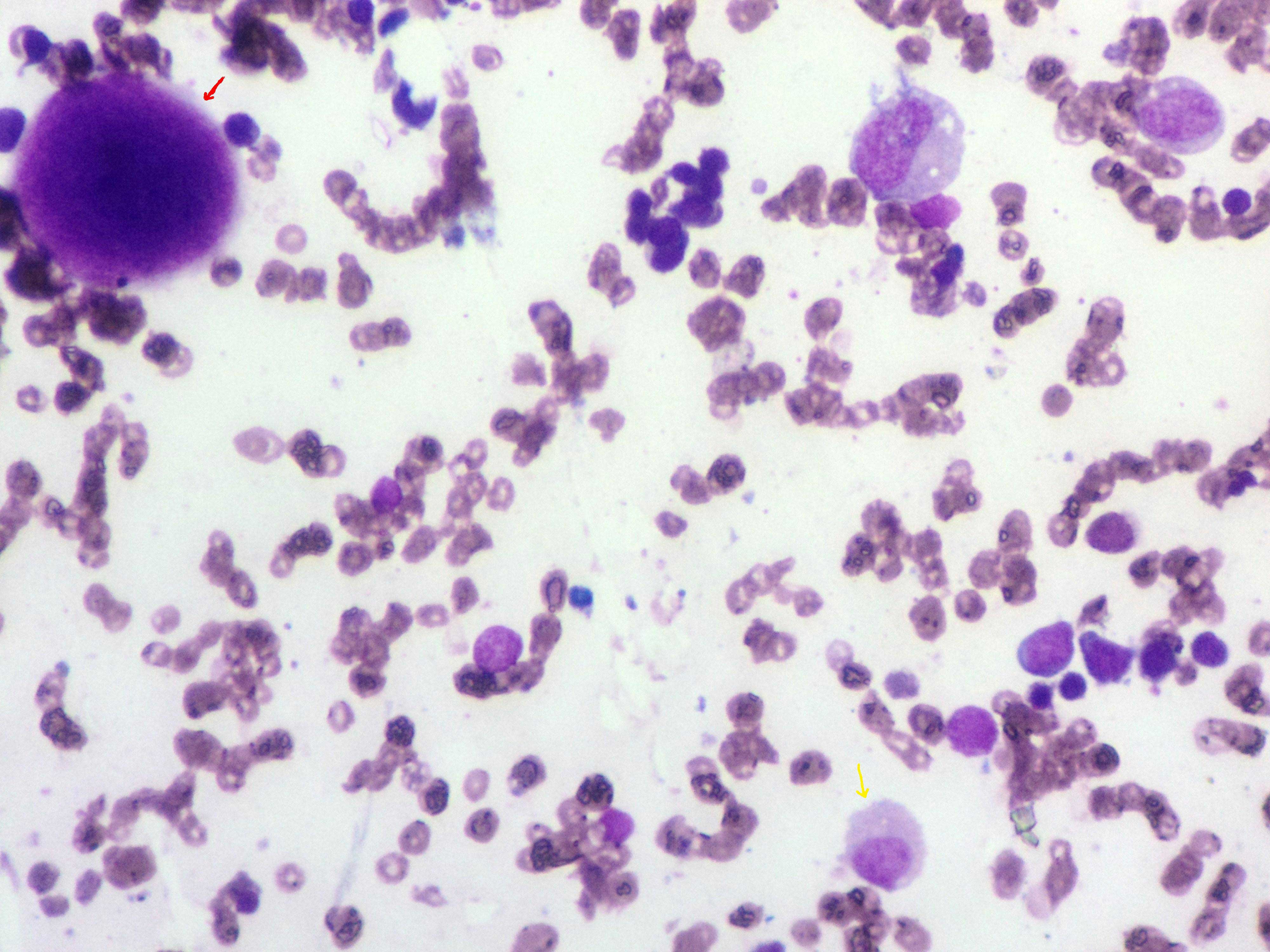
Her haemogram showed a WBC count of 26,000cells/cu mm, a haemoglobin level of 6.8gms/dl, and a platelet count of 75,000cells/cu mm. The differential count showed 19% neutrophils, 10% small lymphocytes, 3% monocytes, 2% eosinophils and 66% abnormal lymphocytes. These abnormal lymphocytes exhibited villous/hairy cytoplasmic projections and small round nuclei with condensed chromatin and a distinct nucleolus. Her peripheral smear was reported as chronic lymphoproliferative disorder (? Splenic Lymphoma with Villous Lymphocytes (SLVL)/Hairy cell leukaemia) [Table/Fig-1]. On bone marrow examination, the differential diagnosis was given as Splenic Lymphoma with Villous Lymphocytes (SLVL) and a prolymphocytic variant of Hairy cell leukaemia [Table/Fig-2].
Peripheral smear showing abnormal lymphocytes with villous/hairy cytoplasmic projections (Red arrows)
Bone marrow smear showing megakaryocyte (Red arrow) and hairy cell (Yellow arrow)
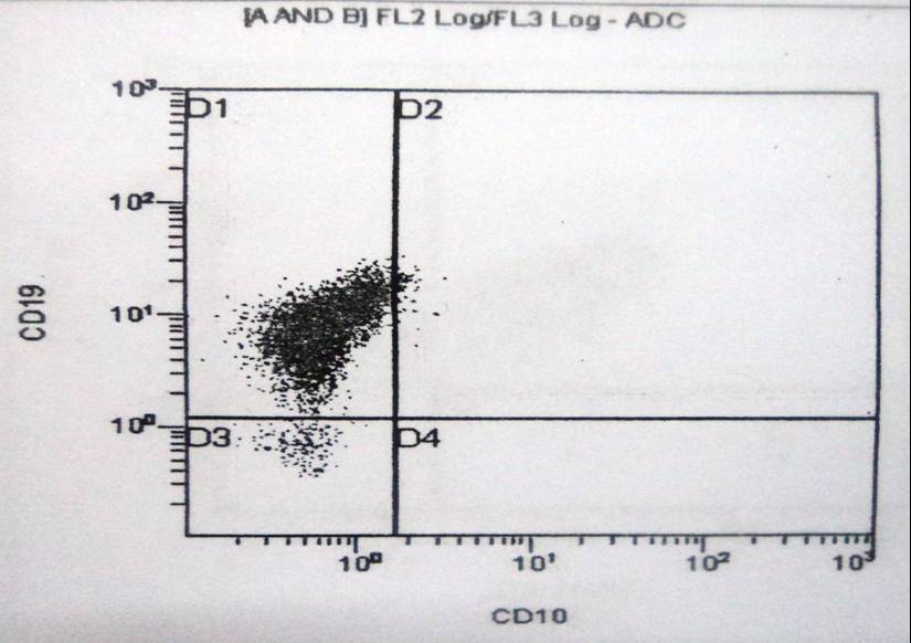
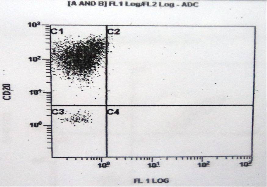
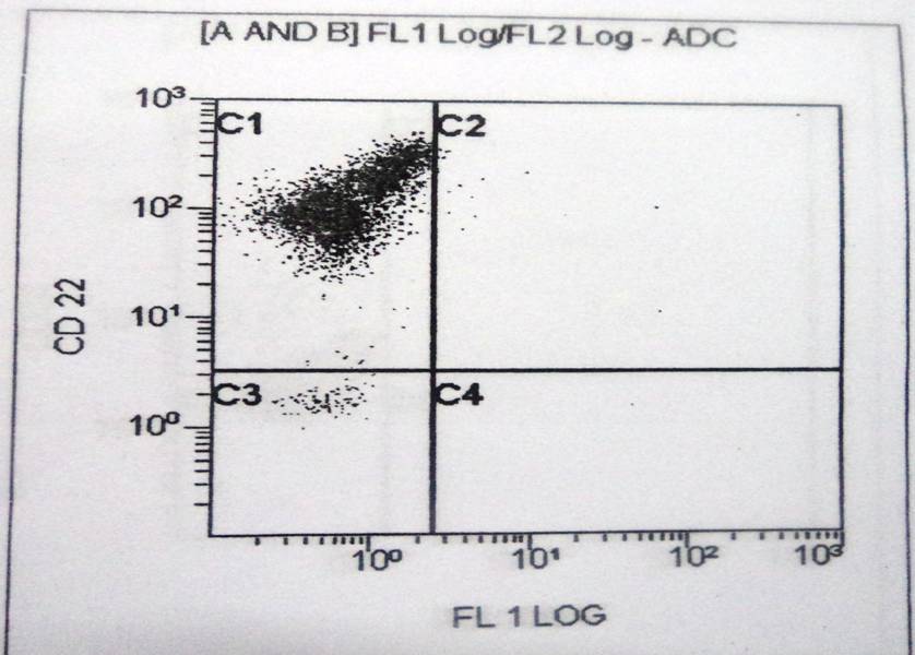
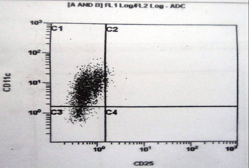
On flow cytometric analysis, these tumour cells were found to be positive for CD11c, CD19, CD20, CD22 [Table/Fig-3,4,5 and 6], FMC7, HLA-DR, SmIgD and the Kappa light chain and to be negative for CD 25.
Flow cytometric dot plots show the existence of an abnormal cells with expression of CD19
Flow cytometric dot plots show the existence of an abnormal cells with expression of CD20.
Flow cytometric dot plots show the existence of an abnormal cells with expression of CD22.
Flow cytometric dot plots show the existence of an abnormal cells with expression of CD11c.
Based on the clinical, peripheral smear, bone marrow and the flow cytometry findings, a diagnosis of a hairy cell leukaemia variant was made. The patient died within 3 months of the diagnosis because of her refusal of the treatment.
DISCUSSION
The HCL-v cases are defined as cases of small B-cell lymphoproliferative disorders which resemble classical HCL but exhibit variant features in the haemogram (leukocytosis without monocytopaenia), cytomorphology (neoplastic cells with prominent nucleoli, with blastic or convoluted nuclei and/or absence of the circumferential shaggy contours), cytochemistry (a weak or a negative reaction for TRAP), immunophenotype (absence of CD25, annexin A1 or TRAP), and resistance to the conventional HCL therapy [2,3].
The bone marrow is typically aspirable in HCl-V, HCl-JV and SLVL. In contrast to these disorders, a “dry tap” is often encountered on bone marrow aspiration in the HCl-C patients. A majority of the HCl-V patients have interstitial bone marrow infiltration, with scattered hairy cells lying within the sinusoids. However, approximately 20% of the patients have a mixed, interstitial and a nodular pattern of the bone marrow. In addition, a pure sinusoidal and an intrasinusoidal bone marrow involvement have also been occasionally reported [4].
The hairy cell leukaemia variant should be differentiated from the classical hairy cell leukaemia, the hairy cell leukaemia Japanese variant and a splenic marginal zone lymphoma [Table/Fig-7], [4].
Differences between classical hairy cell leukemia, hairy cell leukemia variant, hairy cell leukemia Japanese variant and splenic marginal zone lymphoma. 4
| Characteristics | HCL Classical | HCL Variant | HCL Japanese Variant | Splenic Marginal Zone Lymphoma |
|---|
| Median age (y) | 55 | 71 | 60 | 69 |
| WBC count | Low | High | High | High |
| Cytoplasmic projection | Uneven microvilli | Uneven microvilli | Uneven microvilli | Usually polar |
| Nuclei | Oval, indented, bilobed | Round, occasionally bilobed | Round | Round |
| Nucleoli | Indistinct | Prominent (60%) | Inconspicuous | Small (50% distinct) |
| Histology of Bone marrow | Inter sinusoidal | Interstitial, Intersinusoidal | Usually interstitial | Intersinusoidal |
| TRAP activity | Positive | Negative | Absent to week | Weak |
| Immunophenotype of leukemic cells | CD11c+,CD25+, CD103+, CD123+, CD10-/+. | CD11c +/- ,CD 19+, CD 20+, CD 22+, CD25-,CD103+/, CD123-,CD10+/- | CD11c+, CD25-, CD10- | CD11c+, CD25+/-, CD103- |
In the Asian countries, there are limited number of reports on HCL. The largest study was done in Japan, which was reported by Machii T et al., Only 9 cases were of classical HCL, 2 cases were of the HCL prolymphocytic variant and the remainder were of the so called “HCL-Japanese variant”, as they frequently presented with a high WBC count and with a distinct CD25- negative hairy morphology [5].
In an Indian series from New Delhi, 15 cases of HCL, who were aged 32-57 years (median age- 47 years) were reported [6]. The clinical presentations included splenomegaly, hepatomegaly and fatigue. The patients from India were relatively young, in contrast to the older patients of HCL in the western countries.
CONCLUSION
It is important to accurately diagnose SLVL, HCL-C and the Japanese variant of HCL entities, because they have different clinical and biological features, particularly with regards to their response to the purine analogue-based treatment or splenectomy.
[1]. Foucar K, Falini B, Catovsky D, Stein H, Hairy cell lymphoma. In: Swerdlow SH CE, Harris NL, Jaffe ES, Pileri SA, Stein H, Thiele J, Vardiman JW, ed.WHO classification of tumours of haemtopoietic and lymphoid tissues 2008 LyonIARC:188-90. [Google Scholar]
[2]. Piris M, Foucar K, Mollejo M, Campo E, Falini B, Splenic B-cell lymphoma/ leukemia, unclassifiable. In: Swerdlow SH CE, Harris NL, Jaffe ES, Pileri SA, Stein H, Thiele J, Vardiman JW, ed.WHO classification of tumours of haematopoietic and lymphoid tissues 2008 LyonIARC:191-93. [Google Scholar]
[3]. Matutes E, Wotherspoon A, Catovsky D, The variant form of hairy-cell leukaemiaBest Pract Res Clin Haematol 2003 16:41-56. [Google Scholar]
[4]. Tadeusz Robak, Hairy cell leukemia variant: Recent view on diagnosis, biology and treatmentCancer treatment reviews 2011 37:3-10. [Google Scholar]
[5]. Machii T, Tokumine Y, Inoue R, Kitani T, Predominance of a distinct subtype of hairy cell leukemia in JapanLeukemia 1993 7:181-86. [Google Scholar]
[6]. Chatterjee T, Panigrahi I, Mahapatra M, Pati HP, Kumar R, Naithani R, Hairy cell leukemia: clinical, pathological and ultrastructural findings in Asian-IndiansIndian J cancer 2008 45:41-44. [Google Scholar]