Hidrocystomas are rare cystic lesions that form benign tumours of the sweat glands. In this report, a clinical case of multiple apocrine hidrocystomas on both the upper and lower eyelids, and on both the malar regions has been discussed. These lesions are less likely than the eccrine lesions to occur at the periorbital region.
Eyelids, Malar area, Apocrine hidrocystoma
INTRODUCTION
Hidrocystomas are rare, benign, cystic lesions of the skin – they can be either eccrine or apocrine and are often found on the head, neck and the trunk regions [1]. These tumours have also been reported to occur on the penis, axillae, and in the anal region [2].
Apocrine hidrocystomas are often regarded as adenomas, because the secretory cells are not flattened and they have papillary projections which extend into the lumen [3]. They are usually solitary and are found mostly on the head and neck and along the eyelid margins, near the inner canthus. The apocrine lesions are less likely than the eccrine lesions to occur at the periorbital regions [4].
This case report deals with such a rare tumour which presented as multiple cystic lesions on both the upper and the lower eyelids and on both the malar areas.
CASE REPORT
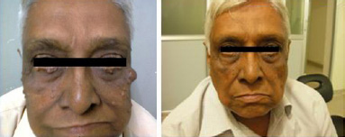
A 75 year old male patient presented with multiple, small, translucent, cystic papules which were filled with a watery fluid, which had been present since five years and he was asymptomatic. On local examination, the lesions were observed on both the upper and lower eyelids and on both the malar areas. The largest cyst measured 2x2cms [Table/Fig-1]. A clinical diagnosis of hidrocystoma was offered. The patient underwent, under local anaesthesia, an excisional biopsy of the mass and the specimen was subjected to a histopathological examination.
Photos (A) showing multiple hidrocystoma lesions on eyelids, malar area & ear lobe.
Photo (B) showing lesions resolved near the eyelids and malar area with good cosmetic appearance 15 days post operatively
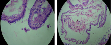
Pathological Findings: The gross examination of this specimen showed a grey white cyst which measured 1.5x1cms. The histopathologic examination of the paraffin sections which were stained with haematoxylin-eosin, revealed the presence of a cyst in the dermis, which was lined by an inner layer of a secretory columnar epithelium, which lay above an outer myoepithelial cell layer. At places, the secretory epithelium showed decapitation [Table/Fig-2], which suggested an apocrine hidrocystoma.
Histopathology
Cyst in the dermis lined by an inner layer of secretory columnar epithelium, which lies above an outer myoepithelial cell layer. At places, secretory epithelium shows decapitation, thus suggesting apocrine hidrocystoma
DISCUSSION
The skin adnexal neoplasms exhibit a morphological differentiation towards one or more types of adnexal structures which are found in the normal skin (the pilosebaceous units, eccrine glands, and the apocrine glands) and they comprise a wide spectrum of benign and malignant tumours [5]. The apocrine glands are most frequently present in the axillae, the groin, the external auditory canal, the eyelids and on the nipple. The apocrine hidrocystomas are benign adenomatous cystic proliferations which are derived from the apocrine sweat glands, which often occur as solitary translucent cystic lesions, most commonly on the head and neck, especially in the periocular tissues [3].
The exact stimulus for the development of an apocrine hidrocystoma is unknown. The occlusion or blockage of the sweat duct apparatus, which results in the retention of sweat, and a dilated cystic structure, are considered to be plausible causes. They are believed to be adenomatous cystic proliferations of the apocrine glands [6].
The literature suggests that apocrine hidrocystomas are relatively common in the United States. There is no predilection for race, sex or geographic region for the apocrine hidrocystomas. They occur in adulthood, although there is no particular age group in which they occur. They are entirely benign and they seldom recur after their removal. The cysts may annoy the patients; however, the symptoms usually are mild or absent. The vision is usually not affected.
They are prevalent in adults who are between 30 to 70 years of age, with a diameter of 3-15mm [1,4]. They can appear as single or multiple cystic lesions. Although a significant number of cases of multiple apocrine hidrocystomas have been reported in the literature, the multiple types are rare in the general population [4].
The presence of multiple hidrocystomas may be a marker of two rare inherited disorders – the Schopf-Schulz-Passarge syndrome and the Goltz-Gorlin syndrome. The Schopf-Schulz-Passarge syndrome is a rare autosomal recessive form of ectodermal dysplasia which is characterized by hypodontia, hypoplastic nails, hypotrichosis, palmoplantar keratoderma, cysts of the eyelid margins and multiple periocular hidrocystomas. The Goltz-Gorlin syndrome is sporadic in nature in most of the cases, but it is assumed to have an X-linked dominance mode of inheritance, as it occurs more commonly in women. It is characterized by its broad spectrum of meso-ectodermal defects which are associated with the skin as well as the eyes, the skeletal system and the teeth. The reported cutaneous associations include linear or reticulated atrophic hypopigmented or hyperpigmented skin lesions, papillomas and periocular multiple hidrocystomas [3,4]. This patient did not have any features which were suggestive of either disorder.
The lesion edges are not well delineated but they blend gradually into the adjacent skin. The walls, although they are translucent, are sufficiently thick that they seldom rupture spontaneously. The cysts are mobile with palpation and they transilluminate.
Clinically, other cystic lesions such as epidermal inclusion cysts, comedones, mucoid cysts, haemangiomas, lymphangiomas, and steatocystoma multiplexes are considered as the differential diagnoses. But all these lesions differ from the apocrine hidrocystoma histologically [1,4]. Very often, the apocrine hidrocystomas present as blue-black coloured nodules and hence, their differentiation from melanomas and basal cell carcinomas is important [1].
The solitary lesions are usually treated by a simple excision. Multiple apocrine hidrocystomas are rare, and a variety of therapeutic techniques have been used. Recent reports have included case studies in which trichloracetic acid, carbon-dioxide lasers, and 1450-nm diode lasers have been employed. Electrosurgery and excisions have also been used. The additional therapies to consider are those which are used for multiple eccrine hidrocystomas, which include the botulinum toxin, atropine and pulsed-dye lasers, and most recently, the 595 nm long-pulsed lasers [7]. The apocrine hidrocystomas grow gradually and they persist indefinitely after attaining their full sizes. They seldom recur after their removal [6].
In conclusion, although multiple apocrine hidrocystomas are rare and typically asymptomatic, they are often of interest, since they closely resemble certain more serious skin disorders such as malignant melanomas and basal cell carcinomas and also, other benign cystic lesions have to be ruled out.
[1]. Shegal S, Agarwal R, Singh S, Goyal P, Fine-needle aspiration cytology of eccrine hidrocystomaCyto Journal February 2012 [Google Scholar]
[2]. Ter Poorten HJ, Apocrine hidrocystoma of the right scapulaArch Dermatol. Dec 1977 113(12):1730[Medline] [Google Scholar]
[3]. Obaidat NA, Ghzarian DM, Bilateral multiple axillary apocrine hidrocystomas associated with benign apocrine hyperplasiaJournal of Clinical Pathology July 2006 59(7):779 [Google Scholar]
[4]. Sarabi K, Khachemoune A, Histiocytomas – A Brief ReviewMedscape General Medicine September 2006 8(3):57 [Google Scholar]
[5]. Ionnidis DG, Drivas EI, Papadakis CE, Feritsian A, Bizakis JG, Skoulakis CE, Histiocytomas of the external auditory canal: a case reportCases Journal January 2009 2:79 [Google Scholar]
[6]. Rapini RP, Apocrine Hidrocystomahttp://www.emedicine.com/derm/topic35.htm [Google Scholar]
[7]. Anandasabapathy N, Multiple apocrine hidrocystomasDermatology Online Journal14(5):12 [Google Scholar]